Redlich–Obersteiner's zone

Last updated

The Redlich–Obersteiner's zone, also known as the root entry zone, is a boundary between the central nervous system (CNS) and the peripheral nervous system (PNS). The Redlich–Obersteiner's zone is located at the point of entry of either between cranial nerves and the brain or spinal nerves and the spinal cord. This narrow zone is identified visually where there is a transition regarding myelin production. It is named after Emil Redlich and Heinrich Obersteiner.

Central nervous system part of the nervous system consisting of the brain and spinal cord

The central nervous system (CNS) is the part of the nervous system consisting of the brain and spinal cord. The CNS is so named because it integrates the received information and coordinates and influences the activity of all parts of the bodies of bilaterally symmetric animals—that is, all multicellular animals except sponges and radially symmetric animals such as jellyfish—and it contains the majority of the nervous system. Many consider the retina and the optic nerve, as well as the olfactory nerves and olfactory epithelium as parts of the CNS, synapsing directly on brain tissue without intermediate ganglia. As such, the olfactory epithelium is the only central nervous tissue in direct contact with the environment, which opens up for therapeutic treatments. The CNS is contained within the dorsal body cavity, with the brain housed in the cranial cavity and the spinal cord in the spinal canal. In vertebrates, the brain is protected by the skull, while the spinal cord is protected by the vertebrae. The brain and spinal cord are both enclosed in the meninges. Within the CNS, the interneuronal space is filled with a large amount of supporting non-nervous cells called neuroglial cells.

Peripheral nervous system part of the nervous system

The peripheral nervous system (PNS) is one of two components that make up the nervous system of bilateral animals, with the other part being the central nervous system (CNS). The PNS consists of the nerves and ganglia outside the brain and spinal cord. The main function of the PNS is to connect the CNS to the limbs and organs, essentially serving as a relay between the brain and spinal cord and the rest of the body. Unlike the CNS, the PNS is not protected by the vertebral column and skull, or by the blood–brain barrier, which leaves it exposed to toxins and mechanical injuries.

Spinal cord long, thin, tubular bundle of nervous tissue and support cells that extends from the brain

The spinal cord is a long, thin, tubular structure made up of nervous tissue, which extends from the medulla oblongata in the brainstem to the lumbar region of the vertebral column. It encloses the central canal of the spinal cord, which contains cerebrospinal fluid. The brain and spinal cord together make up the central nervous system (CNS). In humans, the spinal cord begins at the occipital bone, passing through the foramen magnum and entering the spinal canal at the beginning of the cervical vertebrae. The spinal cord extends down to between the first and second lumbar vertebrae, where it ends. The enclosing bony vertebral column protects the relatively shorter spinal cord. It is around 45 cm (18 in) in men and around 43 cm (17 in) long in women. The diameter of the spinal cord ranges from 13 mm in the cervical and lumbar regions to 6.4 mm in the thoracic area.

As stated above, the Redlich–Obersteiner's zone separates the CNS and PNS. Thus at this point, there is a transition from Schwann cell myelin meet to oligodendrocyte myelin. Of note, in cranial nerves this is often the place of neuro-vascular compression syndromes such as trigeminal neuralgia.

Schwann cell

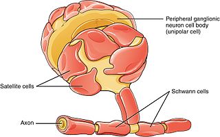
Schwann cells or neurolemmocytes are the principal glia of the peripheral nervous system (PNS). Glial cells function to support neurons and in the PNS, also include satellite cells, olfactory ensheathing cells, enteric glia and glia that reside at sensory nerve endings, such as the Pacinian corpuscle. The two types of Schwann cells are myelinating and nonmyelinating. Myelinating Schwann cells wrap around axons of motor and sensory neurons to form the myelin sheath. The Schwann cell promoter is present in the downstream region of the human dystrophin gene that gives shortened transcript that are again synthesized in a tissue-specific manner.

Oligodendrocyte neural cell type

Oligodendrocytes, or oligodendroglia, are a type of neuroglia whose main functions are to provide support and insulation to axons in the central nervous system of some vertebrates, equivalent to the function performed by Schwann cells in the peripheral nervous system. Oligodendrocytes do this by creating the myelin sheath, which is 80% lipid and 20% protein. A single oligodendrocyte can extend its processes to 50 axons, wrapping approximately 1 μm of myelin sheath around each axon; Schwann cells, on the other hand, can wrap around only one axon. Each oligodendrocyte forms one segment of myelin for several adjacent axons.

Related Research Articles

Ganglion nerve cell cluster or a group of nerve cell bodies located in the autonomic nervous system and sensory system; ganglia house the cells bodies of afferent nerves and efferent nerves

A ganglion is a nerve cell cluster or a group of nerve cell bodies located in the autonomic nervous system and sensory system. Ganglia house the cell bodies of afferent nerves and efferent nerves, or axons.

Myelin fatty white substance, multi-layered membrane, unique to the nervous system, that functions as an insulator to greatly increase the velocity of axonal impulse conduction; it is an outgrowth of a type of glial cell

Myelin is a lipid-rich (fatty) substance formed in the central nervous system (CNS) by glial cells called oligodendrocytes, and in the peripheral nervous system (PNS) by Schwann cells. Myelin insulates nerve cell axons to increase the speed at which information travels from one nerve cell body to another or, for example, from a nerve cell body to a muscle. The myelinated axon can be likened to an electrical wire with insulating material (myelin) around it. However, unlike the plastic covering on an electrical wire, myelin does not form a single long sheath over the entire length of the axon. Rather, each myelin sheath insulates the axon over a single section and, in general, each axon comprises multiple long myelinated sections separated from each other by short gaps called Nodes of Ranvier. Each myelin sheath is formed by the concentric wrapping of an oligodendrocyte or Schwann cell process around the axon.

Nerve enclosed, cable-like bundle of axons in the peripheral nervous system

A nerve is an enclosed, cable-like bundle of nerve fibres called axons, in the peripheral nervous system. A nerve provides a common pathway for the electrochemical nerve impulses called action potentials that are transmitted along each of the axons to peripheral organs or, in the case of sensory nerves, from the periphery back to the central nervous system. Each axon within the nerve is an extension of an individual neuron, along with other supportive cells such as Schwann cells that coat the axons in myelin.

Nervous system Control the whole body function

The nervous system is the part of an animal that coordinates its actions by transmitting signals to and from different parts of its body. The nervous system detects environmental changes that impact the body, then works in tandem with the endocrine system to respond to such events. Nervous tissue first arose in wormlike organisms about 550 to 600 million years ago. In vertebrates it consists of two main parts, the central nervous system (CNS) and the peripheral nervous system (PNS). The CNS consists of the brain and spinal cord. The PNS consists mainly of nerves, which are enclosed bundles of the long fibers or axons, that connect the CNS to every other part of the body. Nerves that transmit signals from the brain are called motor or efferent nerves, while those nerves that transmit information from the body to the CNS are called sensory or afferent. Spinal nerves serve both functions and are called mixed nerves. The PNS is divided into three separate subsystems, the somatic, autonomic, and enteric nervous systems. Somatic nerves mediate voluntary movement. The autonomic nervous system is further subdivided into the sympathetic and the parasympathetic nervous systems. The sympathetic nervous system is activated in cases of emergencies to mobilize energy, while the parasympathetic nervous system is activated when organisms are in a relaxed state. The enteric nervous system functions to control the gastrointestinal system. Both autonomic and enteric nervous systems function involuntarily. Nerves that exit from the cranium are called cranial nerves while those exiting from the spinal cord are called spinal nerves.

Parasympathetic nervous system one of the three divisions of the autonomic nervous system, the others being the sympathetic nervous system and enteric nervous system.

The parasympathetic nervous system (PSNS) is one of the two divisions of the autonomic nervous system, the other being the sympathetic nervous system. The autonomic nervous system is responsible for regulating the body's unconscious actions. The parasympathetic system is responsible for stimulation of "rest-and-digest" or "feed and breed" activities that occur when the body is at rest, especially after eating, including sexual arousal, salivation, lacrimation (tears), urination, digestion and defecation. Its action is described as being complementary to that of the sympathetic nervous system, which is responsible for stimulating activities associated with the fight-or-flight response.

Nervous tissue main component of the nervous system

Nervous tissue, also called neural tissue or nerve tissue, is the main tissue component of the nervous system. The nervous system regulates and controls bodily functions and activity and consists of two parts: the central nervous system (CNS) comprising the brain and spinal cord, and the peripheral nervous system (PNS) comprising the branching peripheral nerves. It is composed of neurons, or nerve cells, which receive and transmit impulses, and neuroglia, also known as glial cells or glia, which assist the propagation of the nerve impulse as well as provide nutrients to the neurons.

Somatic nervous system

The somatic nervous system is the part of the peripheral nervous system associated with the voluntary control of body movements via skeletal muscles.

Afferent nerve fiber

Afferent nerve fibers refer to axonal projections that arrive at a particular brain region ; as opposed to efferent projections that exit the region. These terms have a slightly different meaning in the context of the peripheral nervous system (PNS) and central nervous system (CNS).

Wallerian degeneration

Wallerian degeneration is an active process of degeneration that results when a nerve fiber is cut or crushed and the part of the axon distal to the injury degenerates. A related process of dying back or retrograde degeneration known as 'Wallerian-like degeneration' occurs in many neurodegenerative diseases, especially those where axonal transport is impaired. Primary culture studies suggest that a failure to deliver sufficient quantities of the essential axonal protein NMNAT2 is a key initiating event.

A sensory nerve, also called an afferent nerve, is a nerve that carries sensory information toward the central nervous system (CNS). It is a cable-like bundle of the afferent nerve fibers coming from sensory receptors in the peripheral nervous system (PNS). A motor nerve carries information from the CNS to the PNS, and both types of nerve are called peripheral nerves.

Alpha motor neuron

Alpha (α) motor neurons (also called alpha motoneurons), are large, multipolar lower motor neurons of the brainstem and spinal cord. They innervate extrafusal muscle fibers of skeletal muscle and are directly responsible for initiating their contraction. Alpha motor neurons are distinct from gamma motor neurons, which innervate intrafusal muscle fibers of muscle spindles.

Head and neck anatomy

This article describes the anatomy of the head and neck of the human body, including the brain, bones, muscles, blood vessels, nerves, glands, nose, mouth, teeth, tongue, and throat.

Endoneurium

The endoneurium is a layer of delicate connective tissue around the myelin sheath of each myelinated nerve fiber in the peripheral nervous system. Its component cells are called endoneurial cells. The endoneuria with their enclosed nerve fibers are bundled into groups called nerve fascicles, each fascicle within its own protective sheath called a perineurium. In sufficiently large nerves multiple fascicles, each with its blood supply and fatty tissue, may be bundled within yet another sheath, the epineurium.

Emil Redlich Austrian neurologist

Emil Redlich was an Austrian-Jewish neurologist born in Brünn.

Neuroregeneration refers to the regrowth or repair of nervous tissues, cells or cell products. Such mechanisms may include generation of new neurons, glia, axons, myelin, or synapses. Neuroregeneration differs between the peripheral nervous system (PNS) and the central nervous system (CNS) by the functional mechanisms and especially the extent and speed. When an axon is damaged, the distal segment undergoes Wallerian degeneration, losing its myelin sheath. The proximal segment can either die by apoptosis or undergo the chromatolytic reaction, which is an attempt at repair. In the CNS, synaptic stripping occurs as glial foot processes invade the dead synapse.

Outline of the human nervous system Overview of and topical guide to the human nervous system

The following outline is provided as an overview of and topical guide to the human nervous system: