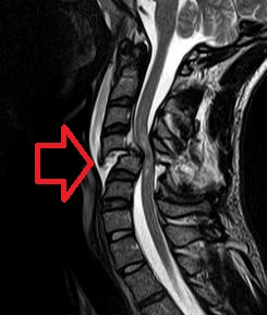
The human leg, in the general meaning, is the entire lower limb of the human body, including the foot, thigh and even the hip or gluteal region. However, the definition in human anatomy refers only to the section of the lower limb extending from the knee to the ankle, also known as the crus.
Legs are used for standing, and all forms of locomotion including recreational such as dancing, and constitute a significant portion of a person's mass. Female legs generally have greater hip anteversion and tibiofemoral angles, but shorter femur and tibial lengths than those in males.

Coxa vara is a deformity of the hip, whereby the angle between the head and the shaft of the femur is reduced to less than 120 degrees. This results in the leg being shortened, and the development of a limp. It is commonly caused by injury, such as a fracture. It can also occur when the bone tissue in the neck of the femur is softer than normal, causing it to bend under the weight of the body. This may either be congenital or the result of a bone disorder. The most common cause of coxa vara is either congenital or developmental. Other common causes include metabolic bone diseases, post-Perthes deformity, osteomyelitis, and post traumatic. Shepherd's Crook deformity is a severe form of coxa vara where the proximal femur is severely deformed with a reduction in the neck shaft angle beyond 90 degrees. It is most commonly a sequela of osteogenesis imperfecta, Pagets disease, osteomyelitis, tumour and tumour-like conditions.

The gluteus minimus, the smallest of the three gluteal muscles, is situated immediately beneath the gluteus medius.
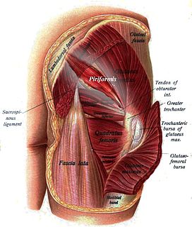
The piriformis is a muscle in the gluteal region of the lower limbs. It is one of the six muscles in the lateral rotator group.

Hip replacement is a surgical procedure in which the hip joint is replaced by a prosthetic implant, that is, a hip prosthesis. Hip replacement surgery can be performed as a total replacement or a hemi (half) replacement. Such joint replacement orthopaedic surgery is generally conducted to relieve arthritis pain or in some hip fractures. A total hip replacement consists of replacing both the acetabulum and the femoral head while hemiarthroplasty generally only replaces the femoral head. Hip replacement is currently one of the most common orthopaedic operations, though patient satisfaction short- and long-term varies widely. Approximately 58% of total hip replacements are estimated to last 25 years. The average cost of a total hip replacement in 2012 was $40,364 in the United States, and about $7,700 to $12,000 in most European countries.

Trendelenburg's sign is found in people with weak or paralyzed abductor muscles of the hip, namely gluteus medius and gluteus minimus. It is named after the German surgeon Friedrich Trendelenburg.

Patrick's test or FABER test is performed to evaluate pathology of the hip joint or the sacroiliac joint.

A hip dislocation a disruption of the joint between the femur and pelvis. Specifically it is when the ball–shaped head of the femur comes out of the cup–shaped acetabulum of the pelvis. Symptoms typically include pain and an inability move the hip. Complications may include avascular necrosis of the hip, injury to the sciatic nerve, or arthritis.
The Trendelenburg gait is an abnormal gait caused by weakness of the abductor muscles of the lower limb, gluteus medius and gluteus minimus. People with a lesion of superior gluteal nerve have weakness of abducting the thigh at the hip.

In human anatomy, the muscles of the hip joint are those muscles that cause movement in the hip. Most modern anatomists define 17 of these muscles, although some additional muscles may sometimes be considered. These are often divided into four groups according to their orientation around the hip joint: the gluteal group; the lateral rotator group; the adductor group; and the iliopsoas group.

Motion, the process of movement, is described using specific anatomical terms. Motion includes movement of organs, joints, limbs, and specific sections of the body. The terminology used describes this motion according to its direction relative to the anatomical position of the joints. Anatomists use a unified set of terms to describe most of the movements, although other, more specialized terms are necessary for describing the uniqueness of the movements such as those of the hands, feet, and eyes.
In medicine, physiotherapy, chiropractic, and osteopathy the hip examination, or hip exam, is undertaken when a patient has a complaint of hip pain and/or signs and/or symptoms suggestive of hip joint pathology. It is a physical examination maneuver.

The acetabular labrum is a ring of cartilage that surrounds the acetabulum of the hip. The anterior portion is most vulnerable when the labrum tears.
Greater trochanteric pain syndrome (GTPS), also known as trochanteric bursitis, is inflammation of the trochanteric bursa, a part of the hip.
The Ortolani test is part of the physical examination for developmental dysplasia of the hip, along with the Barlow maneuver. Specifically, the Ortolani test is positive when a posterior dislocation of the hip is reducible with this maneuver. This is part of the standard infant exam performed preferably in early infancy.
Scissor gait is a form of gait abnormality primarily associated with spastic cerebral palsy. That condition and others like it are associated with an upper motor neuron lesion.
Hoover’s sign of leg paresis is one of two signs named for Charles Franklin Hoover. It is a maneuver aimed to separate organic from non-organic paresis of the leg. The sign relies on the principle of synergistic contraction.
The Thomas test is a physical examination test, named after Dr. Hugh Owen Thomas (1834–1891), a British orthopaedic surgeon, used to rule out hip flexion contracture and psoas syndrome. Often associated with runners, dancers, and gymnasts who complain of hip "stiffness" and reported "snapping" feeling when flexing at the waist.
Ober's test is used in physical examination to identify tightness of the iliotibial band .
During the test, the patient lies on his/her side with the unaffected leg on the bottom with their shoulder and pelvis in line. The lower hip and knee can be in a flexed position to take out any lordosis of the lumbar spine.