Anterior canal may refer to:
| This disambiguation page lists articles associated with the title Anterior canal. If an internal link led you here, you may wish to change the link to point directly to the intended article. |
Anterior canal may refer to:
| This disambiguation page lists articles associated with the title Anterior canal. If an internal link led you here, you may wish to change the link to point directly to the intended article. |

The outer ear, external ear, or auris externa is the external portion of the ear, which consists of the auricle and the ear canal . It gathers sound energy and focuses it on the eardrum.

The inguinal canals are the two passages in the anterior abdominal wall which in males convey the spermatic cords and in females the round ligament of the uterus. The inguinal canals are larger and more prominent in males. There is one inguinal canal on each side of the midline.

The internal carotid artery is located in the inner side of the neck in contrast to the external carotid artery. In human anatomy, they arise from the common carotid arteries where these bifurcate into the internal and external carotid arteries at cervical vertebral level 3 or 4; the internal carotid artery supplies the brain including eyes, while the external carotid nourishes other portions of the head, such as face, scalp, skull, and meninges.

The aqueous humour is a transparent, watery fluid similar to plasma, but containing low protein concentrations. It is secreted from the ciliary epithelium, a structure supporting the lens. It fills both the anterior and the posterior chambers of the eye, and is not to be confused with the vitreous humour, which is located in the space between the lens and the retina, also known as the posterior cavity or vitreous chamber.

The spinal cavity is the cavity that contains the spinal cord within the vertebral column, formed by the vertebrae through which the spinal cord passes. It is a process of the dorsal body cavity. This canal is enclosed within the vertebral foramen of the vertebrae. In the intervertebral spaces, the canal is protected by the ligamentum flavum posteriorly and the posterior longitudinal ligament anteriorly.

The tympanic cavity is a small cavity surrounding the bones of the middle ear. Within it sit the ossicles, three small bones that transmit vibrations used in the detection of sound.

The internal auditory meatus is a canal within the petrous part of the temporal bone of the skull between the posterior cranial fossa and the inner ear.

The anterior ethmoidal artery, also anterior ethmoid artery is an artery of the head.

The adductor canal is an aponeurotic tunnel in the middle third of the thigh, extending from the apex of the femoral triangle to the opening in the adductor magnus, the adductor hiatus.
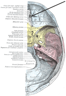
The anterior ethmoidal foramen is a small opening in the ethmoid bone in the skull.

The optic foramen is the opening to the optic canal. The canal is located in the sphenoid bone; it is bounded medially by the body of the sphenoid and laterally by the lesser wing of the sphenoid.

The petrous part of the temporal bone is pyramid-shaped and is wedged in at the base of the skull between the sphenoid and occipital bones. Directed medially, forward, and a little upward, it presents a base, an apex, three surfaces, and three angles, and houses in its interior, the components of the inner ear. The petrous portion is among the most basal elements of the skull and forms part of the endocranium. Petrous comes from the Latin word petrosus, meaning "stone-like, hard". It is one of the densest bones in the body.
The tympanic part of the temporal bone is a curved plate of bone lying below the squamous part of the temporal bone, in front of the mastoid process, and surrounding the external part of the ear canal.

The middle cranial fossa, deeper than the anterior cranial fossa, is narrow medially and widens laterally to the sides of the skull. It is separated from the posterior fossa by the clivus and the petrous crest.

The pterygoid canal is a passage in the sphenoid bone of the skull leading from just anterior to the foramen lacerum in the middle cranial fossa to the pterygopalatine fossa.

In human anatomy of the mouth, the palatine process of maxilla, is a thick, horizontal process of the maxilla. It forms the anterior three quarters of the hard palate, the horizontal plate of the palatine bone making up the rest.

The cervical canal is the spindle-shaped, flattened canal of the cervix, the neck of the uterus.

The siphonal canal is an anatomical feature of the shells of certain groups of sea snails within the clade Neogastropoda. Some sea marine gastropods have a soft tubular anterior extension of the mantle called a siphon through which water is drawn into the mantle cavity and over the gill and which serves as a chemoreceptor to locate food. In certain groups of carnivorous snails, where the siphon is particularly long, the structure of the shell has been modified in order to house and protect the soft structure of the siphon. Thus the siphonal canal is a semi-tubular extension of the aperture of the shell through which the siphon is extended when the animal is active.

The mandibular incisive canal is a bony canal within the anterior mandible that runs bilaterally from the mental foramina usually to the region of the ipsilateral lateral incisor teeth. After branching into the mental nerve that exits the foramen of the same name, the inferior alveolar nerve continues anteriorly within the mandibular incisive canal as the incisive nerve, providing innervation to the mandibular first premolar, canine and lateral and central incisors. The mandibular incisive nerve either terminates as nerve endings within the anterior teeth or adjacent bone, or may join nerve endings that enter through the tiny lingual foramen.
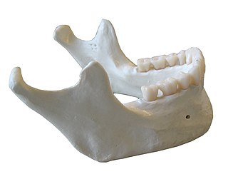
The mandible, lower jaw or jawbone is the largest, strongest and lowest bone in the human face. It forms the lower jaw and holds the lower teeth in place. The mandible sits beneath the maxilla. It is the only movable bone of the skull.