Appendix (anatomy)

Last updated
Appendix
Tractus intestinalis appendix vermiformis.svg
Appendix with surrounding structures
Appendix locations.svg
Variations of the appendix
Details
Precursor Midgut
System Digestive system
Artery Appendicular artery
Vein Appendicular vein
Identifiers
MeSH D001065
TA98 A05.7.02.007
TA2 2976
FMA 14542
Anatomical terminology

The appendix (pl.: appendices or appendixes; also vermiform appendix; cecal (or caecal) appendix; vermix; or vermiform process) is a finger-like, blind-ended tube connected to the cecum, from which it develops in the embryo. The cecum is a pouch-like structure of the large intestine, located at the junction of the small and the large intestines. The term "vermiform" comes from Latin and means "worm-shaped". The appendix was once considered a vestigial organ, but this view has changed since the early 2000s. [1] [2] Research suggests that the appendix may serve an important purpose as a reservoir for beneficial gut bacteria.

Contents

Structure

The human appendix averages 9 cm (3.5 in) in length, ranging from 5 to 35 cm (2.0 to 13.8 in). The diameter of the appendix is 6 mm (0.24 in), and more than 6 mm (0.24 in) is considered a thickened or inflamed appendix. The longest appendix ever removed was 26 cm (10 in) long. [3] The appendix is usually located in the lower right quadrant of the abdomen, near the right hip bone. The base of the appendix is located 2 cm (0.79 in) beneath the ileocecal valve that separates the large intestine from the small intestine. Its position within the abdomen corresponds to a point on the surface known as McBurney's point.

The appendix is connected to the mesentery in the lower region of the ileum, by a short region of the mesocolon known as the mesoappendix. [4]

Variation

Some identical twins—known as mirror image twins—can have a mirror-imaged anatomy, a congenital condition with the appendix located in the lower left quadrant of the abdomen instead of the lower right. [5] [6] Intestinal malrotation may also cause displacement of the appendix to the left side.

While the base of the appendix is typically located 2 cm (0.79 in) below the ileocecal valve, the tip of the appendix can be variably located—in the pelvis, outside the peritoneum or behind the cecum. [7] The prevalence of the different positions varies amongst populations with the retrocecal position being most common in Ghana and Sudan, with 67.3% and 58.3% occurrence respectively, in comparison to Iran and Bosnia where the pelvic position is most common, with 55.8% and 57.7% occurrence respectively. [8] [9] [10] [11]

In very rare cases, the appendix may not be present at all (laparotomies for suspected appendicitis have given a frequency of 1 in 100,000). [12]

Sometimes there is a semi-circular fold of mucous membrane at the opening of the appendix. This valve of the vermiform appendix is also called Gerlach's valve. [4]

Functions

Maintaining gut flora

A possible function of the human appendix is a "safe house" for beneficial bacteria in the recovery from diarrhea Appendix function diagram.svg
A possible function of the human appendix is a "safe house" for beneficial bacteria in the recovery from diarrhea

Although it has been long accepted that the immune tissue surrounding the appendix and elsewhere in the gut—called gut-associated lymphoid tissue—carries out a number of important functions, explanations were lacking for the distinctive shape of the appendix and its apparent lack of specific importance and function as judged by an absence of side effects following its removal. [13] Therefore, the notion that the appendix is only vestigial became widely held.

William Parker, Randy Bollinger, and colleagues at Duke University proposed in 2007 that the appendix serves as a haven for useful bacteria when illness flushes the bacteria from the rest of the intestines. [14] [15] This proposition is based on an understanding that emerged by the early 2000s of how the immune system supports the growth of beneficial intestinal bacteria, [16] [17] in combination with many well-known features of the appendix, including its architecture, its location just below the normal one-way flow of food and germs in the large intestine, and its association with copious amounts of immune tissue. Research performed at Winthrop–University Hospital showed that individuals without an appendix were four times as likely to have a recurrence of Clostridium difficile colitis. [18] The appendix, therefore, may act as a "safe house" for beneficial bacteria. [14] This reservoir of bacteria could then serve to repopulate the gut flora in the digestive system following a bout of dysentery or cholera or to boost it following a milder gastrointestinal illness. [15]

Immune and lymphatic systems

The appendix has been identified as an important component of mammalian mucosal immune function, particularly B cell-mediated immune responses and extrathymically derived T cells. This structure helps in the proper movement and removal of waste matter in the digestive system, contains lymphatic vessels that regulate pathogens, and lastly, might even produce early defences that prevent deadly diseases. Additionally, it is thought that this may provide more immune defences from invading pathogens and getting the lymphatic system's B and T cells to fight the viruses and bacteria that infect that portion of the bowel and training them so that immune responses are targeted and more able to reliably and less dangerously fight off pathogens. [19] In addition, there are different immune cells called innate lymphoid cells that function in the gut in order to help the appendix maintain digestive health. [20] Research also shows a positive correlation between the existence of the appendix and the concentration of cecal lymphoid tissue, which supports the suggestion that not only does the appendix evolve as a complex with the cecum but also has major immune benefits. [21]

Clinical significance

An appendiceal carcinoid tumor Appendiceal carcinoid 1.JPG
An appendiceal carcinoid tumor

Common diseases of the appendix (in humans) are appendicitis and carcinoid tumors (appendiceal carcinoid). [22] Appendix cancer accounts for about 1 in 200 of all gastrointestinal malignancies. In rare cases, adenomas are also present. [23]

Appendicitis

Appendicitis is a condition characterized by inflammation of the appendix. Pain often begins in the center of the abdomen, corresponding to the appendix's development as part of the embryonic midgut. This pain is typically a dull, poorly localized, visceral pain. [24]

As the inflammation progresses, the pain begins to localize more clearly to the right lower quadrant, as the peritoneum becomes inflamed. This peritoneal inflammation, or peritonitis, results in rebound tenderness (pain upon removal of pressure rather than application of pressure). In particular, it presents at McBurney's point, 1/3 of the way along a line drawn from the anterior superior iliac spine to the umbilicus. Typically, point (skin) pain is not present until the parietal peritoneum is inflamed, as well. Fever and an immune system response are also characteristic of appendicitis. [24] Other signs and symptoms may include nausea and vomiting, low-grade fever that may get worse, constipation or diarrhea, abdominal bloating, or flatulence. [25]

Appendicitis usually requires the removal of the inflamed appendix, in an appendectomy either by laparotomy or laparoscopy. Untreated, the appendix may rupture, leading to peritonitis, followed by shock, and, if still untreated, death. [24]

Surgery

The surgical removal of the appendix is called an appendectomy. This removal is normally performed as an emergency procedure when the patient is suffering from acute appendicitis. In the absence of surgical facilities, intravenous antibiotics are used to delay or avoid the onset of sepsis. In some cases, the appendicitis resolves completely; more often, an inflammatory mass forms around the appendix. This is a relative contraindication to surgery.

The appendix is also used for the construction of an efferent urinary conduit, in an operation known as the Mitrofanoff procedure, [26] in people with a neurogenic bladder.

The appendix is also used as a means to access the colon in children with paralysed bowels or major rectal sphincter problems. The appendix is brought out to the skin surface and the child/parent can then attach a catheter and easily wash out the colon (via normal defaecation) using an appropriate solution. [27]

History

Charles Darwin suggested that the appendix was mainly used by earlier hominids for digesting fibrous vegetation, then evolved to take on a new purpose over time. The very long cecum of some herbivorous animals, such as in the horse or the koala, appears to support this hypothesis. The koala's cecum enables it to host bacteria that specifically help to break down cellulose. Human ancestors may have also relied upon this system when they lived on a diet rich in foliage. As people began to eat more easily digested foods, they may have become less reliant on cellulose-rich plants for energy. As the cecum became less necessary for digestion, mutations that were previously deleterious (and would have hindered evolutionary progress) were no longer important, so the mutations survived. It is suggested that these alleles became more frequent and the cecum continued to shrink. After millions of years, the once-necessary cecum degraded to be the appendix of modern humans. [28]

Dr. Heather F. Smith of Midwestern University and colleagues explained:

Recently ... improved understanding of gut immunity has merged with current thinking in biological and medical science, pointing to an apparent function of the mammalian cecal appendix as a safe-house for symbiotic gut microbes, preserving the flora during times of gastrointestinal infection in societies without modern medicine. This function is potentially a selective force for the evolution and maintenance of the appendix. Three morphotypes of cecal-appendices can be described among mammals based primarily on the shape of the cecum: a distinct appendix branching from a rounded or sac-like cecum (as in many primate species), an appendix located at the apex of a long and voluminous cecum (as in the rabbit, greater glider and Cape dune mole rat), and an appendix in the absence of a pronounced cecum (as in the wombat). In addition, long narrow appendix-like structures are found in mammals that either lack an apparent cecum (as in monotremes) or lack a distinct junction between the cecum and appendix-like structure (as in the koala). A cecal appendix has evolved independently at least twice, and apparently represents yet another example of convergence in morphology between Australian marsupials and placentals in the rest of the world. Although the appendix has apparently been lost by numerous species, it has also been maintained for more than 80 million years in at least one clade. [29]

In a 2013 paper, the appendix was found to have independently evolved in different animals at least 32 times (and perhaps as many as 38 times) and to have been lost no more than six times over the course of history. [30] A more recent study using similar methods on an updated database yielded similar, though less spectacular results, with at least 29 gains and at the most 12 losses (all of which were ambiguous), and this is still significantly asymmetrical. [31] This suggests that the cecal appendix has a selective advantage in many situations and argues strongly against its vestigial nature. Given that this organ may have a selective advantage in numerous situations, it appears to have effects that are unique to certain species. For example, in a 2023 study, the protective functions conferred against diarrhea were only observed in humans. [32] This complex evolutionary history of the appendix, along with a great heterogeneity in its evolutionary rate in various taxa, suggests that it is a recurrent trait. [33]

Such a function may be useful in a culture lacking modern sanitation and healthcare practice, where diarrhea may be prevalent. Current epidemiological data on the cause of death in developing countries collected by the World Health Organization in 2001 show that acute diarrhea is now the fourth leading cause of disease-related death in developing countries (data summarized by the Bill and Melinda Gates Foundation). Two of the other leading causes of death are expected to have exerted limited or no selection pressure. [34]

Additional images

See also

Related Research Articles

<span class="mw-page-title-main">Large intestine</span> Last part of the digestive system in vertebrates

The large intestine, also known as the large bowel, is the last part of the gastrointestinal tract and of the digestive system in tetrapods. Water is absorbed here and the remaining waste material is stored in the rectum as feces before being removed by defecation. The colon is the longest portion of the large intestine, and the terms are often used interchangeably but most sources define the large intestine as the combination of the cecum, colon, rectum, and anal canal. Some other sources exclude the anal canal.

<span class="mw-page-title-main">Gastrointestinal tract</span> Organ system within humans and other animals

The gastrointestinal tract is the tract or passageway of the digestive system that leads from the mouth to the anus. The GI tract contains all the major organs of the digestive system, in humans and other animals, including the esophagus, stomach, and intestines. Food taken in through the mouth is digested to extract nutrients and absorb energy, and the waste expelled at the anus as faeces. Gastrointestinal is an adjective meaning of or pertaining to the stomach and intestines.

<span class="mw-page-title-main">Lymphatic system</span> Organ system in vertebrates complementary to the circulatory system

The lymphatic system, or lymphoid system, is an organ system in vertebrates that is part of the immune system, and complementary to the circulatory system. It consists of a large network of lymphatic vessels, lymph nodes, lymphoid organs, lymphoid tissues and lymph. Lymph is a clear fluid carried by the lymphatic vessels back to the heart for re-circulation. The Latin word for lymph, lympha, refers to the deity of fresh water, "Lympha".

<span class="mw-page-title-main">Cecum</span> Pouch in the large intestine

The cecum or caecum is a pouch within the peritoneum that is considered to be the beginning of the large intestine. It is typically located on the right side of the body. The word cecum stems from the Latin caecus meaning blind.

<span class="mw-page-title-main">Ileum</span> Final section of the small intestine

The ileum is the final section of the small intestine in most higher vertebrates, including mammals, reptiles, and birds. In fish, the divisions of the small intestine are not as clear and the terms posterior intestine or distal intestine may be used instead of ileum. Its main function is to absorb vitamin B12, bile salts, and whatever products of digestion that were not absorbed by the jejunum.

<span class="mw-page-title-main">Small intestine</span> Organ in the gastrointestinal tract

The small intestine or small bowel is an organ in the gastrointestinal tract where most of the absorption of nutrients from food takes place. It lies between the stomach and large intestine, and receives bile and pancreatic juice through the pancreatic duct to aid in digestion. The small intestine is about 5.5 metres long and folds many times to fit in the abdomen. Although it is longer than the large intestine, it is called the small intestine because it is narrower in diameter.

<span class="mw-page-title-main">McBurney's point</span> Point over the right side of the abdomen

McBurney's point is the name given to the point over the right side of the abdomen that is one-third of the distance from the anterior superior iliac spine to the umbilicus (navel). This is near the most common location of the appendix.

<span class="mw-page-title-main">Euarchontoglires</span> Superorder of mammals

Euarchontoglires, synonymous with Supraprimates, is a clade and a superorder of mammals, the living members of which belong to one of the five following groups: rodents, lagomorphs, treeshrews, primates, and colugos.

Gut-associated lymphoid tissue (GALT) is a component of the mucosa-associated lymphoid tissue (MALT) which works in the immune system to protect the body from invasion in the gut.

Cecotropes are a nutrient filled package created in the gastointestinal (GI) tract, expelled and eaten by rabbits to get more nutrition out of their food. The first time through the GI tract, small particles of fiber are moved into the cecum, where microbes ferment them. This creates useable nutrients which are stored and expelled in cecotropes. The cecotropes are eaten and the nutrients are absorbed in the small intestine.

<span class="mw-page-title-main">Paneth cell</span> Anti-microbial epithelial cell of the small intestine

Paneth cells are cells in the small intestine epithelium, alongside goblet cells, enterocytes, and enteroendocrine cells. Some can also be found in the cecum and appendix. They are located below the intestinal stem cells in the intestinal glands and the large eosinophilic refractile granules that occupy most of their cytoplasm.

<span class="mw-page-title-main">Human vestigiality</span> Human traits which lost their original function through evolution

In the context of human evolution, human vestigiality involves those traits occurring in humans that have lost all or most of their original function through evolution. Although structures called vestigial often appear functionless, a vestigial structure may retain lesser functions or develop minor new ones. In some cases, structures once identified as vestigial simply had an unrecognized function. Vestigial organs are sometimes called rudimentary organs. Many human characteristics are also vestigial in other primates and related animals.

<span class="mw-page-title-main">Abdominopelvic cavity</span>

The abdominopelvic cavity is a body cavity that consists of the abdominal cavity and the pelvic cavity. The upper portion is the abdominal cavity, and it contains the stomach, liver, pancreas, spleen, gallbladder, kidneys, small intestine, and most of the large intestine. The lower portion is the pelvic cavity, and it contains the urinary bladder, the rest of the large intestine, and the internal reproductive organs.

<i>Trichuris suis</i> Species of roundworm

Trichuris suis is a whipworm; the variations in thickness of the anterior and posterior segments give the parasite the characteristic "whip-like" appearance. Adult females measure 6 to 8 cm and adult males 3 to 4 cm. T. suis eggs are oval and yellow-brown with bipolar plugs. T. suis is also used in helminthic therapy studies.

<span class="mw-page-title-main">Microbial symbiosis and immunity</span>

Long-term close-knit interactions between symbiotic microbes and their host can alter host immune system responses to other microorganisms, including pathogens, and are required to maintain proper homeostasis. The immune system is a host defense system consisting of anatomical physical barriers as well as physiological and cellular responses, which protect the host against harmful microorganisms while limiting host responses to harmless symbionts. Humans are home to 1013 to 1014 bacteria, roughly equivalent to the number of human cells, and while these bacteria can be pathogenic to their host most of them are mutually beneficial to both the host and bacteria.

<span class="mw-page-title-main">Mucosal immunology</span> Field of study

Mucosal immunology is the study of immune system responses that occur at mucosal membranes of the intestines, the urogenital tract, and the respiratory system. The mucous membranes are in constant contact with microorganisms, food, and inhaled antigens. In healthy states, the mucosal immune system protects the organism against infectious pathogens and maintains a tolerance towards non-harmful commensal microbes and benign environmental substances. Disruption of this balance between tolerance and deprivation of pathogens can lead to pathological conditions such as food allergies, irritable bowel syndrome, susceptibility to infections, and more.

Hindgut fermentation is a digestive process seen in monogastric herbivores. Cellulose is digested with the aid of symbiotic bacteria. The microbial fermentation occurs in the digestive organs that follow the small intestine: the large intestine and cecum. Examples of hindgut fermenters include proboscideans and large odd-toed ungulates such as horses and rhinos, as well as small animals such as rodents, rabbits and koalas. In contrast, foregut fermentation is the form of cellulose digestion seen in ruminants such as cattle which have a four-chambered stomach, as well as in sloths, macropodids, some monkeys, and one bird, the hoatzin.

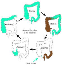
Vermiform (ˈvərməˌfôrm) describes something shaped like a worm. The expression is often employed in biology and anatomy to describe usually soft body parts or animals that are more or less tubular or cylindrical. The word root is Latin, vermes (worms) and formes (shaped). A well known example is the vermiform appendix, a small, blind section of the gut in humans and a number of other mammals.

Innate lymphoid cells (ILCs) are the most recently discovered family of innate immune cells, derived from common lymphoid progenitors (CLPs). In response to pathogenic tissue damage, ILCs contribute to immunity via the secretion of signalling molecules, and the regulation of both innate and adaptive immune cells. ILCs are primarily tissue resident cells, found in both lymphoid, and non- lymphoid tissues, and rarely in the blood. They are particularly abundant at mucosal surfaces, playing a key role in mucosal immunity and homeostasis. Characteristics allowing their differentiation from other immune cells include the regular lymphoid morphology, absence of rearranged antigen receptors found on T cells and B cells, and phenotypic markers usually present on myeloid or dendritic cells.

<span class="mw-page-title-main">Type 3 innate lymphoid cells</span>

Type 3 innate lymphoid cells (ILC3) are immune cells from the lymphoid lineage that are part of the innate immune system. These cells participate in innate mechanisms on mucous membranes, contributing to tissue homeostasis, host-commensal mutualism and pathogen clearance. They are part of a heterogeneous group of innate lymphoid cells, which is traditionally divided into three subsets based on their expression of master transcription factors as well as secreted effector cytokines - ILC1, ILC2 and ILC3.

References

  1. Kooij IA, Sahami S, Meijer SL, Buskens CJ, Te Velde AA (October 2016). "The immunology of the vermiform appendix: a review of the literature". Clinical and Experimental Immunology . 186 (1): 1–9. doi:10.1111/cei.12821. PMC   5011360 . PMID   27271818.
  2. Smith, H. F.; Fisher, R. E.; Everett, M. L.; Thomas, A. D.; Randal Bollinger, R.; Parker, W. (2009). "Comparative anatomy and phylogenetic distribution of the mammalian cecal appendix". Journal of Evolutionary Biology. 22 (10): 1984–1999. doi: 10.1111/j.1420-9101.2009.01809.x . PMID   19678866.
  3. "Largest appendix removed". Guinness World Records. 26 August 2006. Archived from the original on 26 November 2020. Retrieved 22 May 2017.
  4. 1 2 Golalipour, M.J.; Arya, B.; Jahanshahi, M.; Azarhoosh, R. (2003). "Anatomical Variations Of Vermiform Appendix In South-East Caspian Sea (Gorgan-IRAN)" (PDF). J. Anat. Soc. India. Archived (PDF) from the original on 11 July 2020. Retrieved 1 October 2014.
  5. "Unusual Types of Twins". Multiples of America. Archived from the original on 2 May 2014. Retrieved 30 April 2014.
  6. Gedda L, Sciacca A, Brenci G, Villatico S, Bonanni G, Gueli N, Talone C (1984). "Situs viscerum specularis in monozygotic twins". Acta Geneticae Medicae et Gemellologiae. 33 (1): 81–5. doi: 10.1017/S0001566000007546 . PMID   6540028.
  7. Paterson-Brown, S. (2007). "15. The acute abdomen and intestinal obstruction". In Parks, Rowan W.; Garden, O. James; Carter, David John; Bradbury, Andrew W.; Forsythe, John L. R. (eds.). Principles and practice of surgery (5th ed.). Edinburgh: Churchill Livingstone. ISBN   978-0-443-10157-1.
  8. Clegg-Lamptey JN, Armah H, Naaeder SB, Adu-Aryee NA (December 2006). "Position and susceptibility to inflammation of vermiform appendix in Accra, Ghana". East African Medical Journal. 83 (12): 670–3. doi: 10.4314/eamj.v83i12.9498 . PMID   17685212.
  9. Bakheit MA, Warille AA (June 1999). "Anomalies of the vermiform appendix and prevalence of acute appendicitis in Khartoum". East African Medical Journal. 76 (6): 338–40. PMID   10750522.
  10. Ghorbani A, Forouzesh M, Kazemifar AM (2014). "Variation in Anatomical Position of Vermiform Appendix among Iranian Population: An Old Issue Which Has Not Lost Its Importance". Anatomy Research International . 2014: 313575. doi: 10.1155/2014/313575 . PMC   4176911 . PMID   25295193.
  11. Denjalić A, Delić J, Delić-Custendil S, Muminagić S (2009). "[Variations in position and place of formation of appendix vermiformis found in the course of open appendectomy]". Medicinski Arhiv (in Bosnian). 63 (2): 100–1. PMID   19537667.
  12. Zetina-Mejía CA, Alvarez-Cosío JE, Quillo-Olvera J (2009). "Congenital absence of the cecal appendix. Case report". Cirugia y Cirujanos. 77 (5): 407–10. PMID   19944032.
  13. Kumar, Vinay; Robbins, Stanley L.; Cotran, Ramzi S. (1989). Robbins' pathologic basis of disease (4th ed.). Philadelphia: Saunders. pp.  902–3. ISBN   978-0-7216-2302-3.
  14. 1 2 "Scientists may have found appendix's purpose". NBC News . Associated Press. 5 October 2007. Archived from the original on 4 February 2020. Retrieved 24 August 2019.
  15. 1 2 Randal Bollinger R, Barbas AS, Bush EL, Lin SS, Parker W (December 2007). "Biofilms in the large bowel suggest an apparent function of the human vermiform appendix". Journal of Theoretical Biology. 249 (4): 826–31. Bibcode:2007JThBi.249..826R. doi:10.1016/j.jtbi.2007.08.032. PMID   17936308.
  16. Sonnenburg JL, Angenent LT, Gordon JI (June 2004). "Getting a grip on things: how do communities of bacterial symbionts become established in our intestine?". Nature Immunology . 5 (6): 569–73. doi:10.1038/ni1079. PMID   15164016. S2CID   25672527.
  17. Everett M.L.; Palestrant D.; Miller S.E.; Bollinger R.R.; Parker W. (2004). "Immune exclusion and immune inclusion: a new model of host-bacterial interactions in the gut". Clinical and Applied Immunology Reviews. 4 (5): 321–32. doi:10.1016/j.cair.2004.03.001.
  18. Dunn, Rob (January 2, 2012). "Your Appendix Could Save Your Life". Scientific American. Archived from the original on 11 November 2020. Retrieved 22 December 2016.
  19. Zahid A (April 2004). "The vermiform appendix: not a useless organ". Journal of the College of Physicians and Surgeons--Pakistan. 14 (4): 256–8. PMID   15228837.
  20. Rankin LC, Girard-Madoux MJ, Seillet C, Mielke LA, Kerdiles Y, Fenis A, et al. (February 2016). "Complementarity and redundancy of IL-22-producing innate lymphoid cells". Nature Immunology . 17 (2): 179–86. doi:10.1038/ni.3332. PMC   4720992 . PMID   26595889.
  21. Smith H, Parker W, Kotze S, Laurin M (September 2016). "Morphological evolution of the mammalian cecum and cecal appendix". Comptes Rendus Palevol. 16 (1): 39–57. doi: 10.1016/j.crpv.2016.06.001 .
  22. "Miscellaneous conditions of the appendix". Seminars in Diagnostic Pathology. National Institutes of Health. 21 (2): 151–63. 2004. doi:10.1053/j.semdp.2004.11.006. PMID   15807474 . Retrieved 30 December 2020.
  23. "Statistics about Appendix disorder". rightdiagnosis.com. Archived from the original on 2019-10-16. Retrieved 2020-12-30.
  24. 1 2 3 Miller R., Kenneth; Levine, Joseph (2002). Biology. Prentice Hall. pp. 92–98. ISBN   978-0-13-050730-3.
  25. "Appendicitis - Symptoms and causes - Mayo Clinic". mayoclinic.org. Mayo Clinic. Archived from the original on 25 November 2020. Retrieved 29 December 2020.
  26. Mingin GC, Baskin LS (2003). "Surgical management of the neurogenic bladder and bowel". International Braz J Urol. 29 (1): 53–61. doi: 10.1590/S1677-55382003000100012 . PMID   15745470.
  27. "Wellington Children's Hospital : Caring for an ACE or Chait Tube : Healthpoint". Archived from the original on 16 October 2019. Retrieved 22 December 2016.
  28. Darwin, Charles (1871) "Jim's Jesus". The Descent of Man, and Selection in Relation to Sex . John Murray: London.
  29. Smith HF, Fisher RE, Everett ML, Thomas AD, Bollinger RR, Parker W (October 2009). "Comparative anatomy and phylogenetic distribution of the mammalian cecal appendix". Journal of Evolutionary Biology. 22 (10): 1984–99. doi: 10.1111/j.1420-9101.2009.01809.x . PMID   19678866. S2CID   6112969.
  30. Smith H. F.; Parker W.; Kotzé, S. H.; Laurin, M. (2013). "Multiple independent appearances of the cecal appendix in mammalian evolution and an investigation of related ecological and anatomical factors". Comptes Rendus Palevol. 12 (6): 339–354. Bibcode:2013CRPal..12..339S. doi: 10.1016/j.crpv.2012.12.001 .
  31. Smith H. F.; Parker W.; Kotzé, S. H.; Laurin, M. (2017). "Morphological evolution of the mammalian cecum and cecal appendix". Comptes Rendus Palevol. 11 (1): 39–57. Bibcode:2017CRPal..16...39S. doi: 10.1016/j.crpv.2016.06.001 .
  32. Collard, Maxime K.; Bardin, Jérémie; Marquet, Bertille; Laurin, Michel; Ogier-Denis, Éric (2023-09-23). "Correlation between the presence of a cecal appendix and reduced diarrhea severity in primates: new insights into the presumed function of the appendix". Scientific Reports. 13 (1): 15897. Bibcode:2023NatSR..1315897C. doi:10.1038/s41598-023-43070-5. ISSN   2045-2322. PMC   10517977 . PMID   37741857.
  33. Laurin M, Everett ML, Parker W (April 2011). "The cecal appendix: one more immune component with a function disturbed by post-industrial culture". Anatomical Record. 294 (4): 567–79. doi:10.1002/ar.21357. PMID   21370495. S2CID   3237168.
  34. Evolution of the Appendix: A Biological 'Remnant' No More; By Duke Medicine News and Communications; Published: 20 August 2009 Updated: 21 August 2009

Further reading