Aspergillus clavatus

Last updated

Contents

Aspergillus clavatus
Aspergillus clavatus.jpg
Conidial head of Aspergillus clavatus
Scientific classification OOjs UI icon edit-ltr.svg
Domain: Eukaryota
Kingdom: Fungi
Division: Ascomycota
Class: Eurotiomycetes
Order: Eurotiales
Family: Aspergillaceae
Genus: Aspergillus
Species:
A. clavatus
Binomial name
Aspergillus clavatus
Desm. (1834)
Synonyms

Aspergillus pallidus (Samson, 1979)

Aspergillus clavatus is a species of fungus in the genus Aspergillus with conidia dimensions 3–4.5 x 2.5–4.5 μm. It is found in soil and animal manure. The fungus was first described scientifically in 1834 by the French mycologist John Baptiste Henri Joseph Desmazières. [1]

The fungus can produce the toxin patulin, which may be associated with disease in humans and animals. This species is only occasionally pathogenic.

Other sources have identified many species of Aspergillus as producing dry, hydrophobic spores that are easily inhaled by humans and animals. Due to the small size of the spores, about 70% of spores of A. fumigatus are able to penetrate into the trachea and primary bronchi and close to 1% into alveoli. Inhalation of spores of Aspergillus is a health risk. A. clavatus is allergenic, causing the occupational hypersensitivity pneumonitis known as malt-worker's lung.

History and taxonomy

Aspergillus clavatus is a species of Aspergillus and is characterized by elongated club-shaped vesicles, and blue-green uniseriate conidia. [2] The fungus was first described scientifically in 1834 by the French mycologist John Baptiste Henri Joseph Desmazières. [1] It belongs to the Aspergillus section Clavati, (formerly known as the Aspergillus clavatus group) recognized by Charles Thom and Margaret Church (1926), alongside two species, Aspergillus clavatus and Aspergillus giganteus . [3] In the succeeding years, four more species were discovered belonging to the Aspergillus section Clavati, which included Aspergillus rhizopodus , Aspergillus longivesica , Neocarpenteles acanthosporus and Aspergillus clavatonanicus . [3] Later, Aspergillus pallidus was concluded to be a white variant (synonym) of A. clavatus by Samson (1979), which was supported by the identical DNA sequences of the two species. [4] A sexual stage was described in 2018 with a Neocarpenteles teleomorph but under the one fungus-one name convention the original A. clavatus epithet was retained. [5]

Growth and morphology

Aspergillus clavatus undergoes rapid growth, resulting in the formation of a velvety and fairly dense felt that is observed to be bluish-grey green in colour. [6] The emerging conidial heads are large and clavate when very young, quickly splitting into conspicuous and compact divergent columns. [7] The conidia bearing conidiophores are generally coarse, smooth walled, uncoloured, [7] hyaline and can grow to be very long. [6] Elongated club-shaped vesicles [6] clavate, [7] and bear phialides (singular: phialide) over their entire-surface, contributing to its short and densely packed structure. [6] The sterigmata are usually found to be uniseriate, numerous and crowded. [7] Conidia formed in them are elliptical, smooth and comparatively thick-walled. [7] A. clavatus usually express conidiophores 1.5–3.00 mm in length, which arises from specialized and widened hyphal cells that eventually become the branching foot cells. [8] The conidia on A. clavatus has been measured up to 3.0 – 4.5 X 2.5 – 3.5 μm. [8] Cleistothecia are produced in crosses after approximately 4–10 weeks of incubation on suitable growth media at 25 °C. Cleistothecia are yellowish-brown (fawn) to dark brown in colour and range in diameter from 315-700 μm in diameter and have a relatively hard outer wall (peridium). At maturity the cleistothecia contain asci that themselves contain ascospores, which are clear, lenticular (with ridges evident) and between 6.0-7.0 μm in diameter. [5]

Growth on Czapek's solution agar(

Aspergillus clavatus colonies grow rapidly on Czapek's solution agar, reaching 3.0–3.5 cm, in 10 days at 24–26 °C. [7] Growth is usually plane or moderately furrowed, with occasional appearance of floccose strains. But generally, a comparatively thin surface layer of mycelial felt is observed, which produces a copious number of erect conidiophores. [7] The reverse is usually uncoloured but becomes brown with passing time in some strains. [7] While odor is not prominent in some strains, it can be extremely unpleasant in others. [7] Large conidial heads extend from 300 to 400 μm by 150 to 200 μm when young. However, with time, they split into two or more divergent and compressed cordial chains reaching 1.00 mm portraying a colour consisting of artemisia green to slate olive. [7] The observed conidiophores grow up to 1.5–3.00 mm in length with 20–30 μm in diameter. They slowly and ultimately enlarge at the apex into a clavate vesicle, which consists of a fertile area, 200 to 250 μm in length and 40–60 μm wide. [7] The sterigmata usually ranges from 2.5 to 3.5 μm by 2.0 to 3.0 μm at the base of the vesicle, to 7.0 or 8.0 and occasionally 10 μm to 2.5 to 3.0 μm at the apex. [7] The conidia are comparatively thick-walled and measures 3.0 to 4.5 μm by 2.5 to 3.5 μm. While they can be larger in some strains, in others their appearance may be irregular. [7]

Growth on malt extract agar

Aspergillus clavatus colony growing on petri dish Aspergillus clavatus petri dish.jpg
Aspergillus clavatus colony growing on petri dish

On malt extract agar, the structural morphology of A. clavatus appears to be different than in Czapek's solution agar. [7] The typical strains extracted from malt media contain less abundant conidial structures, which could be larger in size. [7] In other (non-typical) strains, the conidial heads increase in number but decrease in size. The conidiophores range from 300 to 500 μm and bear loose, columnar heads. Typical strains may be resembled by strong and unpleasant odor whereas non-typical strains are characterized being odorless. [7] The colonies arising from one conidium on malt extract agar, consisted of 25X10^7 conidia after being observed for six days. [8]

Examination

The phialide development and conidium formation in A. clavatus has been examined using TEM. [8] And by using SEM, it was discovered that the first-formed conidium and phialide share a continuous wall. [8] Additionally recombination with an albino mutant led to the production of heterokaryotic conidial heads with mixed conidial colours. [8] A GC-content of 52.5–55% was also detected upon DNA analysis. [8] And its soluble wall carbohydrates consist of mannitol and arabitol. [8]

Physiology

Light stimulates the elongation of conidiophores in A. clavatus. And the more favourable C sources include starch, dextrin, glycogen and especially fructose. [8] Substantial degree of lipid synthesis occurs, whereas cellulose and usnic acid are degraded. [8] A. clavatus also produces riboflavin, ribonuclease, acid phosphodiesterase and acid phosphatase when in liquid culture. [8]

A. clavatus has the properties to oxidize tryptamine to indole acetic acid. It can absorb and collect hydrocarbons from fuel oil, incorporate metaphosphate and synthesize ethylene, clavatol and kojic acid. [8] It is also responsible for the production of mycotoxins Patulin and sterigmatocystin. [6] And has extremely high capacity for alcohol fermentation. [9]

When it comes to genomics, bioinformatic analysis revealed that A. clavatus contains a full complement of identified euascomycete sex genes. [10] A heterothallic sexual cycle involving outcrossing between MAT1-1 and MAT1-2 isolates was subsequently described . [5] A. clavatus can also be a food source for Collembola and has been found to be parasitized by Fusarium solani. [8]

Habitat and ecology

Aspergillus clavatus is often described as a spoilage organism occurring on dung and in soil and can also grow in strong alkaline conditions. [6] When it comes to geographical distribution, A. clavatus has been spotted in the tropical, subtropical and Mediterranean areas. [8] It has been accounted in low frequencies in the soils of India. And is also found in Bangladesh, Sri Lanka, Hong Kong, Jamaica, Brazil, Argentina, South Africa, the Ivory Coast, Egypt, Libya, Turkey, Greece, Italy, the United States of America, Japan, the USSR and Czechoslovakia. [8] It was tracked in rocks of a carst cave and stratigraphic core samples descending to 1200 m in Central Japan. [8] However, it is usually and solely collected from cultivated soils, including the ones that bear cotton, potatoes, sugar canes, legumes, paddy and Artemisia herba-alba . [8] It has also been garnered from soil under burnt steppe vegetation, desert soils, the rhizospheres of banana, ground-nuts and wheat. [8] A. clavatus has also been detected in the ripe compost of municipal waste, and Nitrogen and NPK fertilizers are found to play an important role in its stimulation process. [8]

A. clavatus is also referred as a cosmopolitan fungus. Other than soil and dung, it can additionally be found in stored products with high levels of entrapped moisture. Such as stored cereals, rice, corn and millet. [3] It has been further isolated from insects, especially from dead adult bees and honeycombs. [8] Moreover, it has been collected from the feathers and droppings from free-living birds. [8] A. clavatus is also common is decomposing materials. [7] Their ability to resist strongly alkaline conditions, allows them to act as decomposition catalysts in situations where other fungus usually do not function. [7]

Applications and medical uses

Weisner in March 1942 first noted the production of an antibiotic by strains of A. clavatus, and the active substance was known as clavatin. [7] Later the antibiotic was named clavacin in August 1942 by Waksman, Horning and Spencer. Clavacin is also known as patulin. [7] Patulin is receiving significant attention in the world today because of its manifestations in apple juices. [10] Clavacin was noted to be valuable in the treatment of common-cold and applies a fungistatic or fungicidal effect on certain dermatophytes. [7] A. clavatus with Phytophthora cryptogea in soil provided protection against damping of tomato seedlings, by decreasing the spreading of pathogens. [7] Reversely, A. clavatus with the addition of glucose, increased the pathogenicity of Verticillium albo-atrum to tomatoes. [7] A. clavatus also produces the following: Cytochalasin E, Cytochalasin K, Tryptoquivaline, Nortryptoquivalone, Nortryptoquivaline, Deoxytryptoquivaline, Deoxynortryptoquivaline, Tryptoquivaline E, and Tryptoquivaline N. [11] Furthermore, A. clavatus isolates produce ribotoxins, which can help develop immunotherapy processes for cancer. [10] A.clavatus has also been used in the formation of extracellular bionanoparticles from silver nitrate solutions. These nanoparticles display antimicrobial properties, which work against MRSA and MRSE. [12]

Pathogenicity

Aspergillus clavatus is known as an agent of allergic aspergillosis [13] and has been implicated in multiple pulmonary infections. [13] It has also been labelled as an opportunistic fungus, as it is responsible for causing aspergillosis in compromised patients. [14] A. clavatus can also cause neurotoxicosis in sheep and otomycosis. [13] In Scotland and elsewhere, A. clavatus is reported for causing the mould allergy "malster's lung" otherwise "maltster's lung". [15] [16]

Extrinsic allergic alveolitis (EAA) is also caused by Aspergillus clavatus with a Type 1 immune reaction. It is described as a true hypersensitivity pneumonia, which usually occurs among malt workers, including symptoms of fever, chills, cough and dyspnea. In severe cases, glucocorticoids are used. [17] Microgranulomatous hypersensitivity pneumonitis, where interstitial granulomatous infiltration occurs, usually in malt workers, is caused by allergy to antigens of Aspergillus clavatus. [18] EAA is caused by allergy to Aspergillus conidia, usually in the non-atopic individual. [19] Such individuals are usually exposed to organic dust heavily packed with conidia and mycelial debris. [19] This condition involves the lung parenchyma. [19]

A strain of A. clavatus has also caused hyperkeratosis in calves. [7] Spore walls of a sputum-derived isolate of Aspergillus clavatus were extracted and treated with ethanol following alkaline hydrolysis. And it yielded mutagens. [20] The extracts were given to unimmunised mice, causing lung reaction and leading to cases of pulmonary mycotoxicosis. A rising incidence of lung tumours were also observed. [20] This study revealed that an isolate of A. clavatus, which is able to convert highly toxic metabolites in bacterial and mammalian cells, will cause inflammatory response in the lungs of unimmunized mice. [20]

Related Research Articles

<span class="mw-page-title-main">Conidium</span> Asexual, non-motile spore of a fungus

A conidium, sometimes termed an asexual chlamydospore or chlamydoconidium, is an asexual, non-motile spore of a fungus. The word conidium comes from the Ancient Greek word for dust, κόνις (kónis). They are also called mitospores due to the way they are generated through the cellular process of mitosis. They are produced exogenously. The two new haploid cells are genetically identical to the haploid parent, and can develop into new organisms if conditions are favorable, and serve in biological dispersal.

<i>Aspergillus terreus</i> Species of fungus

Aspergillus terreus, also known as Aspergillus terrestris, is a fungus (mold) found worldwide in soil. Although thought to be strictly asexual until recently, A. terreus is now known to be capable of sexual reproduction. This saprotrophic fungus is prevalent in warmer climates such as tropical and subtropical regions. Aside from being located in soil, A. terreus has also been found in habitats such as decomposing vegetation and dust. A. terreus is commonly used in industry to produce important organic acids, such as itaconic acid and cis-aconitic acid, as well as enzymes, like xylanase. It was also the initial source for the drug mevinolin (lovastatin), a drug for lowering serum cholesterol.

<i>Purpureocillium lilacinum</i> Species of fungus

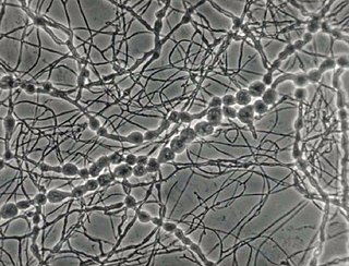
Purpureocillium lilacinum is a species of filamentous fungus in the family Ophiocordycipitaceae. It has been isolated from a wide range of habitats, including cultivated and uncultivated soils, forests, grassland, deserts, estuarine sediments and sewage sludge, and insects. It has also been found in nematode eggs, and occasionally from females of root-knot and cyst nematodes. In addition, it has frequently been detected in the rhizosphere of many crops. The species can grow at a wide range of temperatures – from 8 to 38 °C for a few isolates, with optimal growth in the range 26 to 30 °C. It also has a wide pH tolerance and can grow on a variety of substrates. P. lilacinum has shown promising results for use as a biocontrol agent to control the growth of destructive root-knot nematodes.

Aspergillus ochraceus is a mold species in the genus Aspergillus known to produce the toxin ochratoxin A, one of the most abundant food-contaminating mycotoxins, and citrinin. It also produces the dihydroisocoumarin mellein. It is a filamentous fungus in nature and has characteristic biseriate conidiophores. Traditionally a soil fungus, has now began to adapt to varied ecological niches, like agricultural commodities, farmed animal and marine species. In humans and animals the consumption of this fungus produces chronic neurotoxic, immunosuppressive, genotoxic, carcinogenic and teratogenic effects. Its airborne spores are one of the potential causes of asthma in children and lung diseases in humans. The pig and chicken populations in the farms are the most affected by this fungus and its mycotoxins. Certain fungicides like mancozeb, copper oxychloride, and sulfur have inhibitory effects on the growth of this fungus and its mycotoxin producing capacities.

Aspergillus sydowii is a pathogenic fungus that causes several diseases in humans. It has been implicated in the death of sea fan corals in the Caribbean Sea.

<i>Aspergillus candidus</i> Species of fungus

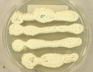
Aspergillus candidus is a white-spored species of fungus in the genus Aspergillus. Despite its lack of pigmentation, it is closely related to the most darkly-pigmented aspergilli in the Aspergillus niger group. It is a common soil fungus worldwide and is known as a contaminant of a wide array of materials from the indoor environment to foods and products. It is an uncommon agent of onychomycosis and aspergillosis. The species epithet candidus (L.) refers to the white pigmentation of colonies of this fungus. It is from the Candidi section. The fungi in the Candidi section are known for their white spores. It has been isolated from wheat flour, djambee, and wheat grain.

<i>Aspergillus glaucus</i> Species of fungus

Aspergillus glaucus is a filamentous fungus which is known to have a wide environmental distribution due to its physiological hardiness under extreme conditions. Like many other fungi belonging to the genus Aspergillus, it can be mildly pathogenic but has a number of useful potential applications in medicine and the production of foodstuffs.

Emmonsia parva is a filamentous, saprotrophic fungus and one of three species within the genus Emmonsia. The fungus is most known for its causal association with the lung disease, adiaspiromycosis which occurs most commonly in small mammals but is also seen in humans. The disease was first described from rodents in Arizona, and the first human case was reported in France in 1964. Since then, the disease has been reported from Honduras, Brazil, the Czech Republic, Russia, the United States of America and Guatemala. Infections in general are quite rare, especially in humans.

<i>Penicillium digitatum</i> Species of fungus

Penicillium digitatum is a mesophilic fungus found in the soil of citrus-producing areas. It is a major source of post-harvest decay in fruits and is responsible for the widespread post-harvest disease in Citrus fruit known as green rot or green mould. In nature, this necrotrophic wound pathogen grows in filaments and reproduces asexually through the production of conidiophores and conidia. However, P. digitatum can also be cultivated in the laboratory setting. Alongside its pathogenic life cycle, P. digitatum is also involved in other human, animal and plant interactions and is currently being used in the production of immunologically based mycological detection assays for the food industry.

Aspergillus unguis is a species of fungus in the genus Aspergillus, and the asexual state (anamorph) of Emericella unguis. Aspergillus unguis is a filamentous soil-borne fungus found on decomposing plant matter and other moist substrates including with building materials and household dust. Aspergillus unguis occurs mainly in tropical and subtropical soils but has also been isolated from various marine and aquatic habitats. The species was first isolated in 1935 by Weill and L. Gaudin. Historically, A. unguis was assigned to the A. nidulans group, a common group of soil-borne fungi due to the resemblance of its ascospores and cleistothecia to those of Emericella nidulans. Aspergillus unguis is distinctive, however, in possessing spicular hyphae. A number of synonyms have been collapsed into this species, including Sterigmatocystis unguis, Aspergillus laokiashanensis and Aspergillus mellinus.

<i>Trichothecium roseum</i> Species of fungus

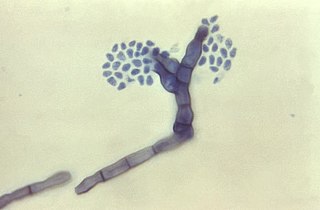
Trichothecium roseum is a fungus in the division Ascomycota first reported in 1809. It is characterized by its flat and granular colonies which are initially white and develop to be light pink in color. This fungus reproduces asexually through the formation of conidia with no known sexual state. Trichothecium roseum is distinctive from other species of the genus Trichothecium in its characteristic zigzag patterned chained conidia. It is found in various countries worldwide and can grow in a variety of habitats ranging from leaf litter to fruit crops. Trichothecium roseum produces a wide variety of secondary metabolites including mycotoxins, such as roseotoxins and trichothecenes, which can infect and spoil a variety of fruit crops. It can act as both a secondary and opportunistic pathogen by causing pink rot on various fruits and vegetables and thus has an economical impact on the farming industry. Secondary metabolites of T. roseum, specifically Trichothecinol A, are being investigated as potential anti-metastatic drugs. Several agents including harpin, silicon oxide, and sodium silicate are potential inhibitors of T. roseum growth on fruit crops. Trichothecium roseum is mainly a plant pathogen and has yet to show a significant impact on human health.

<i>Cladosporium cladosporioides</i> Species of fungus

Cladosporium cladosporioides is a darkly pigmented mold that occurs world-wide on a wide range of materials both outdoors and indoors. It is one of the most common fungi in outdoor air where its spores are important in seasonal allergic disease. While this species rarely causes invasive disease in animals, it is an important agent of plant disease, attacking both the leaves and fruits of many plants. This species produces asexual spores in delicate, branched chains that break apart readily and drift in the air. It is able to grow under low water conditions and at very low temperatures.

<i>Madurella mycetomatis</i> Species of fungus

Madurella mycetomatis is a fungus primarily reported in Central Africa as a cause of mycetoma in humans. It has been misclassified for many years, but with improvement of molecular techniques, its phylogenetic classification has been established. Many methods exist to identify M. mycetomatis, both in lesions and in culture. Histological examination is especially useful, as it has many unique morphological features. Strain-level differences in response to antifungal agents is informative for treatment and laboratory isolation of cultures.

<i>Fonsecaea compacta</i> Species of fungus

Fonsecaea compacta is a saprophytic fungal species found in the family Herpotrichiellaceae. It is a rare etiological agent of chromoblastomycosis, with low rates of correspondence observed from reports. The main active components of F. compacta are glycolipids, yet very little is known about its composition. F. compacta is widely regarded as a dysplastic variety of Fonsecaea pedrosoi, its morphological precursor. The genus Fonsecaea presently contains two species, F. pedrosoi and F. compacta. Over 100 strains of F. pedrosoi have been isolated but only two of F. compacta.

<i>Aspergillus alabamensis</i> Species of fungus

Aspergillus alabamensis is a soil fungus in the division Ascomycota first described in 2009 as a segregated taxon of A. terreus. Originally thought to be a variant of A. terreus, A. alabamensis is situated in a distinctive clade identified by genetic analysis. While A. alabamensis has been found to be morphologically similar to Aspergillus terreus by morphological studies, the two differ significantly in active metabolic pathways, with A. alabamensis producing the mycotoxins citrinin and citreoviridin but lacking mevinolin.

<i>Aspergillus parasiticus</i> Species of fungus

Aspergillus parasiticus is a fungus belonging to the genus Aspergillus. This species is an unspecialized saprophytic mold, mostly found outdoors in areas of rich soil with decaying plant material as well as in dry grain storage facilities. Often confused with the closely related species, A. flavus, A. parasiticus has defined morphological and molecular differences. Aspergillus parasiticus is one of three fungi able to produce the mycotoxin, aflatoxin, one of the most carcinogenic naturally occurring substances. Environmental stress can upregulate aflatoxin production by the fungus, which can occur when the fungus is growing on plants that become damaged due to exposure to poor weather conditions, during drought, by insects, or by birds. In humans, exposure to A. parasiticus toxins can cause delayed development in children and produce serious liver diseases and/or hepatic carcinoma in adults. The fungus can also cause the infection known as aspergillosis in humans and other animals. A. parasiticus is of agricultural importance due to its ability to cause disease in corn, peanut, and cottonseed.

<i>Penicillium spinulosum</i> Species of fungus

Penicillium spinulosum is a non-branched, fast-growing fungus with a swelling at the terminal of the stipe (vesiculate) in the genus Penicillium. P. spinulosum is able to grow and reproduce in environment with low temperature and low water availability, and is known to be acidotolerant. P. spinulosum is ubiquitously distributed, and can often be isolated from soil. Each individual strain of P. spinulosum differs from others in their colony morphology, including colony texture, amount of sporulation and roughness of conidia and conidiophores.

Aspergillus wentii is an asexual, filamentous, endosymbiotic fungus belonging to the mold genus, Aspergillus. It is a common soil fungus with a cosmopolitan distribution, although it is primarily found in subtropical regions. Found on a variety of organic materials, A. wentii is known to colonize corn, cereals, moist grains, peanuts and other ground nut crops. It is also used in the manufacture of biodiesel from lipids and is known for its ability to produce enzymes used in the food industry.

<i>Mariannaea elegans</i> Species of fungus

Mariannaea elegans an anamorphic fungus. It is mainly found on rotting wood and soil. M. elegans is not pathogenic to humans, animals, or plants.

Aspergillus giganteus is a species of fungus in the genus Aspergillus that grows as a mold. It was first described in 1901 by Wehmer, and is one of six Aspergillus species from the Clavati section of the subgenus Fumigati. Its closest taxonomic relatives are Aspergillus rhizopodus and Aspergillus longivescia.

References

  1. 1 2 Desmazières, JBHJ (1834). "Descriptions et figures de six hyphomycètes inédites à ajouter à la flore Française". Annales des Sciences Naturelles Botanique (in French). 2 (2): 69–73.
  2. Howard, Dexter H., ed. (2003). Pathogenic fungi in humans and animals (2. ed.). New York [u.a.]: Dekker. p.  247. ISBN   978-0-8247-0683-8.
  3. 1 2 3 Varga, J.; Due, M.; Frisvad, J.C.; Samson, R.A. (2007). "Taxonomic revision of Aspergillus section Clavati based on molecular, morphological and physiological data". Studies in Mycology. 59: 89–106. doi:10.3114/sim.2007.59.11. PMC   2275193 . PMID   18490946.
  4. Samson, ed. by Robert A.; Pitt, John I. (2000). Integration of modern taxonomic methods for penicillium and aspergillus classification. Amsterdam: Harwood Acad. Publ. ISBN   978-90-5823-159-8.{{cite book}}: |first1= has generic name (help)
  5. 1 2 3 Ojeda-López, M; Chen W; Eagle CE; Gutiérrez G; Jia WL; Swilaiman SS; Huang Z; Park HS; Yu JH; Dyer PS (2018). "Evolution of asexual and sexual reproduction in the aspergilli". Studies in Mycology. 91: 37–59. doi:10.1016/j.simyco.2018.10.002. PMC   6231087 . PMID   30425416.
  6. 1 2 3 4 5 6 Onions, A.H.S.; Allsopp, D.; Eggins, H.O.W. (1981). Smith's introduction to industrial mycology (7th ed.). London, UK: Arnold. ISBN   978-0-7131-2811-6.
  7. 1 2 3 4 5 6 7 8 9 10 11 12 13 14 15 16 17 18 19 20 21 22 23 24 Raper, Kenneth B.; Fennell, Dorothy I. (1965). The Genus Aspergillus. Baltimore: The Williams and Wilkins Company. pp. 137–146.
  8. 1 2 3 4 5 6 7 8 9 10 11 12 13 14 15 16 17 18 19 20 21 Domsch, K.H.; Anderson, Traute-Heidi; Gams, W. (1980). Compendium of Soil Fungi. Academic Press. pp. 86–88.
  9. Harold J. Blumenthal (1965). "10". In Ainsworth, G.C.; Sussman, Alfred S. (eds.). THE FUNGI Volume 1 The Fungal Cell. New York and London: Academic Press. p. 251.
  10. 1 2 3 Machida, edited by Masayuki; Gomi, Katsuya (2010). Aspergillus : molecular biology and genomics. Wymondham, Norfolk, UK: Caister Academic. ISBN   978-1-904455-53-0.{{cite book}}: |first1= has generic name (help)
  11. Jarvis, Richard J. Cole,... Milbra A. Schweikert,... Bruce B. (2003). Handbook of secondary fungal metabolites. Amsterdam [etc.]: Academic press. ISBN   978-0-12-179461-3.{{cite book}}: CS1 maint: multiple names: authors list (link)
  12. Saravanan, M.; Nanda, Anima (June 2010). "Extracellular synthesis of silver bionanoparticles from Aspergillus clavatus and its antimicrobial activity against MRSA and MRSE". Colloids and Surfaces B: Biointerfaces. 77 (2): 214–218. doi:10.1016/j.colsurfb.2010.01.026. PMID   20189360.
  13. 1 2 3 Hoog, G. S. de; Guarro, J.; Gene, J.; Figueras, M. J. (2000). Atlas of clinical fungi (2. ed.). Utrecht: Centraalbureau voor Schimmelcultures [u.a.] ISBN   978-90-7035-143-4.
  14. Al-Doory, Yousef (1980). Laboratory Medical Mycology. Philadelphia: Lea and Febiger.
  15. Ainsworth, G.C. (1986). Introduction to the history of medical and veterinary mycology. Cambridge: Cambridge University Press. ISBN   978-0-521-30715-4.
  16. J E Smith, 1994: Aspergillus (Biotechnology Handbooks 7), p. 226. New York: Springer Science+Business Media
  17. Holmberg, Kenneth; Meyer, Richard D. (1989). Diagnosis and Therapy of Systemic Fungal Infections. New York: Raven Press.
  18. Watts, Francis W. Chandler, John C. (1987). Pathologic diagnosis of fungal infections. Chicago: ASCP Press. ISBN   978-0-89189-252-6.{{cite book}}: CS1 maint: multiple names: authors list (link)
  19. 1 2 3 Rippon, John Willard (1982). Medical mycology : the pathogenic fungi and the pathogenic actinomycetes (2nd ed.). Philadelphia: Saunders. ISBN   978-0-7216-7586-2.
  20. 1 2 3 Blyth W, Hardy JC (1982). "Mutagenic and tumourigenic properties of the spores of Aspergillus clavatus". Br. J. Cancer. 45 (1): 105–17. doi:10.1038/bjc.1982.13. PMC   2010971 . PMID   7059453.