Atypical hemolytic uremic syndrome

Last updated
Atypical hemolytic uremic syndrome
Other namesaHUS
Specialty Hematology   OOjs UI icon edit-ltr-progressive.svg

Atypical hemolytic uremic syndrome (aHUS), also known as complement-mediated hemolytic uremic syndrome, is an extremely rare, life-threatening, progressive disease that frequently has a genetic component. In most cases it can be effectively controlled by interruption of the complement cascade. Particular monoclonal antibodies, discussed later in the article, have proven efficacy in many cases.

Contents

AHUS is usually caused by chronic, uncontrolled activation of the complement system, [1] [2] a branch of the body's immune system that destroys and removes foreign particles. [3] The disease affects both children and adults and is characterized by systemic thrombotic microangiopathy (TMA), the formation of blood clots in small blood vessels throughout the body, which can lead to stroke, heart attack, kidney failure, and death. [1] [4] [5] The complement system activation may be due to mutations in the complement regulatory proteins (factor H, factor I, or membrane cofactor protein), [6] [5] [7] or is occasionally due to acquired neutralizing autoantibody inhibitors of these complement system components, for example anti–factor H antibodies. [8] :1933 Prior to availability of eculizumab (Soliris) and ravulizumab (Ultomiris), an estimated 33–40% of patients died or developed end-stage renal disease (ESRD) (despite the use of supportive care, e.g. plasmapheresis) with the first clinical bout of aHUS. Including subsequent relapses, a total of approximately two-thirds (65%) of patients died, required dialysis, or had permanent renal damage within the first year after diagnosis despite plasma exchange or plasma infusion (PE/PI). [7]

Signs and symptoms

Clinical signs and symptoms of complement-mediated TMA can include abdominal pain, [9] confusion, [9] fatigue, [5] edema (swelling), [10] nausea/vomiting [11] and diarrhea. [12] aHUS often presents with malaise and fatigue, as well as microangiopathic anemia. [8] :1931 However, severe abdominal pain and bloody diarrhea are unusual. [8] :1931 Laboratory tests may also reveal low levels of platelets (cells in the blood that aid in clotting), [1] elevated lactate dehydrogenase (LDH, a chemical released from damaged cells, and which is therefore a marker of cellular damage), [7] decreased haptoglobin (indicative of the breakdown of red blood cells), [7] anemia (low red blood cell count)/schistocytes (damaged red blood cells), [1] [7] elevated creatinine (indicative of kidney dysfunction), [13] and proteinuria (indicative of kidney injury). [14] Patients with aHUS often present with an abrupt onset of systemic signs and symptoms such as acute kidney failure, [1] hypertension (high blood pressure), [5] myocardial infarction (heart attack), [15] stroke, [9] lung complications, [15] pancreatitis (inflammation of the pancreas), [11] liver necrosis (death of liver cells or tissue), [5] encephalopathy (brain dysfunction), [5] seizure, [16] or coma. [17] Failure of neurologic, cardiac, kidney, and gastrointestinal (GI) organs, as well as death, can occur unpredictably at any time, either very quickly or following prolonged symptomatic or asymptomatic disease progression. [1] [14] [18] [19] [20] For example, approximately 1 in 6 patients with aHUS initially will present with proteinuria or hematuria without acute kidney failure. [14] Patients who survive the presenting signs and symptoms endure a chronic thrombotic and inflammatory state, which puts many of them at lifelong elevated risk of sudden blood clotting, kidney failure, other severe complications and premature death. [10] [21]

Comorbidities

Although many patients experience aHUS as a single disease, comorbidities are common. In one study, 25% (47/191) of patients with no known family history of aHUS were found to have a coexisting disease or condition. Comorbidities in this study included malignant hypertension (30%), TMA with a history of transplant (23%), TMA associated with pregnancy (21%), glomerulopathy (17%), systemic disease such as systemic lupus erythematosus (SLE) or progressive systemic sclerosis (PSS) (6%), and malignancy (1%). [5] The presence of mutations in complement regulatory proteins, or of disease-associated variations in the genes encoding these proteins (i.e., in most patients with comorbid conditions as well as in patients with aHUS as a single disease), suggests that deviations from the normal genetic coding of these factors could result in a genetic predisposition to TMA. Individuals so predisposed could have aHUS episodes precipitated by one of the known disease triggers (e.g., infection, pregnancy, surgery, trauma) as well as by other systemic diseases (e.g., malignant hypertension, SLE, cancer).[ citation needed ]

Mechanisms

In healthy individuals, complement is used to attack foreign substances, and the complement system is highly regulated to prevent it from damaging healthy tissues and organs. [1] [18] However, in most patients with aHUS, it has been demonstrated that chronic, uncontrolled, and excessive activation of complement can result from production of anti-factor H autoantibodies or from genetic mutations in any of several complement regulatory proteins (e.g., factor H, factor HR1 or HR3, membrane cofactor protein, factor I, factor B, complement C3, and thrombomodulin). [18] This results in platelet activation, damage to endothelial cells (cells that line the blood vessels), and white blood cell activation, leading to systemic TMA, which manifests as decreased platelet count, hemolysis (breakdown of red blood cells), damage to multiple organs, and often, death. [14] [19] [22]

Diagnosis

aHUS is not the only condition that causes systemic TMA, a fact that makes differential diagnosis essential. Historically, the clinical diagnosis of TMA-causing diseases was grouped into a broad category that (in addition to aHUS) included thrombotic thrombocytopenic purpura (TTP) and Shiga-toxin-producing Escherichia coli hemolytic uremic syndrome (STEC-HUS). [19] [21] However, it is now understood that although aHUS, STEC-HUS, and TTP have similar clinical presentations, they have distinct causes and specific tests can be conducted to differentiate these diseases. In addition, there are other conditions that can cause TMA as a secondary manifestation; these entities include systemic lupus erythematosus (SLE), malignant hypertension, progressive systemic sclerosis (PSS, also known as scleroderma), the pregnancy-associated HELLP (hemolysis, liver dysfunction, and low platelets) syndrome, and toxic drug reaction (e.g., to cocaine, cyclosporine, or tacrolimus). Nevertheless, aHUS should be suspected in patients presenting with systemic TMA, and appropriate diagnostic work-up should be undertaken.[ citation needed ]

The neurological and kidney-related signs and symptoms of aHUS overlap with those of TTP. [13] [23] However, unlike aHUS, TTP is primarily an autoimmune disorder in which the presence of an inhibitory autoantibody results in severe deficiency of ADAMTS13, an enzyme that cleaves von Willebrand factor (vWf), a large protein involved in blood clotting, into smaller pieces. [22] (TTP also can be a genetic disorder characterized by mutations in the ADAMTS13 gene leading to severe ADAMTS13 deficiency. This congenital cause of ADAMTS13 deficiency is called Upshaw-Schülman syndrome. [24] ) A lab test showing ADAMTS13 activity levels of ≤5% is indicative of TTP. [22]

Similarly, the gastrointestinal (GI) signs and symptoms of aHUS overlap with those of STEC-HUS. [12] [25] [26] Stool samples from patients with diarrhea or other GI symptoms should be tested for STEC and the presence of Shiga-toxin. However, a positive identification of Shiga-toxin, which is required to diagnose STEC-HUS, does not rule out aHUS. Nevertheless, in the appropriate clinical setting, a positive identification of Shiga-toxin makes aHUS very unlikely. [13] [26]

aHUS patients report a mean timeline of  29 days for  the overall diagnosis process from first noticing symptoms to receiving an aHUS diagnosis. During which time they report their overall health state drops from 88% of patients feeling good to excellent, to 99% feeling good to very poor and a more than halving of their health status index from 3.8 (on a scale of 1 to 5) pre-illness to 1.4 at diagnosis. [27]

Treatment

Plasma exchange/infusion

Although plasma exchange/infusion (PE/PI) is frequently used, there are no controlled trials of its safety or efficacy in aHUS. Even though PE/PI often partially controls some of the hematological manifestations of aHUS in some patients, its effectiveness has not been demonstrated in terms of inducing total disease remission. PE/PI is associated with significant safety risks, including risk of infection, allergic reactions, thrombosis, loss of vascular access, and poor quality of life. [23] [28] Importantly, terminal complement activation has been shown to be chronically present on the surface of platelets in patients with aHUS who appear to be clinically well while receiving chronic PE/PI. [10] [29]

Guidelines issued by the European Paediatric Study Group for HUS recommend rapid administration of plasma exchange or plasma infusion (PE/PI), intensively administered daily for 5 days and then with reducing frequency. [23] However, the American Society for Apheresis offers a "weak" recommendation for plasma exchange to treat aHUS, due to the "low" or "very low" quality of evidence supporting its use. Although some patients experienced improvements in red blood cell and platelet counts, plasma therapies generally did not result in full remission. [30]

Monoclonal antibody therapy

Eculizumab (Soliris) appears to be useful for atypical hemolytic uremic syndrome (aHUS). [31] In September 2011 the U.S. Food and Drug Administration (FDA) approved it as an orphan drug to treat people with aHUS. [32] This approval was based on two small prospective trials of 17 people and 20 people. [31] In the UK, NICE issued guidance on the use of Eculizumab for treating aHUS, based on five evidence sources, including those used by the FDA [33] No randomised controlled trials were identified. All prospective studies were phase 2, open‑label, non‑randomised, single‑arm studies that included patients with different clinical baseline characteristics. The prospective studies lasted 26 weeks; however, patients were allowed to continue treatment with eculizumab in a long‑term extension study.[ citation needed ]

Ravulizumab-cwvz (Ultomiris) is a second generation monoclonal antibody for aHUS made by Alexion pharmaceuticals, Inc. [34] The target of ravulizumab-cwvz is the same eculizumab (Soliris) with changes to the structure of the antibody resulting in a longer serum half life and therefore reduced dosing regimen. [35]

Dialysis

Patients with aHUS who have ESRD are generally consigned to lifelong dialysis, which carries a 5-year survival rate of 34–38%, [36] [37] with infections accounting for 14% of deaths. [38] These patients also remain at ongoing risk of non-kidney systemic complications of the disease.[ citation needed ]

Kidney transplantation

Despite its history of use in patients with aHUS, kidney transplantation does not address the continued and uncontrolled complement activation that leads to progressive, systemic TMA. As many as 90% of patients with aHUS and who are not treated with Soliris or Ultomiris, experience TMA in the transplanted organ, leading to transplant failure. [12] [39] Patients who have undergone kidney transplantation are still at continued risk of neurological, gastrointestinal, and cardiovascular complications and, importantly, premature mortality. Following kidney transplantation, the ongoing, uncontrolled, chronic complement activation associated with aHUS causes graft loss in 66% of children and 55% of adults, as well as continued inflammatory and TMA insult to other organs. [1] [5] Combined liver-kidney transplantation is only available to very few patients, due to the limited supply of solid organs. In addition, there is a substantial near-term risk of mortality, which many physicians and patients consider excessive. [1] In recent years, some transplant centers have begun to administer eculizumab to patients with TMA who receive a kidney transplant. This strategy has been effective in preventing TMA recurrences in these patients. [40]

Terminal complement inhibition

Patients using either eculizumab or ravulizumab for the treatment of aHUS showed improvements in kidney function even avoiding dialysis and minimizing death. Markers of disease activity in the blood also had a great improvement. However, the only available evidence has substantial bias and low quality and therefore there should be careful considerations for futures studies in treatment duration, adverse outcomes and risk of disease recurrence associated with this treatment. [41]

Historical Prognosis

Prior to the use of monoclonal antibodies(e.g., Soliris, Ultomiris) patients with aHUS had an extremely poor prognosis. Among those with the most commonly identified aHUS genetic mutation, the proportion of patients experiencing negative outcomes (e.g., need for dialysis, permanent kidney damage, death) within the first year rose to 70%. [7] However, sudden morbidity and mortality could occur regardless of mutational status. aHUS can arise at any age, with more than 40% of cases first reported after 18 years of age. [5] The oldest presentation in one study was at age 83. [5] As noted above, kidney transplantation for aHUS patients with ESRD was rarely considered because of a high incidence of graft loss due to TMA recurrence in the transplanted organ in up to 90% of patients. [12] [39] Consequently, most untreated aHUS patients develop ESRD and undergo chronic dialysis, which is associated with significant morbidities and worsened prognosis. [12] Combined liver-kidney transplantation has been attempted in patients with aHUS, although this high-risk procedure has a mortality rate approaching 50%. [42]

Prior to availability and usage of the treatments, quality of life was very poor for patients with aHUS; burdened with fatigue, renal complications, hypertension, neurological impairment, gastrointestinal distress, clotting at the site of venous access, and in worst cases, death. [7] PE/PI is also reported to be associated with significant safety risks and is highly disruptive to patients' lives due to the requirements for extensive vascular access and frequent administration. [13] [43]

Since the approval of eculizumab (Soliris) the prognosis for aHUS patients has improved greatly. [44] Risk of relapse is present after discontinuation of eculizumab treatment and close monitoring is required. [44]

Epidemiology

aHUS can be inherited or acquired, and does not appear to vary by race, gender, or geographic area. [18] As expected with an ultra-rare disease, data on the prevalence of aHUS are extremely limited. A pediatric prevalence of 3.3 cases per million population is documented in one publication of a European hemolytic uremic syndrome (HUS) registry involving 167 pediatric patients. [45] Of aHUS cases, approximately 60 percent have genetically inherited aHUS. [46]

Society and culture

Naming

Atypical hemolytic uremic syndrome (aHUS) has also been referred to as diarrhea-negative hemolytic-uremic syndrome (D HUS). [47] :2170

Research directions

Patient advocacy groups have been helping to determine research priorities. [48]

Related Research Articles

<span class="mw-page-title-main">Hemolysis</span> Rupturing of red blood cells and release of their contents

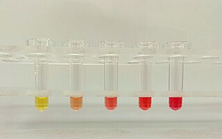
Hemolysis or haemolysis, also known by several other names, is the rupturing (lysis) of red blood cells (erythrocytes) and the release of their contents (cytoplasm) into surrounding fluid. Hemolysis may occur in vivo or in vitro.

<span class="mw-page-title-main">Thrombotic thrombocytopenic purpura</span> Medical condition

Thrombotic thrombocytopenic purpura (TTP) is a blood disorder that results in blood clots forming in small blood vessels throughout the body. This results in a low platelet count, low red blood cells due to their breakdown, and often kidney, heart, and brain dysfunction. Symptoms may include large bruises, fever, weakness, shortness of breath, confusion, and headache. Repeated episodes may occur.

<span class="mw-page-title-main">Uremia</span> Type of kidney disease, urea in the blood

Uremia is the term for high levels of urea in the blood. Urea is one of the primary components of urine. It can be defined as an excess in the blood of amino acid and protein metabolism end products, such as urea and creatinine, which would be normally excreted in the urine. Uremic syndrome can be defined as the terminal clinical manifestation of kidney failure. It is the signs, symptoms and results from laboratory tests which result from inadequate excretory, regulatory, and endocrine function of the kidneys. Both uremia and uremic syndrome have been used interchangeably to denote a very high plasma urea concentration that is the result of renal failure. The former denotation will be used for the rest of the article.

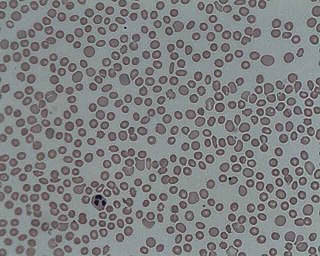
Microangiopathic hemolytic anemia (MAHA) is a microangiopathic subgroup of hemolytic anemia caused by factors in the small blood vessels. It is identified by the finding of anemia and schistocytes on microscopy of the blood film.

<span class="mw-page-title-main">Thrombocytopenia</span> Abnormally low levels of platelets in the blood

In hematology, thrombocytopenia is a condition characterized by abnormally low levels of platelets in the blood. Low levels of platelets in turn may lead to prolonged or excessive bleeding. It is the most common coagulation disorder among intensive care patients and is seen in a fifth of medical patients and a third of surgical patients.

<span class="mw-page-title-main">Hemolytic–uremic syndrome</span> Group of blood disorders related to bacterial infection

Hemolytic–uremic syndrome (HUS) is a group of blood disorders characterized by low red blood cells, acute kidney failure, and low platelets. Initial symptoms typically include bloody diarrhea, fever, vomiting, and weakness. Kidney problems and low platelets then occur as the diarrhea progresses. Children are more commonly affected, but most children recover without permanent damage to their health, although some children may have serious and sometimes life-threatening complications. Adults, especially the elderly, may present a more complicated presentation. Complications may include neurological problems and heart failure.

von Willebrand factor Mammalian protein involved in blood clotting

Von Willebrand factor (VWF) is a blood glycoprotein involved in hemostasis, specifically, platelet adhesion. It is deficient and/or defective in von Willebrand disease and is involved in many other diseases, including thrombotic thrombocytopenic purpura, Heyde's syndrome, and possibly hemolytic–uremic syndrome. Increased plasma levels in many cardiovascular, neoplastic, metabolic, and connective tissue diseases are presumed to arise from adverse changes to the endothelium, and may predict an increased risk of thrombosis.

<span class="mw-page-title-main">Hemoglobinuria</span> Abnormally increased hemoglobin in urine

Hemoglobinuria is a condition in which the oxygen transport protein hemoglobin is found in abnormally high concentrations in the urine. The condition is caused by excessive intravascular hemolysis, in which large numbers of red blood cells (RBCs) are destroyed, thereby releasing free hemoglobin into the plasma. Excess hemoglobin is filtered by the kidneys, which excrete it into the urine, giving urine a purple color. Hemoglobinuria can lead to acute tubular necrosis which is an uncommon cause of a death of uni-traumatic patients recovering in the ICU.

<span class="mw-page-title-main">Plasmapheresis</span> Removal, treatment and return of blood plasma

Plasmapheresis is the removal, treatment, and return or exchange of blood plasma or components thereof from and to the blood circulation. It is thus an extracorporeal therapy, a medical procedure performed outside the body.

<span class="mw-page-title-main">Schistocyte</span> Fragmented portion of a red blood cell

A schistocyte or schizocyte is a fragmented part of a red blood cell. Schistocytes are typically irregularly shaped, jagged, and have two pointed ends.

<span class="mw-page-title-main">Hepatorenal syndrome</span> Human disease

Hepatorenal syndrome is a life-threatening medical condition that consists of rapid deterioration in kidney function in individuals with cirrhosis or fulminant liver failure. HRS is usually fatal unless a liver transplant is performed, although various treatments, such as dialysis, can prevent advancement of the condition.

<span class="mw-page-title-main">Nephritic syndrome</span> Medical condition

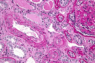
Nephritic syndrome is a syndrome comprising signs of nephritis, which is kidney disease involving inflammation. It often occurs in the glomerulus, where it is called glomerulonephritis. Glomerulonephritis is characterized by inflammation and thinning of the glomerular basement membrane and the occurrence of small pores in the podocytes of the glomerulus. These pores become large enough to permit both proteins and red blood cells to pass into the urine. By contrast, nephrotic syndrome is characterized by proteinuria and a constellation of other symptoms that specifically do not include hematuria. Nephritic syndrome, like nephrotic syndrome, may involve low level of albumin in the blood due to the protein albumin moving from the blood to the urine.

<span class="mw-page-title-main">Thrombotic microangiopathy</span> Medical condition

Thrombotic microangiopathy (TMA) is a pathology that results in thrombosis in capillaries and arterioles, due to an endothelial injury. It may be seen in association with thrombocytopenia, anemia, purpura and kidney failure.

<span class="mw-page-title-main">Calciphylaxis</span> Medical condition

Calciphylaxis, also known as calcific uremic arteriolopathy (CUA) or “Grey Scale”, is a rare syndrome characterized by painful skin lesions. The pathogenesis of calciphylaxis is unclear but believed to involve calcification of the small blood vessels located within the fatty tissue and deeper layers of the skin, blood clots, and eventual death of skin cells due to lack of blood flow. It is seen mostly in people with end-stage kidney disease but can occur in the earlier stages of chronic kidney disease and rarely in people with normally functioning kidneys. Calciphylaxis is a rare but serious disease, believed to affect 1-4% of all dialysis patients. It results in chronic non-healing wounds and indicates poor prognosis, with typical life expectancy of less than one year.

<span class="mw-page-title-main">Eculizumab</span> Pharmaceutical drug

Eculizumab, sold under the brand name Soliris among others, is a recombinant humanized monoclonal antibody used to treat paroxysmal nocturnal hemoglobinuria (PNH), atypical hemolytic uremic syndrome (aHUS), generalized myasthenia gravis, and neuromyelitis optica. In people with PNH, it reduces both the destruction of red blood cells and need for blood transfusion, but does not appear to affect the risk of death. Eculizumab was the first drug approved for each of its uses, and its approval was granted based on small trials. It is given by intravenous infusion.

<span class="mw-page-title-main">Complement factor I</span> Protein

Complement factor I, also known as C3b/C4b inactivator, is a protein that in humans is encoded by the CFI gene. Complement factor I is a protein of the complement system, first isolated in 1966 in guinea pig serum, that regulates complement activation by cleaving cell-bound or fluid phase C3b and C4b. It is a soluble glycoprotein that circulates in human blood at an average concentration of 35 μg/mL.

<span class="mw-page-title-main">C3b</span>

C3b is the larger of two elements formed by the cleavage of complement component 3, and is considered an important part of the innate immune system. C3b is potent in opsonization: tagging pathogens, immune complexes (antigen-antibody), and apoptotic cells for phagocytosis. Additionally, C3b plays a role in forming a C3 convertase when bound to Factor B, or a C5 convertase when bound to C4b and C2b or when an additional C3b molecule binds to the C3bBb complex.

Onconephrology is a specialty in nephrology that deals with the study of kidney diseases in cancer patients. A nephrologist who takes care of patients with cancer and kidney disease is called an onconephrologist. This branch of nephrology encompasses nephrotoxicity associated with existing and novel chemotherapeutics, kidney disease as it pertains to stem cell transplant, paraneoplastic kidney disorders, paraproteinemias, electrolyte disorders associated with cancer, and more as discussed below.

<span class="mw-page-title-main">Upshaw–Schulman syndrome</span> Medical condition

Upshaw–Schulman syndrome (USS) is the recessively inherited form of thrombotic thrombocytopenic purpura (TTP), a rare and complex blood coagulation disease. USS is caused by the absence of the ADAMTS13 protease resulting in the persistence of ultra large von Willebrand factor multimers (ULVWF), causing episodes of acute thrombotic microangiopathy with disseminated multiple small vessel obstructions. These obstructions deprive downstream tissues from blood and oxygen, which can result in tissue damage and death. The presentation of an acute USS episode is variable but usually associated with thrombocytopenia, microangiopathic hemolytic anemia (MAHA) with schistocytes on the peripheral blood smear, fever and signs of ischemic organ damage in the brain, kidney and heart.

Ravulizumab, sold under the brand name Ultomiris, is a humanized monoclonal antibody complement inhibitor medication designed for the treatment of paroxysmal nocturnal hemoglobinuria (PNH) and atypical hemolytic uremic syndrome. It is designed to bind to and prevent the activation of Complement component 5 (C5).

References

  1. 1 2 3 4 5 6 7 8 9 Loirat C, Noris M, Fremeaux-Bacchi V (2008). "Complement and the atypical hemolytic uremia syndrome in children". Pediatr Nephrol. 23 (11): 1957–1972. doi:10.1007/s00467-008-0872-4. PMC   6904381 . PMID   18594873.
  2. Nester, Carla M.; Thomas, Christie P. (8 December 2012). "Atypical hemolytic uremic syndrome: what is it, how is it diagnosed, and how is it treated?". ASH Education Program Book. 2012 (1): 617–625. doi: 10.1182/asheducation.v2012.1.617.3798924 . ISSN   1520-4391. PMID   23233643.
  3. Walport, MJ; Rosen, Fred S.; Walport, Mark J. (2001). "Complement". N Engl J Med. 344 (14): 1058–1066. doi:10.1056/NEJM200104053441406. PMID   11287977.
  4. Hosler GA, Cusumano AM, Hutchins GM (2003). "Thrombotic thrombocytopenic purpura and hemolytic uremic syndrome are distinct pathologic entities. A review of 56 autopsy cases". Arch. Pathol. Lab. Med. 127 (7): 834–9. doi:10.5858/2003-127-834-TTPAHU. PMID   12823037.
  5. 1 2 3 4 5 6 7 8 9 10 Noris M, Caprioli J, Bresin E, Mossali C, Pianetti G, Gamba S, et al. (October 2010). "Relative role of genetic complement abnormalities in sporadic and familial aHUS and their impact on clinical phenotype". Clin J Am Soc Nephrol. 5 (10): 1844–59. doi:10.2215/CJN.02210310. PMC   2974386 . PMID   20595690.
  6. Tzoumas, Nikolaos; Hallam, Dean; Harris, Claire L.; Lako, Majlinda; Kavanagh, David; Steel, David H.W. (November 2020). "Revisiting the role of factor H in age-related macular degeneration: Insights from complement-mediated renal disease and rare genetic variants". Survey of Ophthalmology. 66 (2): 378–401. doi:10.1016/j.survophthal.2020.10.008. ISSN   0039-6257. PMID   33157112. S2CID   226274874.
  7. 1 2 3 4 5 6 7 Caprioli, J; Noris, M; Brioschi, S; Pianetti, G; Castelletti, F; Bettinaglio, P; Mele, C; Bresin, E; Cassis, L; Gamba, S; Porrati, F; Bucchioni, S; Monteferrante, G; Fang, CJ; Liszewski, MK; Kavanagh, D; Atkinson, JP; Remuzzi, G; International Registry of Recurrent Familial HUS/TTP (2006). "Genetics of HUS: the impact of MCP, CFH, and IF mutations on clinical presentation, response to treatment, and outcome". Blood. 108 (4): 1267–1279. doi:10.1182/blood-2005-10-007252. PMC   1895874 . PMID   16621965.
  8. 1 2 3 Hoffman, R; Benz, EJ; Silberstein, LE; Heslop, H; Weitz J; Anastasi, J. (2012). Hematology: Basic Principles and Practice (6th ed.). Elsevier. ISBN   978-1-4377-2928-3.
  9. 1 2 3 Ohanian, M; Cable, C; Halka, K (2011). "Eculizumab safely reverses neurologic impairment and eliminates the need for dialysis in severe atypical hemolytic uremic syndrome". Clin Pharmacol. 5: 687–696. doi: 10.2147/CPAA.S17904 . PMC   3262387 . PMID   22287852.
  10. 1 2 3 Stahl, AL; Vaziri-Sani, F; Heinen, S; Kristoffersson, AC; Gydell, KH; Raafat, R; Gutierrez, A; Beringer, O; Zipfel, PF; Karpman, D. (2008). "Factor H dysfunction in patients with atypical hemolytic uremic syndrome contributes to complement depositionn on platelets and their activation". Blood. 111 (11): 5307–5315. doi: 10.1182/blood-2007-08-106153 . PMID   18268093.
  11. 1 2 Dragon-Durey, M-A; Sethi, SK; Bagga, A; Blanc, C; Blouin, J; Ranchin, B; André, JL; Takagi, N; Cheong, HI; Hari, P.; Le Quintrec, M.; Niaudet, P.; Loirat, C.; Fridman, W. H.; Frémeaux-Bacchi, V. (2010). "Clinical features of anti-factor H autoantibody-associated hemolytic uremic syndrome". J Am Soc Nephrol. 21 (12): 2180–2187. doi:10.1681/ASN.2010030315. PMC   3014031 . PMID   21051740.
  12. 1 2 3 4 5 Zuber, J; Le Quintrec, Moglie; Sberro-Soussan, Rebecca; Loirat, Chantal; Frémeaux-Bacchi, Véronique; Legendre, Christophe (2011). "New insights into postrenal transplant hemolytic uremic syndrome". Nature Reviews Nephrology. 7 (1): 23–25. doi:10.1038/nrneph.2010.155. PMID   21102542. S2CID   2054556.
  13. 1 2 3 4 Ariceta, G; Besbas, N; Johnson, S; Karpman, D; Landau, D; Licht, C; Loirat, C; Pecoraro, C; Taylor, CM; Van De Kar, Nicole; Vandewalle, Johan; Zimmerhackl, Lothar B.; European Paediatric Study Group for HUS (2009). "Guideline for the investigation and initial therapy of diarrhea-negative hemolytic uremic syndrome". Pediatr Nephrol. 24 (4): 687–696. doi: 10.1007/s00467-008-0964-1 . PMID   18800230. S2CID   2967560.
  14. 1 2 3 4 Sellier-Leclerc, AL; Fremeaux-Bacchi, V; Dragon-Durey, MA; Macher, MA; Niaudet, P; Guest, G; Boudailliez, B; Bouissou, F; Deschenes, G; Gie, S.; Tsimaratos, M.; Fischbach, M.; Morin, D.; Nivet, H.; Alberti, C.; Loirat, C.; French Society of Pediatric Nephrology (2007). "Differential impact of complement mutations on clinical characteristics in atypical hemolytic uremic syndrome". J Am Soc Nephrol. 18 (8): 2392–2400. doi: 10.1681/ASN.2006080811 . PMID   17599974.
  15. 1 2 Sallee, M; Daniel, L; Piercecchi, MD; Jaubert, D; Fremeaux-Bacchi, V; Berland, Y; Burtey, S (2010). "Myocardial infarction is a complication of factor H-associated atypical HUS". Nephrol Dial Transplant. 25 (6): 2028–2032. doi: 10.1093/ndt/gfq160 . PMID   20305136.
  16. Neuhaus, TJ; Calonder, S; Leumann, EP (1997). "Heterogeneity of atypical haemolytic uraemic syndromes". Arch Dis Child. 76 (6): 518–521. doi:10.1136/adc.76.6.518. PMC   1717216 . PMID   9245850.
  17. Noris, M; Remuzzi, G (2005). "Hemolytic uremic syndrome". J Am Soc Nephrol. 16 (4): 1035–1050. doi: 10.1681/ASN.2004100861 . PMID   15728781.
  18. 1 2 3 4 Noris, M; Remuzzi, G (2009). "Atypical hemolytic-uremic syndrome". N Engl J Med. 361 (17): 1676–1687. doi:10.1056/NEJMra0902814. PMID   19846853. S2CID   205115628.
  19. 1 2 3 Benz, K; Amann, K (2010). "Thrombotic microangiopathy: new insights". Current Opinion in Nephrology and Hypertension. 19 (3): 242–247. doi:10.1097/MNH.0b013e3283378f25. PMID   20186056. S2CID   25429151.
  20. Mache, CJ; Acham-Roschitz, B.; Fremeaux-Bacchi, V; Kirschfink, M; Zipfel, PF; Roedl, S; Vester, U; Ring, E (2009). "Complement inhibitor eculizumab in atypical hemolytic uremic syndrome". Clinical Journal of the American Society of Nephrology. 4 (8): 1312–1316. doi:10.2215/CJN.01090209. PMC   2723971 . PMID   19556379.
  21. 1 2 Zipfel, PF; Heinen, S; Skerka, C (2010). "Thrombotic microangiopathies:new insights and new challenges". Current Opinion in Nephrology and Hypertension. 19 (4): 372–378. doi:10.1097/MNH.0b013e32833aff4a. PMID   20539230. S2CID   28419.
  22. 1 2 3 Tsai, H-M (2010). "Pathophysiology of thrombotic thrombocytopenic purpura". Int J Hematol. 91 (1): 1–19. doi:10.1007/s12185-009-0476-1. PMC   3159000 . PMID   20058209.
  23. 1 2 3 George, JN (2010). "How I treat patients with thrombotic thrombocytopenic purpura". Blood. 116 (20): 4060–4069. doi: 10.1182/blood-2010-07-271445 . PMID   20686117. S2CID   26844964.
  24. Moake, JL (2004). "von Willebrand factor, ADAMTS-13, and thrombotic thrombocytopenic purpura". Semin Hematol. 41 (1): 4–14. doi:10.1053/j.seminhematol.2003.10.003. PMID   14727254.
  25. Fang CJ, Fremeaux-Bacchi V, Liszewski MK, et al. (2008). "Membrane cofactor protein mutations in atypical hemolytic uremic syndrome (aHUS), fatal Stx-HUS, C3 glomerulonephritis, and the HELLP syndrome". Blood. 111 (2): 624–632. doi:10.1182/blood-2007-04-084533. PMC   2200836 . PMID   17914026.
  26. 1 2 Bitzan, M; Schaefer, Franz; Reymond, Didier (2010). "Treatment of typical (enteropathic) hemolytic uremic syndrome". Semin Thromb Hemost. 36 (6): 594–610. doi:10.1055/s-0030-1262881. PMID   20865636. S2CID   260318768.
  27. "Woodward L, Burke L, Shah K , 2021, Patients' experience and perception of the diagnosis of the rare disease, atypical Hemolytic Uremic Syndrome" (PDF). Archived (PDF) from the original on 2021-09-24.
  28. Michon, B; Moghrabi, A; Winikoff, R; Barrette, S; Bernstein, ML; Champagne, J; David, M; Duval, M; Hume, HA; Robitaille, Nancy; Bélisle, André; Champagne, Martin A. (2007). "Complications of apheresis in children". Transfusion. 47 (10): 1837–1842. doi:10.1111/j.1537-2995.2007.01405.x. PMID   17880609. S2CID   23613105.
  29. Licht, C; Pluthero, FG; Li, L; Christensen, H; Habbig, S; Hoppe, B; Geary, DF; Zipfel, PF; Kahr, WH (2009). "Platelet-associated complement factor H in healthy persons and patients with atypical HUS". Blood. 114 (20): 4538–4545. doi: 10.1182/blood-2009-03-205096 . PMID   19704120. S2CID   206878740.
  30. Szczepiorkowski, ZM; Winters, Jeffrey L.; Bandarenko, Nicholas; Kim, Haewon C.; Linenberger, Michael L.; Marques, Marisa B.; Sarode, Ravindra; Schwartz, Joseph; Weinstein, Robert; Shaz, Beth H.; Apheresis Applications Committee of the American Society for Apheresis (2010). "Guidelines on the use of therapeutic apheresis in clinical practice- evidence based approach from the Apheresis Applications Committee of the American Society of Apheresis". J Clin Apheresis. 25 (3): 83–177. doi:10.1002/jca.20240. PMID   20568098. S2CID   7157740.
  31. 1 2 Keating, GM (December 2013). "Eculizumab: a review of its use in atypical haemolytic uraemic syndrome". Drugs. 73 (18): 2053–66. doi:10.1007/s40265-013-0147-7. PMID   24249647. S2CID   36682579.
  32. "FDA approves Soliris for rare pediatric blood disorder: Orphan drug receives second approval for rare disease", FDA, 23 September 2011, retrieved 25 June 2015
  33. "Eculizumab for treating atypical haemolytic uraemic syndrome | Guidance and guidelines | NICE". nice.org.uk. 28 January 2015.
  34. "aHUS | Alexion". alexion.com. Retrieved 2020-08-19.
  35. "Ultomiris® (ravulizumab-cwvz) | Alexion". alexion.com. Retrieved 2020-08-19.
  36. Collins, AJ; Foley, Robert N.; Herzog, Charles; Chavers, Blanche M; Gilbertson, David; Ishani, Areef; Kasiske, Bertram L.; Liu, Jiannong; Mau, Lih-Wen; McBean, Marshall (2010). "Excerpts from the US Renal Data System 2009 annual report". Am J Kidney Dis. 55 (1 Suppl 1): S1–S7. doi:10.1053/j.ajkd.2009.10.009. PMC   2829836 . PMID   20082919.
  37. "European Renal Association-European Dialysis and Transplant Association Registry". ERA-EDTA Registry Annual Report 2009. The Netherlands: Academic Medical Center, Department of Medical Informatics. 2011.
  38. Klevens, RM; Edwards, JR; Andrus, ML; Peterson, KD; Dudeck, MA; Horan, TC; NHSN Participants in Outpatient Dialysis Surveillance (2008). "Dialysis surveillance report: National Healthcare Safety Network (NHSN)- data summary for 2006". Semin Dial. 21 (1): 24–28. CiteSeerX   10.1.1.397.9342 . doi:10.1111/j.1525-139X.2007.00379.x. PMID   18251954. S2CID   18800012.
  39. 1 2 Kavanagh, D; Richards, A; Goodship, T; Jalanko, H (2010). "Transplantation in atypical hemolytic uremic syndrome". Semin Thromb Hemost. 36 (6): 653–659. doi: 10.1055/s-0030-1262887 . PMID   20865642.
  40. Nester, C; Stewart, Z; Myers, D; Jetton, J; Nair, R; Reed, A; Thomas, C; Smith, R; Brophy, P (2011). "Pre-emptive eculizumab and plasmapheresis for renal transplant in atypical hemolytic syndrome". Clin J Am Soc Nephrol. 6 (6): 1488–1494. doi:10.2215/CJN.10181110. PMC   3109948 . PMID   21617085.
  41. Pugh, Dan; O'Sullivan, Eoin D.; Duthie, Fiona AI; Masson, Philip; Kavanagh, David (2021). "Interventions for atypical haemolytic uraemic syndrome". Cochrane Database of Systematic Reviews. 2021 (3): CD012862. doi:10.1002/14651858.CD012862.pub2. PMC   8078160 . PMID   33783815.
  42. Bresin, E; Daina, E; Noris, M; Castelletti, F; Stefanov, R; Hill, P; Goodship, TH; Remuzzi, G; International Registry of Recurrent Familial HUS/TTP (2006). "Outcome of renal transplantation in patients with non- Shiga toxin-associated hemolytic uremic syndrome: prognostic significance of genetic background" (PDF). Clin J Am Soc Nephrol. 1 (1): 88–99. doi:10.2215/CJN.00050505. PMID   17699195. S2CID   40102561.
  43. Bouts, A; Monnens, Leo; Davin, Jean-Claude; Struijk, Geertrude; Spanjaard, Lodewijk (2011). "Insufficient protection by Neisseria meningitidis vaccination alone during eculizumab therapy". Pediatr Nephrol. 26 (10): 1919–1920. doi:10.1007/s00467-011-1929-3. PMC   3163808 . PMID   21643943.
  44. 1 2 Menne, Jan; Delmas, Yahsou; Fakhouri, Fadi; Licht, Christoph; Lommelé, Åsa; Minetti, Enrico E.; Provôt, François; Rondeau, Eric; Sheerin, Neil S.; Wang, Jimmy; Weekers, Laurent E. (2019-04-10). "Outcomes in patients with atypical hemolytic uremic syndrome treated with eculizumab in a long-term observational study". BMC Nephrology. 20 (1): 125. doi: 10.1186/s12882-019-1314-1 . ISSN   1471-2369. PMC   6456946 . PMID   30971227.
  45. Zimmerhackl, LR; Besbas, N; Jungraithmayr, T; Van De Kar, N; Karch, H; Karpman, D; Landau, D; Loirat, C; Proesmans, W; Prüfer, Friederike; Rizzoni, Gianfranco; Taylor, Mark; European Study Group for Haemolytic Uraemic Syndromes Related Disorders (2006). "Epidemiology, clinical presentation, and pathophysiology of atypical and recurrent hemolytic uremic syndrome". Semin Thromb Hemost. 32 (2): 113–120. doi:10.1055/s-2006-939767. PMID   16575686. S2CID   40037641.
  46. Noris, Marina; Bresin, Elena; Mele, Caterina; Remuzzi, Giuseppe (1993), Adam, Margaret P.; Mirzaa, Ghayda M.; Pagon, Roberta A.; Wallace, Stephanie E. (eds.), "Genetic Atypical Hemolytic-Uremic Syndrome", GeneReviews®, Seattle (WA): University of Washington, Seattle, PMID   20301541 , retrieved 2023-09-30
  47. Kaushansky, K; Lichtman, M; Beutler, E; Kipps, T; Prchal, J; Seligsohn, U. (2010). Williams Hematology (8th ed.). McGraw-Hill. ISBN   978-0071621519.
  48. Woodward, Len (2016). "An innovative and collaborative partnership between patients with rare disease and industry-supported registries: the Global aHUS Registry". Orphanet Journal of Rare Diseases. 11 (1): 154. doi: 10.1186/s13023-016-0537-5 . PMC   5117495 . PMID   27871301.