The brain is an organ that serves as the center of the nervous system in all vertebrate and most invertebrate animals. The brain is located in the head, usually close to the sensory organs for senses such as vision. The brain is the most complex organ in a vertebrate's body. In a human, the cerebral cortex contains approximately 14–16 billion neurons, and the estimated number of neurons in the cerebellum is 55–70 billion. Each neuron is connected by synapses to several thousand other neurons. These neurons communicate with one another by means of long protoplasmic fibers called axons, which carry trains of signal pulses called action potentials to distant parts of the brain or body targeting specific recipient cells.

A nerve is an enclosed, cable-like bundle of nerve fibres called axons, in the peripheral nervous system. A nerve provides a common pathway for the electrochemical nerve impulses called action potentials that are transmitted along each of the axons to peripheral organs or, in the case of sensory nerves, from the periphery back to the central nervous system. Each axon within the nerve is an extension of an individual neuron, along with other supportive cells such as Schwann cells that coat the axons in myelin.

The nervous system is the part of an animal that coordinates its actions by transmitting signals to and from different parts of its body. The nervous system detects environmental changes that impact the body, then works in tandem with the endocrine system to respond to such events. Nervous tissue first arose in wormlike organisms about 550 to 600 million years ago. In vertebrates it consists of two main parts, the central nervous system (CNS) and the peripheral nervous system (PNS). The CNS consists of the brain and spinal cord. The PNS consists mainly of nerves, which are enclosed bundles of the long fibers or axons, that connect the CNS to every other part of the body. Nerves that transmit signals from the brain are called motor or efferent nerves, while those nerves that transmit information from the body to the CNS are called sensory or afferent. Spinal nerves serve both functions and are called mixed nerves. The PNS is divided into three separate subsystems, the somatic, autonomic, and enteric nervous systems. Somatic nerves mediate voluntary movement. The autonomic nervous system is further subdivided into the sympathetic and the parasympathetic nervous systems. The sympathetic nervous system is activated in cases of emergencies to mobilize energy, while the parasympathetic nervous system is activated when organisms are in a relaxed state. The enteric nervous system functions to control the gastrointestinal system. Both autonomic and enteric nervous systems function involuntarily. Nerves that exit from the cranium are called cranial nerves while those exiting from the spinal cord are called spinal nerves.
Electrophysiology is the branch of physiology that studies the electrical properties of biological cells and tissues. It involves measurements of voltage changes or electric current or manipulations on a wide variety of scales from single ion channel proteins to whole organs like the heart. In neuroscience, it includes measurements of the electrical activity of neurons, and, in particular, action potential activity. Recordings of large-scale electric signals from the nervous system, such as electroencephalography, may also be referred to as electrophysiological recordings. They are useful for electrodiagnosis and monitoring.
Behavioral neuroscience, also known as biological psychology, biopsychology, or psychobiology, is the application of the principles of biology to the study of physiological, genetic, and developmental mechanisms of behavior in humans and other animals.
Neural engineering is a discipline within biomedical engineering that uses engineering techniques to understand, repair, replace, enhance, or otherwise exploit the properties of neural systems. Neural engineers are uniquely qualified to solve design problems at the interface of living neural tissue and non-living constructs.

Neural oscillations, or brainwaves, are rhythmic or repetitive patterns of neural activity in the central nervous system. Neural tissue can generate oscillatory activity in many ways, driven either by mechanisms within individual neurons or by interactions between neurons. In individual neurons, oscillations can appear either as oscillations in membrane potential or as rhythmic patterns of action potentials, which then produce oscillatory activation of post-synaptic neurons. At the level of neural ensembles, synchronized activity of large numbers of neurons can give rise to macroscopic oscillations, which can be observed in an electroencephalogram. Oscillatory activity in groups of neurons generally arises from feedback connections between the neurons that result in the synchronization of their firing patterns. The interaction between neurons can give rise to oscillations at a different frequency than the firing frequency of individual neurons. A well-known example of macroscopic neural oscillations is alpha activity.
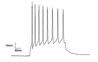
In neuroscience, single-unit recordings provide a method of measuring the electro-physiological responses of single neurons using a microelectrode system. When a neuron generates an action potential, the signal propagates down the neuron as a current which flows in and out of the cell through excitable membrane regions in the soma and axon. A microelectrode is inserted into the brain, where it can record the rate of change in voltage with respect to time. These microelectrodes must be fine-tipped, high-impedance conductors; they are primarily glass micro-pipettes or metal microelectrodes made of platinum or tungsten. Microelectrodes can be carefully placed close to the cell membrane, allowing the ability to record extracellularly.
Sensory neuroscience is a subfield of neuroscience which explores the anatomy and physiology of neurons that are part of sensory systems such as vision, hearing, and olfaction. Neurons in sensory regions of the brain respond to stimuli by firing one or more nerve impulses following stimulus presentation. How is information about the outside world encoded by the rate, timing, and pattern of action potentials? This so-called neural code is currently poorly understood and sensory neuroscience plays an important role in the attempt to decipher it. Looking at early sensory processing is advantageous since brain regions that are "higher up" contain neurons which encode more abstract representations. However, the hope is that there are unifying principles which govern how the brain encodes and processes information. Studying sensory systems is an important stepping stone in our understanding of brain function in general.
A cultured neuronal network is a cell culture of neurons that is used as a model to study the central nervous system, especially the brain. Often, cultured neuronal networks are connected to an input/output device such as a multi-electrode array (MEA), thus allowing two-way communication between the researcher and the network. This model has proved to be an invaluable tool to scientists studying the underlying principles behind neuronal learning, memory, plasticity, connectivity, and information processing.
Microelectrode arrays (MEAs) are devices that contain multiple microelectrodes through which neural signals are obtained or delivered, essentially serving as neural interfaces that connect neurons to electronic circuitry. There are two general classes of MEAs: implantable MEAs, used in vivo, and non-implantable MEAs, used in vitro.

Electroencephalography (EEG) is an electrophysiological monitoring method to record electrical activity of the brain. It is typically noninvasive, with the electrodes placed along the scalp, although invasive electrodes are sometimes used, as in electrocorticography. EEG measures voltage fluctuations resulting from ionic current within the neurons of the brain. Clinically, EEG refers to the recording of the brain's spontaneous electrical activity over a period of time, as recorded from multiple electrodes placed on the scalp. Diagnostic applications generally focus either on event-related potentials or on the spectral content of EEG. The former investigates potential fluctuations time locked to an event, such as 'stimulus onset' or 'button press'. The latter analyses the type of neural oscillations that can be observed in EEG signals in the frequency domain.

The neurotrophic electrode is an intracortical device designed to read the electrical signals that the brain uses to process information. It consists of a small, hollow glass cone attached to several electrically conductive gold wires. The term neurotrophic means "relating to the nutrition and maintenance of nerve tissue" and the device gets its name from the fact that it is coated with Matrigel and nerve growth factor to encourage the expansion of neurites through its tip. It was invented by neurologist Dr. Philip Kennedy and was successfully implanted for the first time in a human patient in 1996 by neurosurgeon Roy Bakay.
A hippocampus prosthesis is a type of cognitive prosthesis. Prosthetic devices replace normal function of a damaged body part; this can be simply a structural replacement or a rudimentary, functional replacement. However, prosthetics involving the brain have some special categories and requirements. "Input" prosthetics, such as retinal or cochlear implant, supply signals to the brain that the patient eventually learns to interpret as sight or sound. "Output" prosthetics use brain signals to drive a bionic arm, hand or computer device, and require considerable training during which the patient learns to generate the desired action via their thoughts. Both of these types of prosthetics rely on the plasticity of the brain to adapt to the requirement of the prosthesis, thus allowing the user to "learn" the use of his new body part. A cognitive or "brain-to-brain" prosthesis involves neither learned input nor output signals, but the native signals used normally by the area of the brain to be replaced. Thus, such a device must be able to fully replace the function of a small section of the nervous system—using that section's normal mode of operation. In order to achieve this, developers require a deep understanding of the functioning of the nervous system. The scope of design must include a reliable mathematical model as well as the technology in order to properly manufacture and install a cognitive prosthesis. The primary goal of an artificial hippocampus is to provide a cure for Alzheimer's disease and other hippocampus—related problems. To do so, the prosthesis has to be able to receive information directly from the brain, analyze the information and give an appropriate output to the cerebral cortex; in other words, it must behave just like a natural hippocampus. At the same time, the artificial organ must be completely autonomous, since any exterior power source will greatly increase the risk of infection.
The evolution of nervous systems dates back to the first development of nervous systems in animals. Neurons developed as specialized electrical signaling cells in multicellular animals, adapting the mechanism of action potentials present in motile single-celled and colonial eukaryotes. Simple nerve nets seen in animals like Cnidaria (jellyfish) evolved first, consisted of polymodal neurons which serve a dual purpose in motor and sensory functions. Cnidarians can be compared to Ctenophores, which although are both jellyfish, have very different nervous systems. Unlike Cnidarians, Ctenophores have neurons that use electrochemical signaling. This was perplexing because the phylum Ctenophora was considered to be more ancient than that of Porifera (sponges), which have no nervous system at all. This led to the rise of two theories which described how the early nervous system came about. One theory stated that the nervous system came about in an ancestor basal to all of these phylum, however was lost in Porifera. The other theory states that the nervous system arose independently twice, one basal to Cnidarians and one basal to Ctenophores. Bilateral animals – ventral nerve cords in invertebrates and dorsal nerve cords supported by a notochord in chordates-- evolved with a central nervous system that was around a central region, a process known as cephalization.

Restorative neurology is a branch of neurology dedicated to improving functions of the impaired nervous system through selective structural or functional modification of abnormal neurocontrol according to underlying mechanisms and clinically unrecognized residual functions. When impaired, the body naturally reconstructs new neurological pathways and redirects activity. The field of restorative neurology works to accentuate these new pathways and primarily focuses on the theory of the plasticity of an impaired nervous system. Its main goal is to take a broken down and disordered nervous system and return it to a state of normal function. Certain treatment strategies are used to augment instead of fully replace any performance of surviving and also improving the potential of motor neuron functions. This rehabilitation of motor neurons allows patients a therapeutic approach to recovery opposed to physical structural reconstruction. It is applied in a wide range of disorders of the nervous system, including upper motor neuron dysfunctions like spinal cord injury, cerebral palsy, multiple sclerosis and acquired brain injury including stroke, and neuromuscular diseases as well as for control of pain and spasticity. Instead of applying a reconstructive neurobiological approach, i.e. structural modifications, restorative neurology relies on improving residual function. While subspecialties like neurosurgery and pharmacology exist and are useful in diagnosing and treating conditions of the nervous system, restorative neurology takes a pathophysiological approach. Instead of heavily relying on neurochemistry or perhaps an anatomical discipline, restorative neurology encompasses many fields and blends them together.
The following outline is provided as an overview of and topical guide to brain mapping:
A chronic electrode implant is an electronic device implanted chronically into the brain or other electrically excitable tissue. It may record electrical impulses in the brain or may stimulate neurons with electrical impulses from an external source.