History of emerging infectious diseases

Last updated

The discovery of disease-causing pathogens is an important activity in the field of medical science. Many viruses, bacteria, protozoa, fungi, helminthes and prions are identified as a confirmed or potential pathogen. In the United States, a Centers for Disease Control program, begun in 1995, identified over a hundred patients with life-threatening illnesses that were considered to be of an infectious cause, but that could not be linked to a known pathogen. [1] The association of pathogens with disease can be a complex and controversial process, in some cases requiring decades or even centuries to achieve.

Contents

Factors impairing identification of pathogens

Factors which have been identified as impeding the identification of pathogens include the following:

1. Lack of animal models: Experimental infection in animals has been used as a criterion to demonstrate a disease-causing ability, but for some pathogens (such as Vibrio cholerae , which cause disease only in humans) animal models do not exist. In cases where animal models were not available, scientists have sometimes infected themselves or others to determine an organism's disease causing ability. [2]
2. Pre-existing theories of disease: Before a pathogen is well-recognized, scientists may attribute the symptoms of infection to other causes, such as toxicological, psychological, or genetic causes. Once a pathogen has been associated with an illness, researchers have reported difficulty displacing these pre-existing theories. [3] [4]
3. Variable pathogenicity: Infection with pathogens can produce varying responses in hosts, complicating the process of showing a relationship between infection and the pathogen. [5] In some infectious diseases, the severity of symptoms has been shown to be dependent on specific genetic traits of the host. [6] [7]
4. Organisms that look alike but behave differently: In some cases a harmless organism exists which looks identical to a disease causing organism with a microscope, which complicates the discovery process. [8]
5. Lack of research effort: Slow progress has been attributed to the small numbers of researchers working on a pathogen. [9]

19th-century discoveries

Vibrio cholera (1849-1884)

Vibrio cholera bacteria are transmitted through contaminated water. [10] Once ingested, the bacteria colonizes the intestinal tract of the host and produces a toxin which causes body fluids to flow across the lining of the intestine. Death can result in 2–3 hours from dehydration if no treatment is provided. [11]

Before the discovery of an infectious cause, the symptoms of cholera were thought to be caused by an excess of bile in the patient; [12] the disease cholera gets its name from the Greek word choler meaning bile. This theory was consistent with humorism, and led to such medical practices as bloodletting. [12] The bacterium was first reported in 1849 by Gabriel Pouchet, who discovered it in stools from patients with cholera, but he did not appreciate the significance of this presence. [13] The first scientist to understand the significance of Vibrio cholerae was the Italian anatomist Filippo Pacini, who published detailed drawings of the organism in "Microscopical observations and pathological deductions on cholera" in 1854. [4] He published further papers in 1866, 1871, 1876 and 1880, which were ignored by the scientific community. [4] He correctly described how the bacteria caused diarrhea, and developed treatments that were found to be effective. [4] Whilst John Snow's edpidemiology maps were well recognised, and led to the removal of the Broad Street pump handle, e.g. 1854 Broad Street cholera outbreak. In 1874, scientific representatives from 21 countries voted unanimously to resolve that cholera was caused by environmental toxins from miasmatas , or clouds of unhealthy substances which float in the air. [14] In 1884, Robert Koch re-discovered Vibrio cholerae as a causal element in cholera. Some scientists opposed the new theory, and even drank cholera cultures to disprove it:

Koch announced his discovery of the cholera vibrio in 1884. His conclusions were based upon the constant finding of the peculiar "comma bacillus" in the stools of cholera patients, and the failure to demonstrate this organism in the feces of other persons. It was not possible to reproduce typical cholera in laboratory animals. At the time the "germ theory" of disease had not yet obtained general acceptance, and Koch's announcement was received with considerable skepticism, particularly after it was found that similar "comma bacilli" could be found at times in the feces of persons not suffering from cholera, and often in all sorts of other environments - well and river waters, cheese, etc. We now know that these were saprotrophic species of Vibrio, which may be differentiated from the cholera vibrio by cultural and immunological methods. But the correctness of Koch's opinion was dramatically demonstrated by von Pettenkofer and Emmerich who, doubting the etiological relationship of Koch's organisms, deliberately drank cultures of it. Von Pettenkofer developed merely a transient diarrhea, but Emmerich suffered from a typical and severe attack of cholera.

by A. T. Henrici, The Biology of Bacteria, DC Heath and Company, 1939. ASIN B00085GABK,

Von Pettenkofer considered his experience proof that Vibrio cholerae was harmless, as he did not develop cholera from consuming the culture. Between 1849 when Pouchet discovered Vibrio cholerae and 1891, over a million people died in cholera epidemics in Europe and Russia. [15] In 1995, researchers published a study in Science explaining why some persons are able to be infected with cholera without symptoms, possibly explaining why Pettenkofer did not get sick. [6] The study showed that a series of genetic mutations in some people provide resistance to cholera toxin; but these mutations come at a price. If too many of them occur in a person, they will develop cystic fibrosis, an incurable and often fatal genetic disorder.

20th-century discoveries

Giardia lamblia (1681-1975)

Giardiasis is a disease caused by infection with the protozoan Giardia lamblia . Infection with Giardia can produce diarrhea, gas, and abdominal pain in some people. If untreated, infection can be chronic. In children, chronic Giardia infection can cause stunting (stunted growth) and lowered intelligence, [16] Infection with Giardia is now universally recognized as a disease, and treated by physicians with anti-protozoal drugs. Since 2002, Giardia cases must be reported to the Center for Disease Control, according to the CDC's Reportable Disease Spreadsheet. [17] The United States National Institutes of Health Gastrointestinal Parasites Lab studies Giardia almost exclusively.

However, Giardia experienced an extraordinarily long term of emergence, from its discovery in 1681, until the 1970s when it was fully accepted that infection with Giardia was a treatable cause of chronic diarrhea:

Giardia lamblia was first discovered by Leeuwenhoeck (1681) who found the parasite in his own {diarrheal} stools. It was long considered to be a harmless commensal organism, but in recent years has been recognized as a cause of intestinal disease often acquired by travelers to foreign countries, persons drinking contaminated water in this country, children in day care nurseries and homosexual males. It is the most common pathogenic intestinal parasite in the United States, being found in 4% of stool specimens submitted to state public health laboratories for parasite examination. Attesting to its increasing importance in the United States, a symposium on Giardiasis, sponsored by the Environmental Protection Agency, was held in the fall of 1978. [5]

by JW Smith, Giardiasis, 1980

Some of the first evidence in modern times of Giardia's pathogenicity came during World War II when soldiers were treated for malaria with the antiprotozoal Quinacrine, and their diarrhea disappeared, as did the Giardia from their stool samples. In 1954, Dr. R.C. Rendtorff performed experiments on prisoner volunteers, infecting them with Giardia . [18] In the experiment, although some prisoners experienced changes in stool habits, he concluded that these could not be conclusively linked to Giardia infection, and also indicated that all prisoners experienced spontaneous clearance of Giardia. His experiments were described in the EPA Symposium on Waterborne Transmission of Giardiasis in 1979:

We also included Giardia lamblia, which at that time was not generally believed to be an invasive pathogenic parasite of man. Giardia was thought in the 1950s to cause occasional problems of diarrhea in children but its appearance was so common and, in adults so lacking in clinical symptomatology, that most considered it a non-pathogen. As a result we felt safe in exposing prisoners to Giardia... [18]

by Dr. RC Rendtorff in an EPA Symposium on the Waterborne Transmission of Giardiasis - in 1979

In 1954–55, an outbreak of Giardia infection occurred in Oregon (US), sickening 50,000 people. [19] This was documented in a communication by Dr. Lyle Veazie, which wasn't published until 15 years later in the New England Journal of Medicine. In the communication, Veazie notes that he was unable to find a publisher for his account of the epidemic. The communication was re-published in the EPA Symposium on Waterborne Transmission of Giardiasis in 1979, and that version included the following quote from the Director of the Oregon State Board of Health, suggesting that diarrhea from Giardia was still being attributed to other causes by health authorities in 1954:

While an unidentified virus seems the most likely etiologic agent, the unusual prevalence of Giardia lamblia cysts in stools of patients seems worthy of record. [19]

by Oregon State Board of Health commenting on 1954-55 outbreak of Giardiasis, as quoted in Veazie, 1979.

Helicobacter pylori (1892-1982)

Infection with the bacteria Helicobacter pylori is the cause of most stomach ulcers. The discovery is generally credited to Australian gastroenterologists Dr. Barry Marshall and Dr. J Robin Warren, who published their findings in 1983. The pair received the Nobel Prize in 2005 for their work. Before this, nobody really knew what caused stomach ulcers, though a popular belief was that the "stress" played a role. Some researchers suggested that ulcers were a psychosomatic illness. [20] [21] [22]

In H Pylori Pioneers, Dr. Marshall noted that other physicians had produced evidence of H. pylori infection as early as 1892. [3] Marshall writes that earlier reports were disregarded because they conflicted with existing belief. The first description of H. Pylori came in 1892 from Giulio Bizzozero, who identified acid-tolerant bacteria living in a dog's stomach. Later, a theory would be developed that no bacteria could live in the stomach. [3] Although the theory has no scientific basis, it would become a stumbling block for scientists, discouraging them for searching for infective causes of stomach ulcers. In 1940, two physicians, Dr. A. Stone Freeberg and Dr. Louis E. Barron published a paper describing a spiral bacteria found in about half of their gastroenterology patients who had stomach ulcers. Dr. John Lykoudis, a Greek physician, was one of the first physicians to treat stomach ulcers as an infectious disease. Between 1960 and 1970, he treated over 10,000 ulcer patients in Athens with antibiotics. [23] Lykoudis tried to publish a paper on his findings, but they conflicted with traditional theory, and his work was never published. [3] Lykoudis' experience was followed in 1975 by a further publication in Gut magazine that included spiral bacteria living on the borders of duodonal ulcers. [24] The medical significance of Steer's findings was disregarded, but he “continued to publish papers on H. Pylori , mostly as a hobby." [3]

H. pylori can infect the stomach of some people without causing stomach ulcers. In investigating asymptomatic carriers of H. pylori , researchers identified a genetic trait called Interleuik-1 beta-31 which causes increased production of stomach acid, resulting in ulcers if patients become infected with H. pylori . Patients without the trait do not develop stomach ulcers in response to H. pylori infection, but instead have increased risk from stomach cancer if they become infected. [7] Investigation into other gastrointestinal infections has also shown that the symptoms are the result of interaction between the infection and specific genetic mutations in the host.

Pathogenic variants of Escherichia coli (1947-1983)

There are different types of E. coli , some of which are found in humans and are harmless. Enterotoxigenic Escherichia coli (ETEC) is a type found to cause illness in humans, possessing gene that allows it to manufacture a substance toxic to humans. Cattle are immune to its effects but when people eat food contaminated with cattle feces, the organism can cause disease. Reports of pathogenic E. coli appear in medical literature as early as 1947. [25] Publications regarding variants of E. coli which cause disease appeared regularly in medical journals throughout the 1950s, 60s, and 70s, [26] [27] [28] [29] [30] with fatalities being reported in humans and infants starting in the 1970s. [31] [32] [33] Despite the earlier reports, pathogenic E. coli did not rise to public prominence until 1983, when a Center for Disease Control researcher published a paper identifying ETEC as the cause of a series of outbreaks of unexplained hemorrhagic gastrointestinal illness. [34] Despite the earlier publication of pathogenic variants of E. coli, researchers encountered significant difficulties in establishing ETEC as a pathogen. [35]

Human immunodeficiency virus (1959-1984)

AIDS was first reported June 5, 1981, when the U.S. Centers for Disease Control and Prevention recorded a cluster of Pneumocystis carinii pneumonia (now still classified as PCP but known to be caused by Pneumocystis jirovecii) in five homosexual men in Los Angeles. [36] The discovery of the virus took several years of research, and was announced in 1984 by Dr. Gallo of the National Cancer Institute, Dr. Luc Montagnier at the Pasteur Institute in Paris, and Dr. Jay Levy at the University of California, San Francisco.

However, HIV existed long before the 1981 CDC report. Three of the earliest known instances of HIV infection are as follows:

  1. A plasma sample taken in 1959 from an adult male living in what is now the Democratic Republic of the Congo. [37]
  2. HIV found in tissue samples from a 15-year-old African-American teenager who died in St. Louis in 1969. [38]
  3. HIV found in tissue samples from a Norwegian sailor who died around 1976. [39]

Two species of HIV infect humans: HIV-1 and HIV-2. HIV-1 is more virulent and more easily transmitted. HIV-1 is the source of the majority of HIV infections throughout the world, while HIV-2 is not as easily transmitted and is largely confined to West Africa. [40] Both HIV-1 and HIV-2 are of primate origin. The origin of HIV-1 is the central common chimpanzee (Pan troglodytes troglodytes) found in southern Cameroon. [41] It is established that HIV-2 originated from the sooty mangabey (Cercocebus atys), an Old World monkey of Guinea Bissau, Gabon, and Cameroon.

It is hypothesized that HIV probably transferred to humans as a result of direct contact with primates, for instance during hunting, butchery, or inter-species sexual contact. [42]

Cyclospora (1995)

Cyclospora is a gastrointestinal pathogen that causes fever, diarrhea, vomiting, and severe weight loss. Outbreaks of the disease occurred in Chicago in 1989 and other areas in the United States. [35] But investigation by the Center for Disease Control could not identify an infectious cause. The discovery of the cause was made by Mr. Ramachandran Rajah, the head of a medical clinic's laboratory in Kathmandu, Nepal. Mr. Rajah was trying to discover why local residents and visitors were becoming ill every summer. [35] He identified an unusual looking organism in stool samples from patients who were sick. But when the clinic sent slides of the organism to the Center for Disease Control, it was identified as blue-green algae, which is harmless. Many pathologists had seen the same thing before, but dismissed it as irrelevant to the patient's disease. [35] Later, the organism would be identified as a special kind of parasite, and treatment would be developed to help patients with the infection. In the United States, Cyclospora infection must be reported to the Center for Disease Control according to the CDC's Reportable Disease Chart

Present day discoveries

The process of identifying new infectious agents continues. One study has suggested there are a large number of pathogens already causing illness in the population, but they have not yet been properly identified. [1]

Gastrointestinal pathogens

Percentage of stool samples from US states found to contain various protozoa in 2000 HISTORY EMERGING AMIN 2000.JPG
Percentage of stool samples from US states found to contain various protozoa in 2000
Number of stool samples from Canadian Lab found to contain various protozoa in 2005 HISTORY EMERGING CMAJ 2005.JPG
Number of stool samples from Canadian Lab found to contain various protozoa in 2005

Many recently emerged pathogens infect the gastrointestinal tract. For example, there are three gastrointestinal protozoal infections which must be reported to the Center for Disease Control. [45] They are Giardia, Cyclospora and Cryptosporidium, and none of them were known to be significant pathogens in the 1970s.

Figure 1 shows the prevalence of gastrointestinal protozoa in studies from the United States and Canada. [44] [43] The most prevalent protozoa in these studies are considered emerging infectious diseases by some researchers, because a consensus does not yet exist in the medical and public health spheres concerning their importance in the role of human disease. [46] [47] Researchers have suggested that their treatment may be complicated by differing opinions regarding pathogenicity, lack of reliable testing procedures, and lack of reliable treatments. [48] As with newly discovered pathogens before them, researchers are reporting that these organisms may be responsible for illnesses for which no clear cause has been found, such as irritable bowel syndrome. [49] [50] [51]

Dientamoeba fragilis

Dientamoeba fragilis is a single-celled parasite which infects the large intestine causing diarrhea, gas, and abdominal pain. An Australian study identified patients with symptoms of IBS who were actually infected with Dientamoeba fragilis. [50] Their symptoms resolved following treatment. A study in Denmark identified a high incidence Dientamoeba fragilis infection in a group of patients suspected of having gastrointestinal illness of an infectious nature. [52] The study also suggested special methods may be required to identify infection.

Blastocystis

Blastocystis is a single-celled protozoan which infects the large intestine. Physicians report that patients with infection show symptoms of abdominal pain, constipation, and diarrhea. [53] [54] [55] One study found that 43% of IBS patients were infected with Blastocystis versus 7% of controls. [48] An additional study found that many IBS patients from whom Blastocystis could not be identified showed a strong antibody reaction to the organism, which is a type of test used to diagnose certain difficult-to-detect infections. [51] Other researchers have also reported that special testing techniques may be necessary to identify the infection in some people. [56] While some scientists believe the finding that IBS patients carry a protozoal infection to be significant, other researchers have reported their belief that the presence of the infection is not medically significant. [46] Researchers report that the infection can be resistant to common protozoal treatments in laboratory culture study, [48] and in experience with patients,; [46] therefore, identifying Blastocystis infection may not be of immediate help to a patient. A 2006 study of gastrointestinal infections in the United States suggested that Blastocystis infection has become the leading cause of protozoal diarrhea in that country. [43] Blastocystis was the most frequently identified protozoal infection found in patients in a 2006 Canadian study. [44]

See also

Related Research Articles

<i>Campylobacter</i> Genus of Gram-negative bacteria

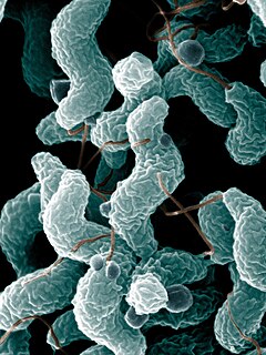
Campylobacter is a genus of Gram-negative bacteria. Campylobacter typically appear comma- or s-shaped, and are motile.

Kochs postulates Four criteria showing a causal relationship between a causative microbe and a disease

Koch's postulates are four criteria designed to establish a causative relationship between a microbe and a disease. The postulates were formulated by Robert Koch and Friedrich Loeffler in 1884, based on earlier concepts described by Jakob Henle, and refined and published by Koch in 1890. Koch applied the postulates to describe the etiology of cholera and tuberculosis, but they have been controversially generalized to other diseases. These postulates were generated before modern concepts in microbial pathogenesis that cannot be examined using Koch's postulates, including viruses and asymptomatic carriers. They have largely been supplanted by other criteria such as the Bradford Hill criteria for infectious disease causality in modern public health.

<i>Helicobacter pylori</i> Species of bacteria

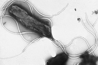
Helicobacter pylori, previously known as Campylobacter pylori, is a gram-negative, helically-shaped, microaerophilic bacterium usually found in the stomach. Its helical shape is thought to have evolved in order to penetrate the mucoid lining of the stomach and thereby establish infection. The bacterium was first identified in 1982 by Australian doctors Barry Marshall and Robin Warren. H. pylori has been associated with the mucosa-associated lymphoid tissue in the stomach, esophagus, colon, rectum, or tissues around the eye, and of lymphoid tissue in the stomach.

<i>Helicobacter</i>

Helicobacter is a genus of Gram-negative bacteria possessing a characteristic helical shape. They were initially considered to be members of the genus Campylobacter, but in 1989, Goodwin et al. published sufficient reasons to justify the new genus name Helicobacter. The genus Helicobacter contains about 35 species.

Virulence is a pathogen's or microbe's ability to infect or damage a host.

Giardiasis Parasitic disease that results in diarrhea

Giardiasis, popularly known as beaver fever, is a parasitic disease caused by Giardia duodenalis. About 10% of those infected have no symptoms. When symptoms occur they may include diarrhea, abdominal pain, and weight loss. Vomiting, blood in the stool, and fever are less common. Symptoms usually begin 1 to 3 weeks after exposure and without treatment may last up to six weeks.

Gastritis

Gastritis is inflammation of the lining of the stomach. It may occur as a short episode or may be of a long duration. There may be no symptoms but, when symptoms are present, the most common is upper abdominal pain. Other possible symptoms include nausea and vomiting, bloating, loss of appetite and heartburn. Complications may include stomach bleeding, stomach ulcers, and stomach tumors. When due to autoimmune problems, low red blood cells due to not enough vitamin B12 may occur, a condition known as pernicious anemia.

Travelers diarrhea Disease

Travelers' diarrhea (TD) is a stomach and intestinal infection. TD is defined as the passage of unformed stool while traveling. It may be accompanied by abdominal cramps, nausea, fever, and bloating. Occasionally bloody diarrhea may occur. Most travelers recover within four days with little or no treatment. About 10% of people may have symptoms for a week.

Dientamoebiasis is a medical condition caused by infection with Dientamoeba fragilis, a single-cell parasite that infects the lower gastrointestinal tract of humans. It is an important cause of traveler's diarrhea, chronic abdominal pain, chronic fatigue, and failure to thrive in children.

Timeline of peptic ulcer disease and <i>Helicobacter pylori</i>

This is a timeline of the events relating to the discovery that peptic ulcer disease and some cancers are caused by H. pylori. In 2005, Barry Marshall and Robin Warren were awarded the Nobel Prize in Physiology or Medicine for their discovery that peptic ulcer disease (PUD) was primarily caused by Helicobacter pylori, a bacterium with affinity for acidic environments, such as the stomach. As a result, PUD that is associated with H. pylori is currently treated with antibiotics used to eradicate the infection. For decades prior to their discovery, it was widely believed that PUD was caused by excess acid in the stomach. During this time, acid control was the primary method of treatment for PUD, to only partial success. Among other effects, it is now known that acid suppression alters the stomach milieu to make it less amenable to H. pylori infection.

Saccharomyces boulardii is a tropical species of yeast first isolated from lychee and mangosteen fruit in 1923 by French scientist Henri Boulard. Although early reports described distinct taxonomic, metabolic, and genetic properties, S. boulardii is a strain of S. cerevisiae, sharing >99% genomic relatedness, giving the synonym S. cerevisiae var boulardii. A type strain is Hansen CBS 5926.

<i>Blastocystis</i>

Blastocystis is a genus of single-celled heterokont parasites belonging to a group of organisms that are known as the Stramenopiles that includes algae, diatoms, and water molds. Blastocystis consists of several species, living in the gastrointestinal tracts of species as diverse as humans, farm animals, birds, rodents, reptiles, amphibians, fish, and cockroaches. Blastocystis exhibits low host specificity, and many different species of Blastocystis can infect humans, and by current convention, any of these species would be identified as Blastocystis hominis.

Blastocystosis medical condition caused by infection with Blastocystis

Blastocystosis refers to a medical condition caused by infection with Blastocystis. Blastocystis is a protozoal, single-celled parasite that inhabits the gastrointestinal tracts of humans and other animals. Many different types of Blastocystis exist, and they can infect humans, farm animals, birds, rodents, amphibians, reptiles, fish, and even cockroaches. Blastocystosis has been found to be a possible risk factor for development of irritable bowel syndrome.

In biology, a pathogen in the oldest and broadest sense, is any organism that can produce disease. A pathogen may also be referred to as an infectious agent, or simply a germ.

The host–pathogen interaction is defined as how microbes or viruses sustain themselves within host organisms on a molecular, cellular, organismal or population level. This term is most commonly used to refer to disease-causing microorganisms although they may not cause illness in all hosts. Because of this, the definition has been expanded to how known pathogens survive within their host, whether they cause disease or not.

Helicobacter felis is a bacterium species in the Helicobacteraceae family, Campylobacterales order, Helicobacter genus. This bacterium is Gram-negative, microaerophilic, urease-positive, and spiral-shaped. Its type strain is CS1T. It can be pathogenic.

Helicobacter salomonis is a species within the Helicobacter genus of Gram negative bacteria. Helicobacter pylori is by far the best known Helicobacter species primarily because humans infected with it may develop gastrointestinal tract diseases such as stomach inflammation, stomach ulcers, doudenal ulcers, stomach cancers of the non-lymphoma type, and various subtypes of extranodal marginal zone lymphomass, e.g. those of the stomach, small intestines, large intestines, and rectumn. H. pylori is also associated with the development of bile duct cancer and has been associated with a wide range of other diseases although its role in the development of many of these other diseases requires further study. Humans infected with H. salomonis may develop some of the same gastrointestinal diseases viz., stomach inflammation, stomach ulcers, duodenum ulcers, stomach cancers that are not lymphomas, and extrnodal marginal B cell lymphomas of the stomach. Other non-H. pylori Helicobacter species that are known to be associated with these gastrointestinal diseases are Helicobacter bizzozeronii, Helicobacter suis, Helicobacter felis, and Helicobacter heilmannii s.s Because of their disease associations, these four Helicobacter species plus H. salomonis are often group together and termed Helicobacter heilmannii sensu lato.

Helicobacter bizzozeronii is a species within the Helicobacter genus of Gram negative bacteria. Helicobacter pylori is by far the best known Helicobacter species primarily because humans infected with it may develop gastrointestinal tract diseases such as stomach inflammation, stomach ulcers, doudenal ulcers, stomach cancers of the non-lymphoma type, and various subtypes of extranodal marginal zone lymphomass, e.g. those of the stomach, small intestines, large intestines, and rectumn. H. pylori is also associated with the development of bile duct cancer and has been associated with a wide range of other diseases although its role in the development of many of these other diseases requires further study. Humans infected with H. bizzozeronii are prone to develop some of the same gastrointestinal diseases viz., stomach inflammation, stomach ulcers, duodenum ulcers, stomach cancers that are not lymphomas, and extrnodal marginal B cell lymphomas of the stomach. Other non-H. pylori Helicobacter species that are known to be associated with these gastrointestinal diseases are Helicobacter felis, Helicobacter salomonis, Helicobacter suis, and Helicobacter heilmannii s.s. Because of their disease associations, these four Helicobacter species plus H. bizzozeronii are often group together and termed Helicobacter heilmannii sensu lato.

Helicobacter suis is a species within the Helicobacter genus of Gram negative bacteria. Helicobacter pylori is by far the best known Helicobacter species primarily because humans infected with it may develop gastrointestinal tract diseases such as stomach inflammation, stomach ulcers, doudenal ulcers, stomach cancers of the non-lymphoma type, and various subtypes of extranodal marginal zone lymphomass, e.g. those of the stomach, small intestines, large intestines, and rectumn. H. pylori is also associated with the development of bile duct cancer and has been associated with a wide range of other diseases although its role in the development of many of these other diseases requires further study. Humans infected with H. suis may develop some of the same gastrointestinal diseases viz., stomach inflammation, stomach ulcers, duodenum ulcers, stomach cancers that are not lymphomas, and extrnodal marginal B cell lymphomas of the stomach. Other non-H. pylori Helicobacter species that are known to be associated with these gastrointestinal diseases are Helicobacter bizzozeronii, Helicobacter salomonis, Helicobacter felis, and Helicobacter heilmannii s.s. Because of their disease associations, these four Helicobacter species plus H. suis are often group together and termed Helicobacter heilmannii sensu lato.

Helicobacter heilmannii s.s. is a species within the Helicobacter genus of Gram negative bacteria. Helicobacter pylori is by far the best known Helicobacter species primarily because humans infected with it may develop gastrointestinal tract diseases such as stomach inflammation, stomach ulcers, doudenal ulcers, stomach cancers of the non-lymphoma type, and various subtypes of extranodal marginal zone lymphomass, e.g. those of the stomach, small intestines, large intestines, and rectumn. H. pylori is also associated with the development of bile duct cancer and has been associated with a wide range of other diseases although its role in the development of many of these other diseases requires further study. Humans infected with H. heilmannii s.s. may develop some of the same gastrointestinal diseases viz., stomach inflammation, stomach ulcers, duodenum ulcers, stomach cancers that are not lymphomas, and extranodal marginal B cell lymphomas of the stomach. Other non-H. pylori Helicobacter species that are known to be associated with these gastrointestinal diseases are Helicobacter bizzozeronii, Helicobacter suis, Helicobacter felis, and Helicobacter salomonis. Because of their disease associations, these four Helicobacter species plus H. heilmannii s.s. are often group together and termed Helicobacter heilmannii sensu lato.

References

  1. 1 2 Day M (June 7, 1997). "Stalking the diseases with no name". New Scientist.
  2. Barry, Marshall (2005). "Autobiography". Nobel Foundation . Retrieved 2007-01-28.
  3. 1 2 3 4 5 Marshall, Barry (May 1, 2002). Helicobacter Pioneers: Firsthand Accounts from the Scientists who Discovered Helicobacters, 1892-1982. Blackwell Publishing Limited. ISBN   978-0-86793-035-1.
  4. 1 2 3 4 Bentivoglio M, Pacini P (1995). "Filippo Pacini: a determined observer". Brain Res. Bull. 38 (2): 161–5. CiteSeerX   10.1.1.362.6850 . doi:10.1016/0361-9230(95)00083-Q. PMID   7583342.
  5. 1 2 Smith JW, Wolfe MS (1980). "Giardiasis". Annu. Rev. Med. 31 (1): 373–83. doi:10.1146/annurev.me.31.020180.002105. PMID   6994619.
  6. 1 2 Gabriel SE, Brigman KN, Koller BH, Boucher RC, Stutts MJ (1994). "Cystic fibrosis heterozygote resistance to cholera toxin in the cystic fibrosis mouse model". Science. 266 (5182): 107–9. Bibcode:1994Sci...266..107G. doi:10.1126/science.7524148. PMID   7524148.
  7. 1 2 El-Omar EM, Carrington M, Chow WH, et al. (2000). "Interleukin-1 polymorphisms associated with increased risk of gastric cancer". Nature. 404 (6776): 398–402. Bibcode:2000Natur.404..398E. doi:10.1038/35006081. PMID   10746728.
  8. Kolata, Gina (January 6, 1998). "Detective Work and Science Reveals a New Lethal Bacteria". New York Times. Retrieved 2007-08-08.
  9. Tan KS (2004). "Blastocystis in humans and animals: new insights using modern methodologies". Vet. Parasitol. 126 (1–2): 121–44. doi:10.1016/j.vetpar.2004.09.017. PMID   15567582.
  10. WHO. "WOrld Health Organisation factsheet".
  11. Ryan, KJ (2004). Sherris Medical Microbiology, 4th ed. McGraw Hill. pp. 276–7. ISBN   978-0-8385-8529-0.
  12. 1 2 Shapin, S (2006-11-06). "Sick City:Maps and Morbidity in the Time of CHolera". New Yorker.
  13. Bulloch, William (1979). The History of Bacteriology. Nature. 142. pp. 771–773. Bibcode:1938Natur.142..771D. doi:10.1038/142771a0. ISBN   978-0-486-23761-9.
  14. Howard-Jones N (1984). "Robert Koch and the cholera vibrio: a centenary". British Medical Journal (Clinical Research Ed.). 288 (6414): 379–81. doi:10.1136/bmj.288.6414.379. PMC   1444283 . PMID   6419937.
  15. Stitt, Edward (1922). The Diagnosis and Treatment of Tropical Diseases. P. Blakiston's Son, Philadelphia.
  16. Ali SA, Hill DR (2003). "Giardia intestinalis". Curr. Opin. Infect. Dis. 16 (5): 453–60. doi:10.1097/00001432-200310000-00012. PMID   14501998.
  17. "CDC Reportable Disease Spreadsheet" (PDF). Archived from the original (PDF) on 2007-06-13. Retrieved 2007-09-08.
  18. 1 2 Rendtorff, RC (1954). "The experimental transmission of human intestinal protozoan parasites. II. Giardia lamblia cysts given in capsules". American Journal of Hygiene. 59 (2): 209–20. doi:10.1093/oxfordjournals.aje.a119634. PMID   13138586.
  19. 1 2 Veazie L (1969). "Epidemic giardiasis". N. Engl. J. Med. 281 (15): 853. doi:10.1056/NEJM196910092811521. PMID   5809527.
  20. Paulley JW (1975). "Cultural influences on the incidence and pattern of disease". Psychotherapy and Psychosomatics. 26 (1): 2–11. doi:10.1159/000286889. PMID   1178798.
  21. Kellner R (1975). "Psychotherapy in psychosomatic disorders". Arch. Gen. Psychiatry. 32 (8): 1021–8. doi:10.1001/archpsyc.1975.01760260085007. PMID   1156110.
  22. Aitken C, Cay E (1975). "Clinical psychosomatic research". International Journal of Psychiatry in Medicine. 6 (1–2): 29–41. doi:10.2190/NTKP-0HDK-YUAW-7XN4. PMID   773858.
  23. Rigas B, Feretis C, Papavassiliou ED (1999). "John Lykoudis: an unappreciated discoverer of the cause and treatment of peptic ulcer disease". Lancet. 354 (9190): 1634–5. doi:10.1016/S0140-6736(99)06034-1. PMID   10560691.
  24. Steer HW, Colin-Jones DG (1975). "Mucosal changes in gastric ulceration and their response to carbenoxolone sodium". Gut. 16 (8): 590–7. doi:10.1136/gut.16.8.590. PMC   1411007 . PMID   810394.
  25. Ruchman I, Dodd K (1947). "The Isolation of a Strain of Escherichia coli Pathogenic for the Rabbit's Eye from a Patient with Diarrhea". J. Bacteriol. 53 (5): 653–6. PMC   518366 . PMID   16561320.
  26. MACQUEEN RL (1954). "Isolation of Bact. coli O.26.B.6 from a child with recurrent diarrhoea". British Medical Journal. 1 (4877): 1475–6. doi:10.1136/bmj.1.4877.1475-a. PMC   2085403 . PMID   13160505.
  27. McCLURE WB (1955). "A severe nursery epidemic of diarrhoea associated with Esch. coli type 111 B4". Canadian Medical Association Journal. 72 (2): 83–5. PMC   1825495 . PMID   13230990.
  28. GRONROOS JA (1957). "Investigations on Escherichia coli O groups 1-25, 44 and 78 and serotypes 26:B6, 55:B5, 86:B7, 111:B4, 125:B15 and 126:B16; occurrence in faeces of healthy and diarrhoeal infants". Annales Medicinae Experimentalis et Biologiae Fenniae. 35 (Suppl 2): 1–35. PMID   13444875.
  29. COWART GS, THOMASON BM; Thomason (1965). "Immunofluorescent Detection of Escherichia Coli". Am. J. Dis. Child. 110 (2): 131–6. doi:10.1001/archpedi.1965.02090030141004. PMID   14320761.
  30. Linde K, Köditz H, Hasenjäger H (1966). "[Treatment of infantile diarrhea caused by pathogenic Escherichia coli strains with oral large doses of benzyl procaine penicillin]". Pediatria Polska (in Polish). 41 (8): 905–12. PMID   5341176.
  31. Glantz PJ (1970). "Unclassified Escherichia coli serogroup OXI isolated from fatal diarrhea of rabbits". Can. J. Comp. Med. 34 (1): 47–9. PMC   1319419 . PMID   4246003.
  32. Drucker MM, Polliack A, Yeivin R, Sacks TG (1970). "Immunofluorescent demonstration of enteropathogenic Escherichia coli in tissues of infants dying with enteritis". Pediatrics. 46 (6): 855–64. PMID   4099362.
  33. Smith HW, Gyles CL (1970). "The relationship between two apparently different enterotoxins produced by enteropathogenic strains of Escherichia coli of porcine origin". J. Med. Microbiol. 3 (3): 387–401. doi: 10.1099/00222615-3-3-387 . PMID   4919579.
  34. Riley LW, Remis RS, Helgerson SD, et al. (1983). "Hemorrhagic colitis associated with a rare Escherichia coli serotype". N. Engl. J. Med. 308 (12): 681–5. doi:10.1056/NEJM198303243081203. PMID   6338386.
  35. 1 2 3 4 Goleman, Daniel (January 3, 1995). "Tiny Clinic in Katmandy Solves Mystery". New York Times. Retrieved 2007-08-08.
  36. CDC (1981). "Pneumocystis Pneumonia Los Angeles". CDC. Retrieved 2006-01-17.
  37. Zhu T, Korber BT, Nahmias AJ, Hooper E, Sharp PM, Ho DD (1998). "An African HIV-1 Sequence from 1959 and Implications for the Origin of the Epidemic". Nature. 391 (6667): 594–597. Bibcode:1998Natur.391..594Z. doi:10.1038/35400. PMID   9468138.
  38. Kolata, G. (1987-10-28). "Boy's 1969 death suggests AIDS invaded U.S. several times". The New York Times. Retrieved 2006-06-19.
  39. Hooper, E. (1997). "Sailors and star-bursts, and the arrival of HIV". BMJ. 315 (7123): 1689–1691. doi:10.1136/bmj.315.7123.1689. PMC   2128008 . PMID   9448543.
  40. Reeves, J. D.; Doms, R. W (2002). "Human Immunodeficiency Virus Type 2". J. Gen. Virol. 83 (Pt 6): 1253–1265. doi: 10.1099/0022-1317-83-6-1253 . PMID   12029140.
  41. Keele BF, van Heuverswyn F, Li YY, Bailes E, Takehisa J, Santiago ML, Bibollet-Ruche F, Chen Y, Wain LV, Liegois F, Loul S, Mpoudi Ngole E, Bienvenue Y, Delaporte E, Brookfield JF, Sharp PM, Shaw GM, Peeters M, Hahn BH (2006-05-25). "Chimpanzee Reservoirs of Pandemic and Nonpandemic HIV-1". Science. 313 (5786): 523–6. Bibcode:2006Sci...313..523K. doi:10.1126/science.1126531. PMC   2442710 . PMID   16728595.
  42. Cohen, J. (2000). "Vaccine Theory of AIDS Origins Disputed at Royal Society". Science. 289 (5486): 1850–1851. doi:10.1126/science.289.5486.1850. PMID   11012346.
  43. 1 2 3 Amin OM (2002). "Seasonal prevalence of intestinal parasites in the United States during 2000". Am. J. Trop. Med. Hyg. 66 (6): 799–803. doi: 10.4269/ajtmh.2002.66.799 . PMID   12224595.
  44. 1 2 3 Lagacé-Wiens PR, VanCaeseele PG, Koschik C (2006). "Dientamoeba fragilis: an emerging role in intestinal disease". Canadian Medical Association Journal. 175 (5): 468–9. doi:10.1503/cmaj.060265. PMC   1550747 . PMID   16940260.
  45. "Nationally Notifiable Infectious Diseases - 2007". Center for Disease Control. 2007. Archived from the original on 2007-06-24. Retrieved 2007-08-08.
  46. 1 2 3 Markell EK, Udkow MP (1990). "Association of Blastocystis hominis with human disease?". J. Clin. Microbiol. 28 (5): 1085–6. doi:10.1128/JCM.28.5.1085-1086.1990. PMC   267874 . PMID   2351728.
  47. Zierdt CH (1991). "Blastocystis hominis--past and future". Clin. Microbiol. Rev. 4 (1): 61–79. doi:10.1128/CMR.4.1.61. PMC   358179 . PMID   2004348.
  48. 1 2 3 Yakoob J, Jafri W, Jafri N, Islam M, Asim Beg M (2004). "In vitro susceptibility of Blastocystis hominis isolated from patients with irritable bowel syndrome". Br. J. Biomed. Sci. 61 (2): 75–7. doi:10.1080/09674845.2004.11732647. PMID   15250669.
  49. Yakoob J, Jafri W, Jafri N, et al. (2004). "Irritable bowel syndrome: in search of an etiology: role of Blastocystis hominis". Am. J. Trop. Med. Hyg. 70 (4): 383–5. CiteSeerX   10.1.1.484.928 . doi:10.4269/ajtmh.2004.70.383. PMID   15100450.
  50. 1 2 Borody T, Warren E, Wettstein A, et al. (2002). "Eradication of Dientamoeba fragilis can resolve IBS-like symptoms". J Gastroenterol Hepatol. 17 (Suppl): A103.
  51. 1 2 Hussain R, Jaferi W, Zuberi S, et al. (1997). "Significantly increased IgG2 subclass antibody levels to Blastocystis hominis in patients with irritable bowel syndrome". Am. J. Trop. Med. Hyg. 56 (3): 301–6. doi:10.4269/ajtmh.1997.56.301. PMID   9129532.
  52. Stensvold CR, Arendrup MC, Mølbak K, Nielsen HV (2007). "The prevalence of Dientamoeba fragilis in patients with suspected enteroparasitic disease in a metropolitan area in Denmark". Clin. Microbiol. Infect. 13 (8): 839–42. doi:10.1111/j.1469-0691.2007.01760.x. PMID   17610603.
  53. Qadri SM, al-Okaili GA, al-Dayel F (1989). "Clinical significance of Blastocystis hominis". J. Clin. Microbiol. 27 (11): 2407–9. doi:10.1128/JCM.27.11.2407-2409.1989. PMC   267045 . PMID   2808664.
  54. Grossman I, Weiss LM, Simon D, Tanowitz HB, Wittner M (1992). "Blastocystis hominis in hospital employees". Am. J. Gastroenterol. 87 (6): 729–32. PMID   1590309.
  55. Zdero M, Cabrera G, Ponce de León P, Nocito I, Echenique C (1997). "[Parasitosis in an adult population with chronic gastrointestinal disorders]". Acta Gastroenterol. Latinoam. (in Spanish). 27 (2): 67–73. PMID   9412130.
  56. Stensvold R, Brillowska-Dabrowska A, Nielsen HV, Arendrup MC (2006). "Detection of Blastocystis hominis in unpreserved stool specimens by using polymerase chain reaction". J. Parasitol. 92 (5): 1081–7. doi:10.1645/GE-840R.1. PMID   17152954.