Integrin

Last updated
Integrin alphaVbeta3 extracellular domains
PDB 1jv2 EBI.jpg
Structure of the extracellular segment of integrin alpha Vbeta3. [1]
Identifiers
SymbolIntegrin_alphaVbeta3
Pfam PF08441
Pfam clan CL0159
InterPro IPR013649
SCOP2 1jv2 / SCOPe / SUPFAM
OPM superfamily 176
OPM protein 2knc
Membranome 13
Available protein structures:
Pfam   structures / ECOD  
PDB RCSB PDB; PDBe; PDBj
PDBsum structure summary
Integrin alpha cytoplasmic region
PDB 1dpk EBI.jpg
Structure of chaperone protein PAPD. [2]
Identifiers
SymbolIntegrin_alpha
Pfam PF00357
InterPro IPR000413
PROSITE PDOC00215
SCOP2 1dpk / SCOPe / SUPFAM
Available protein structures:
Pfam   structures / ECOD  
PDB RCSB PDB; PDBe; PDBj
PDBsum structure summary
Integrin, beta chain (vWA)
Integrinalpha.png
Identifiers
SymbolIntegrin_beta
Pfam PF00362
InterPro IPR002369
SMART SM00187
PROSITE PDOC00216
SCOP2 1jv2 / SCOPe / SUPFAM
Available protein structures:
Pfam   structures / ECOD  
PDB RCSB PDB; PDBe; PDBj
PDBsum structure summary
PDB 1jv2 , 1kup , 1kuz , 1l3y , 1l5g , 1m1x , 1m8o , 1s4x , 1txv , 1ty3 , 1ty5 , 1ty6 , 1ty7 , 1tye , 1u8c
Integrin beta 7 cytoplasmic domain: complex with filamin
PDB 2brq EBI.jpg
crystal structure of the filamin a repeat 21 complexed with the integrin beta7 cytoplasmic tail peptide
Identifiers
SymbolIntegrin_b_cyt
Pfam PF08725
InterPro IPR014836
SCOP2 1m8O / SCOPe / SUPFAM
Available protein structures:
Pfam   structures / ECOD  
PDB RCSB PDB; PDBe; PDBj
PDBsum structure summary

Integrins are transmembrane receptors that help cell-cell and cell-extracellular matrix (ECM) adhesion. [3] Upon ligand binding, integrins activate signal transduction pathways that mediate cellular signals such as regulation of the cell cycle, organization of the intracellular cytoskeleton, and movement of new receptors to the cell membrane. [4] The presence of integrins allows rapid and flexible responses to events at the cell surface (e.g. signal platelets to initiate an interaction with coagulation factors).

Contents

Several types of integrins exist, and one cell generally has multiple different types on its surface. Integrins are found in all animals while integrin-like receptors are found in plant cells. [3]

Integrins work alongside other proteins such as cadherins, the immunoglobulin superfamily cell adhesion molecules, selectins and syndecans, to mediate cell–cell and cell–matrix interaction. Ligands for integrins include fibronectin, vitronectin, collagen and laminin.

Structure

Integrins are obligate heterodimers composed of α and β subunits. Several genes code for multiple isoforms of these subunits, which gives rise to an array of unique integrins with varied activity. In mammals, integrins are assembled from eighteen α and eight β subunits, [5] in Drosophila five α and two β subunits, and in Caenorhabditis nematodes two α subunits and one β subunit. [6] The α and β subunits are both class I transmembrane proteins, so each penetrates the plasma membrane once, and can possess several cytoplasmic domains. [7]

alpha (mammal)
geneproteinsynonyms
ITGA1 CD49a VLA1
ITGA2 CD49b VLA2
ITGA3 CD49c VLA3
ITGA4 CD49d VLA4
ITGA5 CD49e VLA5
ITGA6 CD49f VLA6
ITGA7 ITGA7 FLJ25220
ITGA8 ITGA8
ITGA9 ITGA9 RLC
ITGA10 ITGA10 PRO827
ITGA11 ITGA11 HsT18964
ITGAD CD11D FLJ39841
ITGAE CD103 HUMINAE
ITGAL CD11a LFA1A
ITGAM CD11b MAC-1
ITGAV CD51 VNRA, MSK8
ITGA2B CD41 GPIIb
ITGAX CD11c
beta (mammal)
geneproteinsynonyms
ITGB1 CD29 FNRB, MSK12, MDF2
ITGB2 CD18 LFA-1, MAC-1, MFI7
ITGB3 CD61 GP3A, GPIIIa
ITGB4 CD104
ITGB5 ITGB5 FLJ26658
ITGB6 ITGB6
ITGB7 ITGB7
ITGB8 ITGB8

Variants of some subunits are formed by differential RNA splicing; for example, four variants of the beta-1 subunit exist. Through different combinations of the α and β subunits, 24 unique mammalian integrins are generated, excluding splice- and glycosylation variants. [8]

Integrin subunits span the cell membrane and have short cytoplasmic domains of 40–70 amino acids. The exception is the beta-4 subunit, which has a cytoplasmic domain of 1,088 amino acids, one of the largest of any membrane protein. Outside the cell membrane, the α and β chains lie close together along a length of about 23  nm; the final 5 nm N-termini of each chain forms a ligand-binding region for the ECM. They have been compared to lobster claws, although they don't actually "pinch" their ligand, they chemically interact with it at the insides of the "tips" of their "pinchers".

The molecular mass of the integrin subunits can vary from 90  kDa to 160 kDa. Beta subunits have four cysteine-rich repeated sequences. Both α and β subunits bind several divalent cations. The role of divalent cations in the α subunit is unknown, but may stabilize the folds of the protein. The cations in the β subunits are more interesting: they are directly involved in coordinating at least some of the ligands that integrins bind.

Integrins can be categorized in multiple ways. For example, some α chains have an additional structural element (or "domain") inserted toward the N-terminal, the alpha-A domain (so called because it has a similar structure to the A-domains found in the protein von Willebrand factor; it is also termed the α-I domain). Integrins carrying this domain either bind to collagens (e.g. integrins α1 β1, and α2 β1), or act as cell-cell adhesion molecules (integrins of the β2 family). This α-I domain is the binding site for ligands of such integrins. Those integrins that don't carry this inserted domain also have an A-domain in their ligand binding site, but this A-domain is found on the β subunit.

In both cases, the A-domains carry up to three divalent cation binding sites. One is permanently occupied in physiological concentrations of divalent cations, and carries either a calcium or magnesium ion, the principal divalent cations in blood at median concentrations of 1.4 mM (calcium) and 0.8 mM (magnesium). The other two sites become occupied by cations when ligands bind—at least for those ligands involving an acidic amino acid in their interaction sites. An acidic amino acid features in the integrin-interaction site of many ECM proteins, for example as part of the amino acid sequence Arginine-Glycine-Aspartic acid ("RGD" in the one-letter amino acid code).

Structure

Despite many years of effort, discovering the high-resolution structure of integrins proved to be challenging, as membrane proteins are classically difficult to purify, and as integrins are large, complex and highly glycosylated with many sugar 'trees' attached to them. Low-resolution images of detergent extracts of intact integrin GPIIbIIIa, obtained using electron microscopy, and even data from indirect techniques that investigate the solution properties of integrins using ultracentrifugation and light scattering, were combined with fragmentary high-resolution crystallographic or NMR data from single or paired domains of single integrin chains, and molecular models postulated for the rest of the chains.

The X-ray crystal structure obtained for the complete extracellular region of one integrin, αvβ3, [1] shows the molecule to be folded into an inverted V-shape that potentially brings the ligand-binding sites close to the cell membrane. Perhaps more importantly, the crystal structure was also obtained for the same integrin bound to a small ligand containing the RGD-sequence, the drug cilengitide. [9] As detailed above, this finally revealed why divalent cations (in the A-domains) are critical for RGD-ligand binding to integrins. The interaction of such sequences with integrins is believed to be a primary switch by which ECM exerts its effects on cell behaviour.

The structure poses many questions, especially regarding ligand binding and signal transduction. The ligand binding site is directed towards the C-terminal of the integrin, the region where the molecule emerges from the cell membrane. If it emerges orthogonally from the membrane, the ligand binding site would apparently be obstructed, especially as integrin ligands are typically massive and well cross-linked components of the ECM. In fact, little is known about the angle that membrane proteins subtend to the plane of the membrane; this is a problem difficult to address with available technologies. The default assumption is that they emerge rather like little lollipops, but there is little evidence for this. The integrin structure has drawn attention to this problem, which may have general implications for how membrane proteins work. It appears that the integrin transmembrane helices are tilted (see "Activation" below), which hints that the extracellular chains may also not be orthogonal with respect to the membrane surface.

Although the crystal structure changed surprisingly little after binding to cilengitide, the current hypothesis is that integrin function involves changes in shape to move the ligand-binding site into a more accessible position, away from the cell surface, and this shape change also triggers intracellular signaling. There is a wide body of cell-biological and biochemical literature that supports this view. Perhaps the most convincing evidence involves the use of antibodies that only recognize integrins when they have bound to their ligands, or are activated. As the "footprint" that an antibody makes on its binding target is roughly a circle about 3 nm in diameter, the resolution of this technique is low. Nevertheless, these so-called LIBS (Ligand-Induced-Binding-Sites) antibodies unequivocally show that dramatic changes in integrin shape routinely occur. However, how the changes detected with antibodies look on the structure is still unknown.

Activation

When released into the cell membrane, newly synthesized integrin dimers are speculated to be found in the same "bent" conformation revealed by the structural studies described above. One school of thought claims that this bent form prevents them from interacting with their ligands, although bent forms can predominate in high-resolution EM structures of integrin bound to an ECM ligand. Therefore, at least in biochemical experiments, integrin dimers must apparently not be 'unbent' in order to prime them and allow their binding to the ECM. In cells, the priming is accomplished by a protein talin, which binds to the β tail of the integrin dimer and changes its conformation. [10] [11] The α and β integrin chains are both class-I transmembrane proteins: they pass the plasma membrane as single transmembrane alpha-helices. Unfortunately, the helices are too long, and recent studies suggest that, for integrin gpIIbIIIa, they are tilted with respect both to one another and to the plane of the membrane. Talin binding alters the angle of tilt of the β3 chain transmembrane helix in model systems and this may reflect a stage in the process of inside-out signalling which primes integrins. [12] Moreover, talin proteins are able to dimerize [13] and thus are thought to intervene in the clustering of integrin dimers which leads to the formation of a focal adhesion. Recently, the Kindlin-1 and Kindlin-2 proteins have also been found to interact with integrin and activate it. [14]

Function

Integrins have two main functions, attachment of the cells to the ECM and signal transduction from the ECM to the cells. [15] They are also involved in a wide range of other biological activities, including extravasation, cell-to-cell adhesion, cell migration, and as receptors for certain viruses, such as adenovirus, echovirus, hantavirus, and foot-and-mouth disease, polio virus and other viruses. Recently, the importance of integrins in the progress of autoimmune disorders is also gaining attention of the scientists. These mechanoreceptors seem to regulate autoimmunity by dictating various intracellular pathways to control immune cell adhesion to endothelial cell layers followed by their trans-migration. This process might or might not be dependent on the sheer force faced by the extracellular parts of different integrins. [16]

A prominent function of the integrins is seen in the molecule GpIIb/IIIa, an integrin on the surface of blood platelets (thrombocytes) responsible for attachment to fibrin within a developing blood clot. This molecule dramatically increases its binding affinity for fibrin/fibrinogen through association of platelets with exposed collagens in the wound site. Upon association of platelets with collagen, GPIIb/IIIa changes shape, allowing it to bind to fibrin and other blood components to form the clot matrix and stop blood loss.

Attachment of cell to the ECM

Integrins couple the cell-extracellular matrix (ECM) outside a cell to the cytoskeleton (in particular, the microfilaments) inside the cell. Which ligand in the ECM the integrin can bind to is defined by which α and β subunits the integrin is made of. Among the ligands of integrins are fibronectin, vitronectin, collagen, and laminin. The connection between the cell and the ECM may help the cell to endure pulling forces without being ripped out of the ECM. The ability of a cell to create this kind of bond is also of vital importance in ontogeny.

Cell attachment to the ECM is a basic requirement to build a multicellular organism. Integrins are not simply hooks, but give the cell critical signals about the nature of its surroundings. Together with signals arising from receptors for soluble growth factors like VEGF, EGF, and many others, they enforce a cellular decision on what biological action to take, be it attachment, movement, death, or differentiation. Thus integrins lie at the heart of many cellular biological processes. The attachment of the cell takes place through formation of cell adhesion complexes, which consist of integrins and many cytoplasmic proteins, such as talin, vinculin, paxillin, and alpha-actinin. These act by regulating kinases such as FAK (focal adhesion kinase) and Src kinase family members to phosphorylate substrates such as p130CAS thereby recruiting signaling adaptors such as CRK. These adhesion complexes attach to the actin cytoskeleton. The integrins thus serve to link two networks across the plasma membrane: the extracellular ECM and the intracellular actin filamentous system. Integrin α6β4 is an exception: it links to the keratin intermediate filament system in epithelial cells. [17]

Focal adhesions are large molecular complexes, which are generated following interaction of integrins with ECM, then their clustering. The clusters likely provide sufficient intracellular binding sites to permit the formation of stable signaling complexes on the cytoplasmic side of the cell membrane. So the focal adhesions contain integrin ligand, integrin molecule, and associate plaque proteins. Binding is propelled by changes in free energy. [18] As previously stated, these complexes connect the extracellular matrix to actin bundles. Cryo-electron tomography reveals that the adhesion contains particles on the cell membrane with diameter of 25 +/- 5 nm and spaced at approximately 45 nm. [19] Treatment with Rho-kinase inhibitor Y-27632 reduces the size of the particle, and it is extremely mechanosensitive. [20]

One important function of integrins on cells in tissue culture is their role in cell migration. Cells adhere to a substrate through their integrins. During movement, the cell makes new attachments to the substrate at its front and concurrently releases those at its rear. When released from the substrate, integrin molecules are taken back into the cell by endocytosis; they are transported through the cell to its front by the endocytic cycle, where they are added back to the surface. In this way they are cycled for reuse, enabling the cell to make fresh attachments at its leading front. [21] The cycle of integrin endocytosis and recycling back to the cell surface is important also for not migrating cells and during animal development. [22]

Signal transduction

Integrins play an important role in cell signaling by modulating the cell signaling pathways of transmembrane protein kinases such as receptor tyrosine kinases (RTK). While the interaction between integrin and receptor tyrosine kinases originally was thought of as uni-directional and supportive, recent studies indicate that integrins have additional, multi-faceted roles in cell signaling. Integrins can regulate the receptor tyrosine kinase signaling by recruiting specific adaptors to the plasma membrane. For example, β1c integrin recruits Gab1/Shp2 and presents Shp2 to IGF1R, resulting in dephosphorylation of the receptor. [23] In a reverse direction, when a receptor tyrosine kinase is activated, integrins co-localise at focal adhesion with the receptor tyrosine kinases and their associated signaling molecules.

The repertoire of integrins expressed on a particular cell can specify the signaling pathway due to the differential binding affinity of ECM ligands for the integrins. The tissue stiffness and matrix composition can initiate specific signaling pathways regulating cell behavior. Clustering and activation of the integrins/actin complexes strengthen the focal adhesion interaction and initiate the framework for cell signaling through assembly of adhesomes. [24]

Depending on the integrin's regulatory impact on specific receptor tyrosine kinases, the cell can experience:

Knowledge of the relationship between integrins and receptor tyrosine kinase has laid a foundation for new approaches to cancer therapy. Specifically, targeting integrins associated with RTKs is an emerging approach for inhibiting angiogenesis. [26]

Integrins are localised at the growth cone of regenerating neurons. Figure 1 - Nieuwenhuis et al 2018 - Integrins promote axonal regeneration after injury of the nervous system - Biological Reviews - doi 10.1111-brv.12398.jpg
Integrins are localised at the growth cone of regenerating neurons.

Integrins and nerve repair

Integrins have an important function in neuroregeneration after injury of the peripheral nervous system (PNS). [27] Integrins are present at the growth cone of damaged PNS neurons and attach to ligands in the ECM to promote axon regeneration. It is unclear whether integrins can promote axon regeneration in the adult central nervous system (CNS). There are two obstacles that prevent integrin-mediated regeneration in the CNS: 1) integrins are not localised in the axon of most adult CNS neurons and 2) integrins become inactivated by molecules in the scar tissue after injury. [27]

Vertebrate integrins

The following are 16 of the ~24 integrins found in vertebrates:

NameSynonymsDistributionLigands
α1β1VLA-1Many Collagens, laminins [28]
α2β1VLA-2ManyCollagens, laminins [28]
α3β1VLA-3ManyLaminin-5
α4β1 VLA-4 [28] Hematopoietic cells Fibronectin, VCAM-1 [28]
α4β7 LPAM-1T cellsMAD-CAM1 [29] [30]
α5β1 VLA-5; fibronectin receptorwidespreadfibronectin [28] and proteinases
α6β1 VLA-6; laminin receptorwidespreadlaminins
α7β1 muscle, gliomalaminins
αLβ2 LFA-1 [28] T-lymphocytes ICAM-1, ICAM-2 [28]
αMβ2 Mac-1, CR3 [28] Neutrophils and monocytes Serum proteins, ICAM-1 [28]
αIIbβ3 Fibrinogen receptor; gpIIbIIIa [31] Platelets [28] fibrinogen, fibronectin [28]
αVβ1 neurological tumors vitronectin, osteopontin, [32] fibrinogen
αVβ3 vitronectin receptor [33] activated endothelial cells, melanoma, glioblastoma vitronectin, [33] fibronectin, fibrinogen, osteopontin, [32] Cyr61, thyroxine, [34] TETRAC
αVβ5 widespread, esp. fibroblasts, epithelial cells vitronectin, osteopontin, [32] and adenovirus
αVβ6 proliferating epithelia, esp. lung and mammary gland fibronectin; TGFβ1+3
αVβ8 neural tissue; peripheral nerve fibronectin; TGFβ1+3
α6β4 Epithelial cells [28] Laminin [28]

Beta-1 integrins interact with many alpha integrin chains. Gene knockouts of integrins in mice are not always lethal, which suggests that during embryonal development, one integrin may substitute its function for another in order to allow survival. Some integrins are on the cell surface in an inactive state, and can be rapidly primed, or put into a state capable of binding their ligands, by cytokines. Integrins can assume several different well-defined shapes or "conformational states". Once primed, the conformational state changes to stimulate ligand binding, which then activates the receptors — also by inducing a shape change — to trigger outside-in signal transduction.

See also

Related Research Articles

<span class="mw-page-title-main">G protein-coupled receptor</span> Class of cell surface receptors coupled to G-protein-associated intracellular signaling

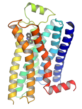
G protein-coupled receptors (GPCRs), also known as seven-(pass)-transmembrane domain receptors, 7TM receptors, heptahelical receptors, serpentine receptors, and G protein-linked receptors (GPLR), form a large group of evolutionarily related proteins that are cell surface receptors that detect molecules outside the cell and activate cellular responses. They are coupled with G proteins. They pass through the cell membrane seven times in the form of six loops of amino acid residues, which is why they are sometimes referred to as seven-transmembrane receptors. Ligands can bind either to the extracellular N-terminus and loops or to the binding site within transmembrane helices. They are all activated by agonists, although a spontaneous auto-activation of an empty receptor has also been observed.

<span class="mw-page-title-main">Signal transduction</span> Cascade of intracellular and molecular events for transmission/amplification of signals

Signal transduction is the process by which a chemical or physical signal is transmitted through a cell as a series of molecular events. Most commonly, protein phosphorylation is catalyzed by protein kinases, ultimately resulting in a cellular response. Proteins responsible for detecting stimuli are generally termed receptors, although in some cases the term sensor is used. The changes elicited by ligand binding in a receptor give rise to a biochemical cascade, which is a chain of biochemical events known as a signaling pathway.

<span class="mw-page-title-main">Tyrosine kinase</span> Class hi residues

A tyrosine kinase is an enzyme that can transfer a phosphate group from ATP to the tyrosine residues of specific proteins inside a cell. It functions as an "on" or "off" switch in many cellular functions.

<span class="mw-page-title-main">Cell adhesion</span> Process of cell attachment

Cell adhesion is the process by which cells interact and attach to neighbouring cells through specialised molecules of the cell surface. This process can occur either through direct contact between cell surfaces such as cell junctions or indirect interaction, where cells attach to surrounding extracellular matrix, a gel-like structure containing molecules released by cells into spaces between them. Cells adhesion occurs from the interactions between cell-adhesion molecules (CAMs), transmembrane proteins located on the cell surface. Cell adhesion links cells in different ways and can be involved in signal transduction for cells to detect and respond to changes in the surroundings. Other cellular processes regulated by cell adhesion include cell migration and tissue development in multicellular organisms. Alterations in cell adhesion can disrupt important cellular processes and lead to a variety of diseases, including cancer and arthritis. Cell adhesion is also essential for infectious organisms, such as bacteria or viruses, to cause diseases.

<span class="mw-page-title-main">Receptor (biochemistry)</span> Protein molecule receiving signals for a cell

In biochemistry and pharmacology, receptors are chemical structures, composed of protein, that receive and transduce signals that may be integrated into biological systems. These signals are typically chemical messengers which bind to a receptor and produce physiological responses such as change in the electrical activity of a cell. For example, GABA, an inhibitory neurotransmitter, inhibits electrical activity of neurons by binding to GABAA receptors. There are three main ways the action of the receptor can be classified: relay of signal, amplification, or integration. Relaying sends the signal onward, amplification increases the effect of a single ligand, and integration allows the signal to be incorporated into another biochemical pathway.

<span class="mw-page-title-main">Insulin receptor</span> Mammalian protein found in Homo sapiens

The insulin receptor (IR) is a transmembrane receptor that is activated by insulin, IGF-I, IGF-II and belongs to the large class of receptor tyrosine kinase. Metabolically, the insulin receptor plays a key role in the regulation of glucose homeostasis; a functional process that under degenerate conditions may result in a range of clinical manifestations including diabetes and cancer. Insulin signalling controls access to blood glucose in body cells. When insulin falls, especially in those with high insulin sensitivity, body cells begin only to have access to lipids that do not require transport across the membrane. So, in this way, insulin is the key regulator of fat metabolism as well. Biochemically, the insulin receptor is encoded by a single gene INSR, from which alternate splicing during transcription results in either IR-A or IR-B isoforms. Downstream post-translational events of either isoform result in the formation of a proteolytically cleaved α and β subunit, which upon combination are ultimately capable of homo or hetero-dimerisation to produce the ≈320 kDa disulfide-linked transmembrane insulin receptor.

Cell adhesion molecules (CAMs) are a subset of cell surface proteins that are involved in the binding of cells with other cells or with the extracellular matrix (ECM), in a process called cell adhesion. In essence, CAMs help cells stick to each other and to their surroundings. CAMs are crucial components in maintaining tissue structure and function. In fully developed animals, these molecules play an integral role in generating force and movement and consequently ensuring that organs are able to execute their functions normally. In addition to serving as "molecular glue", CAMs play important roles in the cellular mechanisms of growth, contact inhibition, and apoptosis. Aberrant expression of CAMs may result in a wide range of pathologies, ranging from frostbite to cancer.

<span class="mw-page-title-main">Focal adhesion</span>

In cell biology, focal adhesions are large macromolecular assemblies through which mechanical force and regulatory signals are transmitted between the extracellular matrix (ECM) and an interacting cell. More precisely, focal adhesions are the sub-cellular structures that mediate the regulatory effects of a cell in response to ECM adhesion.

Biological crosstalk refers to instances in which one or more components of one signal transduction pathway affects another. This can be achieved through a number of ways with the most common form being crosstalk between proteins of signaling cascades. In these signal transduction pathways, there are often shared components that can interact with either pathway. A more complex instance of crosstalk can be observed with transmembrane crosstalk between the extracellular matrix (ECM) and the cytoskeleton.

In biology, cell signaling is the process by which a cell interacts with itself, other cells and the environment. Cell signaling is a fundamental property of all cellular life in prokaryotes and eukaryotes.

<span class="mw-page-title-main">B-cell receptor</span> Transmembrane protein on the surface of a B cell

The B-cell receptor (BCR) is a transmembrane protein on the surface of a B cell. A B-cell receptor is composed of a membrane-bound immunoglobulin molecule and a signal transduction moiety. The former forms a type 1 transmembrane receptor protein, and is typically located on the outer surface of these lymphocyte cells. Through biochemical signaling and by physically acquiring antigens from the immune synapses, the BCR controls the activation of the B cell. B cells are able to gather and grab antigens by engaging biochemical modules for receptor clustering, cell spreading, generation of pulling forces, and receptor transport, which eventually culminates in endocytosis and antigen presentation. B cells' mechanical activity adheres to a pattern of negative and positive feedbacks that regulate the quantity of removed antigen by manipulating the dynamic of BCR–antigen bonds directly. Particularly, grouping and spreading increase the relation of antigen with BCR, thereby proving sensitivity and amplification. On the other hand, pulling forces delinks the antigen from the BCR, thus testing the quality of antigen binding.

<span class="mw-page-title-main">Receptor tyrosine kinase</span> Class of enzymes

Receptor tyrosine kinases (RTKs) are the high-affinity cell surface receptors for many polypeptide growth factors, cytokines, and hormones. Of the 90 unique tyrosine kinase genes identified in the human genome, 58 encode receptor tyrosine kinase proteins. Receptor tyrosine kinases have been shown not only to be key regulators of normal cellular processes but also to have a critical role in the development and progression of many types of cancer. Mutations in receptor tyrosine kinases lead to activation of a series of signalling cascades which have numerous effects on protein expression. Receptor tyrosine kinases are part of the larger family of protein tyrosine kinases, encompassing the receptor tyrosine kinase proteins which contain a transmembrane domain, as well as the non-receptor tyrosine kinases which do not possess transmembrane domains.

<span class="mw-page-title-main">Integrin beta 1</span> Mammalian protein found in Homo sapiens

Integrin beta-1 (ITGB1), also known as CD29, is a cell surface receptor that in humans is encoded by the ITGB1 gene. This integrin associates with integrin alpha 1 and integrin alpha 2 to form integrin complexes which function as collagen receptors. It also forms dimers with integrin alpha 3 to form integrin receptors for netrin 1 and reelin. These and other integrin beta 1 complexes have been historically known as very late activation (VLA) antigens.

Talin is a high-molecular-weight cytoskeletal protein concentrated at regions of cell–substratum contact and, in lymphocytes, at cell–cell contacts. Discovered in 1983 by Keith Burridge and colleagues, talin is a ubiquitous cytosolic protein that is found in high concentrations in focal adhesions. It is capable of linking integrins to the actin cytoskeleton either directly or indirectly by interacting with vinculin and α-actinin.

Platelet membrane glycoproteins are surface glycoproteins found on platelets (thrombocytes) which play a key role in hemostasis. When the blood vessel wall is damaged, platelet membrane glycoproteins interact with the extracellular matrix.

<span class="mw-page-title-main">Signal-regulatory protein alpha</span> Protein-coding gene in the species Homo sapiens

Signal regulatory protein α (SIRPα) is a regulatory membrane glycoprotein from SIRP family expressed mainly by myeloid cells and also by stem cells or neurons.

Collagen receptors are membrane proteins that bind the extracellular matrix protein collagen, the most abundant protein in mammals. They control mainly cell proliferation, migration and adhesion, coagulation cascade activation and they affect ECM structure by regulation of MMP.

<span class="mw-page-title-main">Cell surface receptor</span> Class of ligand activated receptors localized in surface of plama cell membrane

Cell surface receptors are receptors that are embedded in the plasma membrane of cells. They act in cell signaling by receiving extracellular molecules. They are specialized integral membrane proteins that allow communication between the cell and the extracellular space. The extracellular molecules may be hormones, neurotransmitters, cytokines, growth factors, cell adhesion molecules, or nutrients; they react with the receptor to induce changes in the metabolism and activity of a cell. In the process of signal transduction, ligand binding affects a cascading chemical change through the cell membrane.

Rap1 is a small GTPase, which are small cytosolic proteins that act like cellular switches and are vital for effective signal transduction. There are two isoforms of the Rap1 protein, each encoded by a separate gene, RAP1A and RAP1B. Rap1 belongs to Ras-related protein family.

<span class="mw-page-title-main">Integrin-like receptors</span>

Integrin-like receptors (ILRs) are found in plants and carry unique functional properties similar to true integrin proteins. True homologs of integrins exist in mammals, invertebrates, and some fungi but not in plant cells. Mammalian integrins are heterodimer transmembrane proteins that play a large role in bidirectional signal transduction. As transmembrane proteins, integrins connect the extracellular matrix (ECM) to the plasma membrane of the animal cell. The extracellular matrix of plant cells, fungi, and some protist is referred to as the cell wall. The plant cell wall is composed of a tough cellulose polysaccharide rather than the collagen fibers of the animal ECM. Even with these differences, research indicates that similar proteins involved in the interaction between the ECM and animals cells are also involved in the interaction of the cell wall and plant cells.

References

  1. 1 2 Xiong JP, Stehle T, Diefenbach B, Zhang R, Dunker R, Scott DL, Joachimiak A, Goodman SL, Arnaout MA (October 2001). "Crystal structure of the extracellular segment of integrin alpha Vbeta3". Science. 294 (5541): 339–45. Bibcode:2001Sci...294..339X. doi:10.1126/science.1064535. PMC   2885948 . PMID   11546839.
  2. Sauer FG, Fütterer K, Pinkner JS, Dodson KW, Hultgren SJ, Waksman G (August 1999). "Structural basis of chaperone function and pilus biogenesis". Science. 285 (5430): 1058–61. doi:10.1126/science.285.5430.1058. PMID   10446050.
  3. 1 2 Hynes RO (September 2002). "Integrins: bidirectional, allosteric signaling machines". Cell. 110 (6): 673–87. doi: 10.1016/s0092-8674(02)00971-6 . PMID   12297042. S2CID   30326350.
  4. Giancotti FG, Ruoslahti E (August 1999). "Integrin signaling". Science. 285 (5430): 1028–32. doi:10.1126/science.285.5430.1028. PMID   10446041.
  5. Bruce A, Johnson A, Lewis J, Raff M, Roberts K, Walter P (2002). "Integrins". Molecular Biology of the Cell (4th ed.). New York: Garland Science.
  6. Humphries MJ (2000). "Integrin structure". Biochemical Society Transactions. 28 (4): 311–39. doi:10.1042/0300-5127:0280311. PMID   10961914.
  7. Nermut MV, Green NM, Eason P, Yamada SS, Yamada KM (December 1988). "Electron microscopy and structural model of human fibronectin receptor". The EMBO Journal. 7 (13): 4093–9. doi:10.1002/j.1460-2075.1988.tb03303.x. PMC   455118 . PMID   2977331.
  8. Hynes RO (September 2002). "Integrins: bidirectional, allosteric signaling machines". Cell. 110 (6): 673–87. doi: 10.1016/S0092-8674(02)00971-6 . PMID   12297042. S2CID   30326350.
  9. Smith JW (June 2003). "Cilengitide Merck". Current Opinion in Investigational Drugs. 4 (6): 741–5. PMID   12901235.
  10. Calderwood DA (June 2004). "Talin controls integrin activation". Biochemical Society Transactions. 32 (Pt3): 434–7. doi:10.1042/BST0320434. PMID   15157154.
  11. Calderwood DA, Zent R, Grant R, Rees DJ, Hynes RO, Ginsberg MH (October 1999). "The Talin head domain binds to integrin beta subunit cytoplasmic tails and regulates integrin activation". The Journal of Biological Chemistry. 274 (40): 28071–4. doi: 10.1074/jbc.274.40.28071 . PMID   10497155.
  12. Shattil SJ, Kim C, Ginsberg MH (April 2010). "The final steps of integrin activation: the end game". Nature Reviews. Molecular Cell Biology. 11 (4): 288–300. doi:10.1038/nrm2871. PMC   3929966 . PMID   20308986.
  13. Goldmann WH, Bremer A, Häner M, Aebi U, Isenberg G (1994). "Native talin is a dumbbell-shaped homodimer when it interacts with actin". Journal of Structural Biology. 112 (1): 3–10. doi:10.1006/jsbi.1994.1002. PMID   8031639.
  14. Harburger DS, Bouaouina M, Calderwood DA (April 2009). "Kindlin-1 and -2 directly bind the C-terminal region of beta integrin cytoplasmic tails and exert integrin-specific activation effects". The Journal of Biological Chemistry. 284 (17): 11485–97. doi: 10.1074/jbc.M809233200 . PMC   2670154 . PMID   19240021.
  15. Yamada KM, Miyamoto S (October 1995). "Integrin transmembrane signaling and cytoskeletal control". Current Opinion in Cell Biology. 7 (5): 681–9. doi:10.1016/0955-0674(95)80110-3. PMID   8573343.
  16. Banerjee, S; Nara, R; Chakraborty, S; Chowdhury, D; Haldar, S (2022). "Integrin Regulated Autoimmune Disorders: Understanding the Role of Mechanical Force in Autoimmunity". Frontiers in Cell and Developmental Biology. 10: 852878. doi: 10.3389/fcell.2022.852878 . PMC   8971850 . PMID   35372360.
  17. Wilhelmsen K, Litjens SH, Sonnenberg A (April 2006). "Multiple functions of the integrin alpha6beta4 in epidermal homeostasis and tumorigenesis". Molecular and Cellular Biology. 26 (8): 2877–86. doi:10.1128/MCB.26.8.2877-2886.2006. PMC   1446957 . PMID   16581764.
  18. Olberding JE, Thouless MD, Arruda EM, Garikipati K (August 2010). Buehler MJ (ed.). "The non-equilibrium thermodynamics and kinetics of focal adhesion dynamics". PLOS ONE. 5 (8): e12043. Bibcode:2010PLoSO...512043O. doi: 10.1371/journal.pone.0012043 . PMC   2923603 . PMID   20805876.
  19. Patla I, Volberg T, Elad N, Hirschfeld-Warneken V, Grashoff C, Fässler R, Spatz JP, Geiger B, Medalia O (September 2010). "Dissecting the molecular architecture of integrin adhesion sites by cryo-electron tomography". Nature Cell Biology. 12 (9): 909–15. doi:10.1038/ncb2095. PMID   20694000. S2CID   20775305.
  20. Gullingsrud J, Sotomayor M. "Mechanosensitive channels". Theoretical and Computational Biophysics Group, Beckman Institute for Advanced Science and Technology: University of Illinois at Urbana-Champaign. Archived from the original on 2010-12-02.
  21. Paul NR, Jacquemet G, Caswell PT (November 2015). "Endocytic Trafficking of Integrins in Cell Migration". Current Biology. 25 (22): R1092-105. doi: 10.1016/j.cub.2015.09.049 . PMID   26583903.
  22. Moreno-Layseca P, Icha J, Hamidi H, Ivaska J (February 2019). "Integrin trafficking in cells and tissues". Nature Cell Biology. 21 (2): 122–132. doi:10.1038/s41556-018-0223-z. PMC   6597357 . PMID   30602723.
  23. Goel HL, Breen M, Zhang J, Das I, Aznavoorian-Cheshire S, Greenberg NM, Elgavish A, Languino LR (August 2005). "beta1A integrin expression is required for type 1 insulin-like growth factor receptor mitogenic and transforming activities and localization to focal contacts". Cancer Research. 65 (15): 6692–700. doi:10.1158/0008-5472.CAN-04-4315. PMID   16061650.
  24. Kim SH, Turnbull J, Guimond S (May 2011). "Extracellular matrix and cell signalling: the dynamic cooperation of integrin, proteoglycan and growth factor receptor". The Journal of Endocrinology. 209 (2): 139–51. doi:10.1530/JOE-10-0377. PMID   21307119.
  25. 1 2 3 Bostwick DG, Cheng L (2020-01-01). "9 - Neoplasms of the Prostate". In Cheng L, MacLennan GT, Bostwick DG (eds.). Urologic Surgical Pathology (Fourth ed.). Philadelphia: Content Repository Only!. pp. 415–525.e42. ISBN   978-0-323-54941-7.
  26. Carbonell WS, DeLay M, Jahangiri A, Park CC, Aghi MK (May 2013). "β1 integrin targeting potentiates antiangiogenic therapy and inhibits the growth of bevacizumab-resistant glioblastoma". Cancer Research. 73 (10): 3145–54. doi:10.1158/0008-5472.CAN-13-0011. PMC   4040366 . PMID   23644530.
  27. 1 2 3 Nieuwenhuis B, Haenzi B, Andrews MR, Verhaagen J, Fawcett JW (February 2018). "Integrins promote axonal regeneration after injury of the nervous system". Biological Reviews of the Cambridge Philosophical Society. 93 (3): 1339–1362. doi:10.1111/brv.12398. PMC   6055631 . PMID   29446228.
  28. 1 2 3 4 5 6 7 8 9 10 11 12 13 Krieger M, Scott MP, Matsudaira PT, Lodish HF, Darnell JE, Zipursky L, Kaiser C, Berk A (2004). Molecular cell biology (fifth ed.). New York: W.H. Freeman and CO. ISBN   978-0-7167-4366-8.
  29. Wang, Caihong; McDonough, Jacquelyn S.; McDonald, Keely G.; Huang, Conway; Newberry, Rodney D. (2008-09-15). "Alpha4beta7/MAdCAM-1 interactions play an essential role in transitioning cryptopatches into isolated lymphoid follicles and a nonessential role in cryptopatch formation". Journal of Immunology. 181 (6): 4052–4061. doi:10.4049/jimmunol.181.6.4052. ISSN   1550-6606. PMC   2778276 . PMID   18768861.
  30. Wagner, N.; Löhler, J.; Kunkel, E. J.; Ley, K.; Leung, E.; Krissansen, G.; Rajewsky, K.; Müller, W. (1996-07-25). "Critical role for beta7 integrins in formation of the gut-associated lymphoid tissue". Nature. 382 (6589): 366–370. doi:10.1038/382366a0. ISSN   0028-0836. PMID   8684468.
  31. Elangbam CS, Qualls CW, Dahlgren RR (January 1997). "Cell adhesion molecules--update". Veterinary Pathology. 34 (1): 61–73. doi:10.1177/030098589703400113. PMID   9150551.
  32. 1 2 3 Kazanecki, CC; Uzwiak, DJ; Denhxxardt, DT (1 November 2007). "Control of osteopontin signaling and function by post-translational phosphorylation and protein folding". Journal of Cellular Biochemistry. 102 (4): 912–24. doi:10.1002/jcb.21558. PMID   17910028. S2CID   24240459.
  33. 1 2 Hermann P, Armant M, Brown E, Rubio M, Ishihara H, Ulrich D, Caspary RG, Lindberg FP, Armitage R, Maliszewski C, Delespesse G, Sarfati M (February 1999). "The vitronectin receptor and its associated CD47 molecule mediates proinflammatory cytokine synthesis in human monocytes by interaction with soluble CD23". The Journal of Cell Biology. 144 (4): 767–75. doi:10.1083/jcb.144.4.767. PMC   2132927 . PMID   10037797.
  34. Bergh JJ, Lin HY, Lansing L, Mohamed SN, Davis FB, Mousa S, Davis PJ (July 2005). "Integrin alphaVbeta3 contains a cell surface receptor site for thyroid hormone that is linked to activation of mitogen-activated protein kinase and induction of angiogenesis". Endocrinology. 146 (7): 2864–71. doi: 10.1210/en.2005-0102 . PMID   15802494.

Commons-logo.svg Media related to Integrins at Wikimedia Commons