Loa loa

Last updated

Loa loa
Loa loa in blood Giemsa stain.jpg
Loa loa microfilaria found in blood film.
(Giemsa stain)
Scientific classification OOjs UI icon edit-ltr.svg
Domain: Eukaryota
Kingdom: Animalia
Phylum: Nematoda
Class: Chromadorea
Order: Rhabditida
Family: Onchocercidae
Genus: Loa
Species:
L. loa
Binomial name
Loa loa
(Cobbold, 1864)
Synonyms
  • Dracunculus loaCobbold, 1864 [1]
  • Filaria loa(Cobbold, 1864)

Loa loa is a filarial (arthropod-borne) nematode (roundworm) that causes Loa loa filariasis. Loa loa actually means "worm worm", but is commonly known as the "eye worm", as it localizes to the conjunctiva of the eye. Loa loa is commonly found in Africa. [2] It mainly inhabits rain forests in West Africa and has native origins in Ethiopia. [3] The disease caused by Loa loa is called loiasis and is one of the neglected tropical diseases. [4]

Contents

L. loa is one of three parasitic filarial nematodes that cause subcutaneous filariasis in humans. The other two are Mansonella streptocerca and Onchocerca volvulus (causes river blindness).

Maturing larvae and adults of the "eye worm" occupy the subcutaneous layer of the skin – the fat layer – of humans, causing disease. The L. loa adult worm which travels under the skin can survive up to 10–15 years, causing inflammations known as Calabar swellings. The adult worm travels under the skin, where the female deposits the microfilariae which can develop in the host’s blood within 5 to 6 months and can survive up to 17 years. The young larvae, or microfilariae, develop in horseflies of the genus Chrysops (deer flies, yellow flies), including the species C. dimidiata and C. silacea , which infect humans by biting them. After bites from these infected flies, the microfilariae are unique in that they travel in the peripheral blood during the day and migrate into the lungs at night. [5]

Morphology

Whole blood with microfilaria worm, giemsa stain Loa Loa.jpg
Whole blood with microfilaria worm, giemsa stain

L. loa worms have a simple structure consisting of a head (which lacks lips), a body, and a blunt tail. The outer body of the worm is composed of a cuticle with 3 main layers made up of collagen and other compounds which aid in protecting the nematodes while they are inside the digestive system of their host. Juveniles have a similar appearance to adult worms, but are significantly smaller. [6] Male adults range from 20 to 34 mm long and 350 to 430 μm wide. Female adults range from 20 to 70 mm long and can be about 425 μm wide. They vary in color. [2]

Lifecycle

The human is the definitive host, in which the parasitic worms attain sexual maturity, mate, and produce microfilariae. The flies serve as intermediate hosts in which the microfilariae undergo part of their morphological development, and then are borne to the next definitive host. [7]

Two species of Chrysops deerflies, C. silacea and C. dimidiata, are the main vectors for this filariasis. [8]

  1. A fly bearing third-stage filarial larvae in its proboscis infects the human host through the bite wound.
  2. After entering the human host, the larvae mature into adults, commonly in subcutaneous tissue. Adult females measure about 40 to 70 mm in length and 0.5 mm in diameter. Males measure some 30 to 34 mm by 0.35 to 0.43 mm.
  3. The adult female produces large numbers of microfilariae, about 250 to 300 μm in length and 6-8 μm in width. She continues to do so continuously for her lifetime, which typically spans several years.
  4. Microfilariae tend to reside within spinal fluids, urine, and sputum; by day, they also circulate in the bloodstream. Apart from their presence in bodily fluids, however, microfilariae in the noncirculating phase also occur in the lungs.
  5. The vector fly ingests microfilariae while feeding on the host's blood.
  6. Once inside the vector, the microfilaria sheds its sheaths and escapes through the walls of the midgut into the fly’s haemocoel.
  7. It then migrates through the hemolymph into the wing muscles in the fly's thorax.
  8. In the thoracic muscles, the microfilaria develops successively into a first-stage larva, second-stage larva, and finally into the infectious third-stage larva.
  9. The third-stage larva migrates to the fly’s proboscis.
  10. Once the larva is established in the proboscis and the fly takes its next human blood meal, the cycle of infection continues.

Disease

Signs and symptoms

Usually, about five months are needed for larvae (transferred from a fly) to mature into adult worms, which they can only do inside the human body. The most common display of infection is the localized allergic inflammations called Calabar or Cameroon swellings that signify the migration of the adult worm in the tissues away from the injection site by the vector. The migration does not cause significant damage to the host and is referred to as benign. However, these swellings can be painful, as they are mostly found near the joints. [9]

Although most infections with L. loa are asymptomatic, symptoms generally do not appear until years, or even more than a decade, after the bite from an infected fly. However, symptoms can appear as early as four months after a bite. [10] These parasites have a diurnal periodicity in which they circulate in the peripheral blood during the daytime, but migrate to vascular parts of the lungs during the night, where they are considered non circulatory. Therefore, the appearing and disappearing characteristics of this parasite can cause recurrent swelling that can cause painful enlargements of cysts in the connective tissue surrounding tendons. Additionally, chronic abscesses can be caused by the dying worms. [5]

The most visual sign of an adult worm infections is when the worm crosses the sclera of the eye, which causes significant pain to the host and is usually associated with inflammation and less likely, blindness. Eye worms typically cause little eye damage and last a few hours to a week. [9] Other tissues in which this worm can be found includes: the penis, testes, nipples, bridge of the nose, kidneys, and heart. The worms in these locations are not always externally visible. [11]

Risk factors

People at the highest risk for acquiring loiasis are those who live in the rainforests of West or Central Africa. Furthermore, the probability of getting bitten by a deerfly increases during the day and during rainy seasons. The flies are also attracted to smoke from wood fires. These flies do not commonly enter houses, but they are attracted to the houses that are well lit, so will congregate outside. [9]

Travelers can be infected in less than 30 days after arriving in an affected area, although they are more likely to be infected whilst being bitten by multiple deerflies over the course of many months. Men are more susceptible than women due to their increased exposure to the vectors during activities such as farming, hunting, and fishing. [12]

Diagnosis

The main methods of diagnosis include the presence of microfilariae in the blood, the presence of a worm in the eye, and the presence of skin swellings. However, in cases where none of those is the case, a blood count can be done. Patients with infections have a higher number of blood cells, namely eosinophils, as well as high IgE levels that indicate an active infection with helminth parasites. Due to the migration of microfilariae during the day, the accuracy of a blood test can be increased when samples are taken between 10 am and 2 pm. [11] A Giemsa stain is the most commonly used diagnostic test that uses a thick blood smear to count the microfilariae. Other than blood, microfilariae can also be observed in urine and saliva samples. [12]

Treatment

Adult worms found in the eye can be surgically removed with forceps after being paralyzed with a topical anesthesia. The worm is not paralyzed completely, so if it is not extracted quickly, it can vanish upon attempting extraction. [12]

Ivermectin has become the most common antiparasitic agent used worldwide, but can lead to residual microfilarial load when given in the management of loiasis. Treatment with ivermectin has shown to produce severe adverse neurological consequences in some cases. These treatment complications can be increased in individuals co-infected with onchocerciasis. [11] Some of these patients experienced cases of coma and resultant encephalopathy, parkinsonism, and death. After about 12 hours, the first signs start to appear and include fatigue, pain in joints, mutism, and incontinence. Severe disorders of the consciousness start to develop after about a day. [13]

Although Ivermectin is a common treatment for loiasis, the Centers for Disease Control (CDC) recommends treatment with diethylcarbamazine (DEC). Symptoms may be resolved with as little as 1–2 courses of DEC. DEC is chosen over Ivermectin because evidence supports its ability to kill both the adult worms and the microfilariae, which are the main cause of the severe neurological problems mentioned above. In some cases, albendazole may also be an effective treatment used to reduce the microfilariae prior to treatment with DEC. The body's response to albendazole is slow, so the patient being treated must be monitored closely and frequently to ensure it is effective. [14]

Prevention

Currently, no control programs or vaccines for loiasis are available. However, diethylcarbamazine treatment is suggested to the reduce risk of infection. Avoiding areas where the vectors, deerflies, are found also reduces risk. This includes swamps, bogs, and shaded areas near rivers or near wood fires. Fly bites can be reduced by using insect repellents such as DEET and wearing long sleeves and pants during the daytime. Permethrin treatment on clothes is an additional repellent that could be used. Also, using malaria nets can reduce the number of fly bites acquired. [9]

Epidemiology

Reports of microfilaremia have been made in Angola, Benin, Cameroon, Central African Republic, Congo, the Democratic Republic of Congo, Equatorial Guinea, Gabon, Nigeria, and Sudan, and possibly rare cases in Chad, Ghana, Guinea, Liberia, Uganda, and Zambia. [11] Of the 10 countries that have high rates of infection, about 40% of the people who live in the area have reported being infected with the worm in the past. The population in high-risk areas is about 14.4 million; in addition, 15.2 million people live in areas where around 20–40% of people admitted to having the worm in the past. [9]

Epidemiological studies have been emphasized in the western part of Africa. In this area, the disease is considered endemic. A study conducted by the Research Foundation in Tropical Diseases and Environment in 2002 had a sample of 1458 persons, spanning 16 different villages, and found Loa loa presence in these villages ranging from 2.22 to 19.23% of the population. The disease was found to be slightly more prevalent in men. [15]

In a different country in western Africa, a cross-sectional community survey was conducted in Gabon, Africa. The study was performed by the department of Tsamba-Magotsi from August 2008 to February 2009. The study of 1,180 subjects evaluated the presence of microfilaria using microscopy. The carriage rate of L. loa in the subjects tested was 5%. This rate falls within the range of the study listed above. [16]

In the western part of Africa, an increase in prevalence has been associated with the distribution of ivermectin, which is used to prevent the infection of onchocerciasis, which is also very prevalent in the same region. Patients with L. loa who are treated with ivermectin have extreme adverse effects, including death. Therefore, a prevalence mapping system was created called REMO. REMO is used to determine which areas to distribute the ivermectin based on lower levels of L. loa prevalence. The area discovered to be the most overlapping was where Cameroon and the Democratic Republic of Congo overlap. [3]

A study performed to review reported cases of L. loa in nonendemic countries in the past 25 years reviewed 102 cases of imported loiasis, 61 of them from Europe and 31 from the US. Three-quarters of the infestations were acquired in three countries considered endemic: Cameroon, Nigeria, and Gabon. In the subjects viewed, peripheral blood microfilariae were detected 61.4% of the time, eye worm migration 53.5% of the time, and Calabar swellings 41.6% of the time. A trend appeared in the symptoms of the patients where Calabar swellings and eosinophilia were more common among travelers. African immigrants tended to have microfilaremia. Eye worm migration was observed in a similar proportion between the two groups. Only 35 of the patients underwent clinical follow-up. The researchers concluded that L. loa would end up migrating to Europe and the United States, due to increased travel to already endemic regions. [17]

See also

Related Research Articles

<i>Loa loa</i> filariasis Medical condition

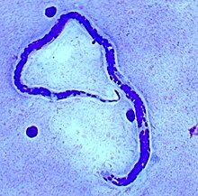
Loa loa filariasis is a skin and eye disease caused by the nematode worm Loa loa. Humans contract this disease through the bite of a deer fly or mango fly, the vectors for Loa loa. The adult Loa loa filarial worm migrates throughout the subcutaneous tissues of humans, occasionally crossing into subconjunctival tissues of the eye where it can be easily observed. Loa loa does not normally affect one's vision but can be painful when moving about the eyeball or across the bridge of the nose. The disease can cause red itchy swellings below the skin called "Calabar swellings". The disease is treated with the drug diethylcarbamazine (DEC), and when appropriate, surgical methods may be employed to remove adult worms from the conjunctiva. Loiasis belongs to the so-called neglected diseases.

<span class="mw-page-title-main">Diethylcarbamazine</span> Chemical compound

Diethylcarbamazine is a medication used in the treatment of filariasis including lymphatic filariasis, tropical pulmonary eosinophilia, and loiasis. It may also be used for prevention of loiasis in those at high risk. While it has been used for onchocerciasis, ivermectin is preferred. It is taken by mouth.

<span class="mw-page-title-main">Onchocerciasis</span> Human helminthiasis (infection by parasite)

Onchocerciasis, also known as river blindness, is a disease caused by infection with the parasitic worm Onchocerca volvulus. Symptoms include severe itching, bumps under the skin, and blindness. It is the second-most common cause of blindness due to infection, after trachoma.

<span class="mw-page-title-main">Filariasis</span> Parasitic disease caused by a family of nematode worms

Filariasis is a parasitic disease caused by an infection with roundworms of the Filarioidea type. These are spread by blood-feeding insects such as black flies and mosquitoes. They belong to the group of diseases called helminthiases.

<i>Wuchereria bancrofti</i> Species of parasitic worm

Wuchereria bancrofti is a filarial (arthropod-borne) nematode (roundworm) that is the major cause of lymphatic filariasis. It is one of the three parasitic worms, together with Brugia malayi and B. timori, that infect the lymphatic system to cause lymphatic filariasis. These filarial worms are spread by a variety of mosquito vector species. W. bancrofti is the most prevalent of the three and affects over 120 million people, primarily in Central Africa and the Nile delta, South and Central America, the tropical regions of Asia including southern China, and the Pacific islands. If left untreated, the infection can develop into lymphatic filariasis. In rare conditions, it also causes tropical pulmonary eosinophilia. No vaccine is commercially available, but high rates of cure have been achieved with various antifilarial regimens, and lymphatic filariasis is the target of the World Health Organization Global Program to Eliminate Lymphatic Filariasis with the aim to eradicate the disease as a public-health problem by 2020. However, this goal was not met by 2020.

Gnathostomiasis, also known as larva migrans profundus, is the human infection caused by the nematode Gnathostoma spinigerum and/or Gnathostoma hispidum, which infects vertebrates.

<i>Brugia malayi</i> Medical condition

Brugia malayi is a filarial (arthropod-borne) nematode (roundworm), one of the three causative agents of lymphatic filariasis in humans. Lymphatic filariasis, also known as elephantiasis, is a condition characterized by swelling of the lower limbs. The two other filarial causes of lymphatic filariasis are Wuchereria bancrofti and Brugia timori, which both differ from B. malayi morphologically, symptomatically, and in geographical extent.

<i>Dirofilaria immitis</i> Species of worm that causes parasitic disease in animals

Dirofilaria immitis, also known as heartworm or dog heartworm, is a parasitic roundworm that is a type of filarial worm, a small thread-like worm, that causes dirofilariasis. It is spread from host to host through the bites of mosquitoes. There are four genera of mosquitoes that transmit dirofilariasis, Aedes, Culex, Anopheles, and Mansonia. The definitive host is the dog, but it can also infect cats, wolves, coyotes, jackals, foxes, ferrets, bears, seals, sea lions and, under rare circumstances, humans.

<i>Onchocerca volvulus</i> Nematode

Onchocerca volvulus is a filarial (arthropod-borne) nematode (roundworm) that causes onchocerciasis, and is the second-leading cause of blindness due to infection worldwide after trachoma. It is one of the 20 neglected tropical diseases listed by the World Health Organization, with elimination from certain countries expected by 2025.

Acanthocheilonema is a genus within the family Onchocercidae which comprises mainly tropical parasitic worms. Cobbold created the genus Acanthocheilonema with only one species, Acanthocheilonema dracunculoides, which was collected from aardwolf in the region of South Africa in the nineteenth century. These parasites have a wide range of mammalian species as hosts, including members of Carnivora, Macroscelidea, Rodentia, Pholidota, Edentata, and Marsupialia. Many species among several genera of filarioids exhibit a high degree of endemicity in studies done on mammalian species in Japan. However, no concrete evidence has confirmed any endemic species in the genus Acanthocheilonema.

Acanthocheilonemiasis is a rare tropical infectious disease caused by a parasite known as Acanthocheilonema perstans. It can cause skin rashes, abdominal and chest pains, muscle and joint pains, neurological disorders and skin lumps. It is mainly found in Africa. The parasite is transmitted through the bite of small flies. Studies show that there are elevated levels of white blood cells.

<span class="mw-page-title-main">Lymphatic filariasis</span> Medical condition

Lymphatic filariasis is a human disease caused by parasitic worms known as filarial worms. Usually acquired in childhood, it is a leading cause of permanent disability worldwide, impacting over a hundred million people and manifesting itself in a variety of severe clinical pathologies While most cases have no symptoms, some people develop a syndrome called elephantiasis, which is marked by severe swelling in the arms, legs, breasts, or genitals. The skin may become thicker as well, and the condition may become painful. Affected people are often unable to work and are often shunned or rejected by others because of their disfigurement and disability.

<i>Mansonella perstans</i> Species of roundworm

Mansonella perstans is a filarial (arthropod-borne) nematode (roundworm), transmitted by tiny blood-sucking flies called midges. Mansonella perstans is one of two filarial nematodes that causes serous cavity filariasis in humans. The other filarial nematode is Mansonella ozzardi. M. perstans is widespread in many parts of sub-Saharan Africa, parts of Central and South America, and the Caribbean.

Mansonelliasis is the condition of infection by the nematode Mansonella. The disease exists in Africa and tropical Americas, spread by biting midges or blackflies. It is usually asymptomatic.

<span class="mw-page-title-main">Microfilaria</span> Early stage in the life cycle of certain parasitic nematodes in the family Onchocercidae

The microfilaria is an early stage in the life cycle of certain parasitic nematodes in the family Onchocercidae. In these species, the adults live in a tissue or the circulatory system of vertebrates. They release microfilariae into the bloodstream of the vertebrate host. The microfilariae are taken up by blood-feeding arthropod vectors. In the intermediate host the microfilariae develop into infective larvae that can be transmitted to a new vertebrate host.

Mansonella ozzardi is a filarial (arthropod-borne) nematode (roundworm). This filarial nematode is one of two that causes serous cavity filariasis in humans. The other filarial nematode that causes it in humans is Mansonella perstans. M. ozzardi is an endoparasite that inhabits the serous cavity of the abdomen in the human host. It lives within the mesenteries, peritoneum, and in the subcutaneous tissue.

<i>Dirofilaria repens</i> Species of roundworm

Dirofilaria repens is a filarial nematode that affects dogs and other carnivores such as cats, wolves, coyotes, foxes, and sea lions, as well as muskrats. It is transmitted by mosquitoes. Although humans may become infected as aberrant hosts, the worms fail to reach adulthood while infecting a human body.

<span class="mw-page-title-main">Filarioidea</span> Superfamily of roundworms

The Filarioidea are a superfamily of highly specialised parasitic nematodes. Species within this superfamily are known as filarial worms or filariae. Infections with parasitic filarial worms cause disease conditions generically known as filariasis. Drugs against these worms are known as filaricides.

Mansonella streptocerca,, is a filarial (arthropod-borne) nematode (roundworm) causing the disease streptocerciasis. It is a common parasite in the skin of humans in the rain forests of Africa, where it is thought to be a parasite of chimpanzees, as well.

<i>Brugia</i> Genus of roundworms

Brugia is a genus for a group of small roundworms. They are among roundworms that cause the parasitic disease filariasis. Specifically, of the three species known, Brugia malayi and Brugia timori cause lymphatic filariasis in humans; and Brugia pahangi and Brugia patei infect domestic cats, dogs and other animals. They are transmitted by the bite of mosquitos.

References

  1. Cobbold, T. S. (1864). Entozoa, an introduction to the study of helminthology, with reference more particularly to the internal parasites of man. London: Groombridge and Sons. doi: 10.5962/bhl.title.46771 .
  2. 1 2 Schmidt, Gerald et al. "Foundations of Parasitology". 7th ed. McGraw Hill, New York, NY, 2005.
  3. 1 2 Thomson, MC; Obsomer, V; Dunne, M; Connor, SJ; Molyneux, DH (2000). "Satellite mapping of Loa loa prevalence in relation to ivermectin use in west and central Africa". The Lancet. 356 (9235): 1077–1078. doi:10.1016/s0140-6736(00)02733-1. PMID   11009145. S2CID   11743223.
  4. Metzger, Wolfram Gottfried; Mordmüller, Benjamin (April 2014). "Loa loa—does it deserve to be neglected?". The Lancet Infectious Diseases. 14 (4): 353–357. doi:10.1016/S1473-3099(13)70263-9. PMID   24332895.
  5. 1 2 Turkington, C., & Ashby, B. (2007). Encyclopedia of Infectious Diseases. New York: Facts on File.
  6. Harris, Michael. "Loa loa". Animal Diversity Web. Retrieved 2019-04-26.
  7. Prevention, CDC – Centers for Disease Control and. "CDC – Loiasis – Biology". www.cdc.gov. Retrieved 2016-11-28.
  8. Whittaker, C; Walker, M; Pion, SDS; Chesnais, CB; Boussinesq, M; Basáñez, MG (April 2018). "The Population Biology and Transmission Dynamics of Loa loa". Trends in Parasitology. 34 (4): 335–350. doi: 10.1016/j.pt.2017.12.003 . hdl: 10044/1/55863 . PMID   29331268. S2CID   4545820.
  9. 1 2 3 4 5 Centers for Disease Control and Prevention (CDC). (2015 January 20). Parasites – Loiasis. Global Health – Division of Parasitic Diseases. Retrieved from: https://www.cdc.gov/parasites/loiasis/
  10. Agbolade, O.M.; Akinboye, D.O.; Ogunkola, O. (2005). "Loa loa and Mansonella perstans: neglected human infections that need control in Nigeria". Afr. J. Biotechnol. 4: 1554–1558.
  11. 1 2 3 4 Padgett, J. J.; Jacobsen, K. H. (2008). "Loiasis: African eye worm". Transactions of the Royal Society of Tropical Medicine and Hygiene. 102 (10): 983–989. doi:10.1016/j.trstmh.2008.03.022. PMID   18466939.
  12. 1 2 3 Zierhut, M., Pavesio, C., Ohno, S., Oréfice, F., Rao, N. A. (2014). Intraocular Inflammation. Springer Dordrecht Heidelberg London New York.
  13. Holmes, D (2013). "Loa loa: neglected neurology and nematodes". The Lancet. Neurology. 12 (7): 631–632. doi:10.1016/S1474-4422(13)70139-X. PMID   23769594. S2CID   140206733.
  14. Prevention, CDC-Centers for Disease Control and (2019-04-18). "CDC - Loiasis - Resources for Health Professionals". www.cdc.gov. Retrieved 2019-04-26.
  15. Wanji, S.; Tendongfor, N.; Esum, M.; Ndindeng, S.; Enyong, P. (2003). "Epidemiology of concomitant infections due to Loa loa, Mansonella perstans, and Onchocerca volvulus in rain forest villages of Cameroon". Medical Microbiology and Immunology. 192 (1): 15–21. doi:10.1007/s00430-002-0154-x. PMID   12592559. S2CID   42438020.
  16. Manego, R.; Mombo-Ngoma, Witte; Held, Gmeiner; Gebru (2017). "Demography, maternal health and the epidemiology of malaria and other major infectious diseases in the rural department Tsamba-Magotsi, Ngounie Province, in central African Gabon". BMC Public Health. 17 (1): 130. doi: 10.1186/s12889-017-4045-x . PMC   5273856 . PMID   28129759.
  17. Antinori, S.; Schifanella, Million; Galimberti, Ferraris; Mandia (2012). "Imported Loa loa filariasis: Three cases and a review of cases reported in nonendemic countries in the past 25 years. International Journal of Infectious Diseases IJID : Official Publication of the International Society for Infectious Diseases". International Journal of Infectious Diseases : Ijid : Official Publication of the International Society for Infectious Diseases. 16 (9): E649–E662. doi: 10.1016/j.ijid.2012.05.1023 . PMID   22784545.