
The medulla oblongata is a long stem-like structure which makes up part of the brainstem. It is anterior and partially inferior to the cerebellum. It is a cone-shaped neuronal mass responsible for autonomic (involuntary) functions ranging from vomiting to sneezing. The medulla contains the cardiac, respiratory, vomiting and vasomotor centers and therefore deals with the autonomic functions of breathing, heart rate and blood pressure as well as the sleep wake cycle.

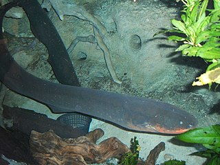
The electric eel is a South American electric fish. Until 2019, it was classified as the only species in its genus. Despite the name, it is not an eel, but rather a knifefish.

In the human brainstem, the solitary nucleus(SN) is a series of purely sensory nuclei forming a vertical column of grey matter embedded in the medulla oblongata. Through the center of the SN runs the solitary tract, a white bundle of nerve fibers, including fibers from the facial, glossopharyngeal and vagus nerves, that innervate the SN. The SN projects to, among other regions, the reticular formation, parasympathetic preganglionic neurons, hypothalamus and thalamus, forming circuits that contribute to autonomic regulation. Cells along the length of the SN are arranged roughly in accordance with function; for instance, cells involved in taste are located In the rostrum part, while those receiving information from cardio-respiratory and gastrointestinal processes are found in the caudal part.
An electric fish is any fish that can generate electric fields. A fish that can generate electric fields is said to be electrogenic while a fish that has the ability to detect electric fields is said to be electroreceptive. Most electrogenic fish are also electroreceptive. Electric fish species can be found both in the ocean and in freshwater rivers of South America (Gymnotiformes) and Africa (Mormyridae). Many fish such as sharks, rays and catfishes can detect electric fields and are thus electroreceptive, but they are not classified as electric fish because they cannot generate electricity. Most common bony fish (teleosts), including most fish kept in aquaria or caught for food, are neither electrogenic nor electroreceptive.

The reticular formation is a set of interconnected nuclei that are located throughout the brainstem. The reticular formation is not anatomically well defined because it includes neurons located in different parts of the brain. The neurons of the reticular formation make up a complex set of networks in the core of the brainstem that extend from the upper part of the midbrain to the lower part of the medulla oblongata. The reticular formation includes ascending pathways to the cortex in the ascending reticular activating system (ARAS) and descending pathways to the spinal cord via the reticulospinal tracts.

The thalamic reticular nucleus is part of the ventral thalamus that forms a capsule around the thalamus laterally. However, recent evidence from mice and fish question this statement and define it as dorsal thalamic structure. It is separated from the thalamus by the external medullary lamina. Reticular cells are GABAergic, and have discoid dendritic arbors in the plane of the nucleus.

The dentate nucleus is a cluster of neurons, or nerve cells, in the central nervous system that has a dentate – tooth-like or serrated – edge. It is located within the deep white matter of each cerebellar hemisphere, and it is the largest single structure linking the cerebellum to the rest of the brain. It is the largest and most lateral, or farthest from the midline, of the four pairs of deep cerebellar nuclei, the others being the fastigial nucleus and the globose and emboliform nuclei which together are referred to as the interposed nucleus. The dentate nucleus is responsible for the planning, initiation and control of voluntary movements. The dorsal region of the dentate nucleus contains output channels involved in motor function, which is the movement of skeletal muscle, while the ventral region contains output channels involved in nonmotor function, such as conscious thought and visuospatial function.

The flocculus is a small lobe of the cerebellum at the posterior border of the middle cerebellar peduncle anterior to the biventer lobule. Like other parts of the cerebellum, the flocculus is involved in motor control. It is an essential part of the vestibulo-ocular reflex, and aids in the learning of basic motor skills in the brain.

The tegmentum is a general area within the brainstem. The tegmentum is the ventral part of the midbrain and the tectum is the dorsal part of the midbrain. It is located between the ventricular system and distinctive basal or ventral structures at each level. It forms the floor of the midbrain (mesencephalon) whereas the tectum forms the ceiling. It is a multisynaptic network of neurons that is involved in many subconscious homeostatic and reflexive pathways. It is a motor center that relays inhibitory signals to the thalamus and basal nuclei preventing unwanted body movement. The tegmentum area includes various different structures, such as the "rostral (=frontal/cranial/oral) end of the reticular formation, several nuclei controlling eye movements, the periaqueductal gray matter, the red nucleus, the substantia nigra, and the ventral tegmental area".

The vestibulospinal tract is a neural tract in the central nervous system. Specifically, it is a component of the extrapyramidal system and is classified as a component of the medial pathway. Like other descending motor pathways, the vestibulospinal fibers of the tract relay information from nuclei to motor neurons. The vestibular nuclei receive information through the vestibulocochlear nerve about changes in the orientation of the head. The nuclei relay motor commands through the vestibulospinal tract. The function of these motor commands is to alter muscle tone, extend, and change the position of the limbs and head with the goal of supporting posture and maintaining balance of the body and head.

The cochlear nuclear (CN) complex comprises two cranial nerve nuclei in the human brainstem, the ventral cochlear nucleus (VCN) and the dorsal cochlear nucleus (DCN). The ventral cochlear nucleus is unlayered whereas the dorsal cochlear nucleus is layered. Auditory nerve fibers, fibers that travel through the auditory nerve carry information from the inner ear, the cochlea, on the same side of the head, to the nerve root in the ventral cochlear nucleus. At the nerve root the fibers branch to innervate the ventral cochlear nucleus and the deep layer of the dorsal cochlear nucleus. All acoustic information thus enters the brain through the cochlear nuclei, where the processing of acoustic information begins. The outputs from the cochlear nuclei are received in higher regions of the auditory brainstem.

The nucleus raphe pallidus receives afferent connections from the periaqueductal gray, the Paraventricular nucleus of hypothalamus, central nucleus of the amygdala, lateral hypothalamic area, and parvocellular reticular nucleus.

In biology, the electric organ is an organ common to all electric fish used for the purposes of creating an electric field. The electric organ is derived from modified nerve or muscle tissue. The electric discharge from this organ is used for navigation, communication, mating, defense and also sometimes for the incapacitation of prey.

The lateral hypothalamus(LH), also called the lateral hypothalamic area, contains the primary orexinergic nucleus within the hypothalamus that widely projects throughout the nervous system; this system of neurons mediates an array of cognitive and physical processes, such as promoting feeding behavior and arousal, reducing pain perception, and regulating body temperature, digestive functions, and blood pressure, among many others. Clinically significant disorders that involve dysfunctions of the orexinergic projection system include narcolepsy, motility disorders or functional gastrointestinal disorders involving visceral hypersensitivity, and eating disorders.
The reticulotegmental nucleus, tegmental pontine reticular nucleus is an area within the floor of the midbrain. This area is known to affect the cerebellum with its axonal projections.

The median preoptic nucleus is located dorsal to the other three nuclei of the preoptic area of the anterior hypothalamus. The hypothalamus is located just beneath the thalamus, the main sensory relay station of the nervous system, and is considered part of the limbic system, which also includes structures such as the hippocampus and the amygdala. The hypothalamus is highly involved in maintaining homeostasis of the body, and the median preoptic nucleus is no exception, contributing to regulation of blood composition, body temperature, and non-REM sleep.
In neuroethology and the study of learning, anti-Hebbian learning describes a particular class of learning rule by which synaptic plasticity can be controlled. These rules are based on a reversal of Hebb's postulate, and therefore can be simplistically understood as dictating reduction of the strength of synaptic connectivity between neurons following a scenario in which a neuron directly contributes to production of an action potential in another neuron.

Jamming avoidance response (JAR) is a behavior performed by some species of weakly electric fish. The JAR occurs when two electric fish with wave discharges meet – if their discharge frequencies are very similar, each fish will shift its discharge frequency to increase the difference between the two fish's discharge frequencies. By doing this, both fish prevent jamming of their sense of electroreception.

Electrocommunication is the communication method used by weakly electric fishes. Weakly electric fishes are a group of animals that utilize a communicating channel that is "invisible" to most other animals: electric signaling. Electric fishes communicate electrically by one fish generating an electric field and a second individual receiving that electric field with its electroreceptors. The receiving side will interpret the signal frequencies, waveforms, and delay, etc. The best studied species are two freshwater lineages- the African Mormyridae and the South American Gymnotiformes. While weakly electric fish are the only group that have been identified to carry out both generation and reception of electric fields, other species either generate signals or receive them, but not both. Animals that either generate or receive electric fields are found only in aquatic environments due to large resistance of all other media. So far, communication between electric fish has been identified mainly to serve the purpose of conveying information in
The parabrachial nuclei, also known as the parabrachial complex, are a group of nuclei in the dorsolateral pons that surrounds the superior cerebellar peduncle as it enters the brainstem from the cerebellum. They are named from the Latin term for the superior cerebellar peduncle, the brachium conjunctivum. In the human brain, the expansion of the superior cerebellar peduncle expands the parabrachial nuclei, which form a thin strip of grey matter over most of the peduncle. The parabrachial nuclei are typically divided along the lines suggested by Baxter and Olszewski in humans, into a medial parabrachial nucleus and lateral parabrachial nucleus. These have in turn been subdivided into a dozen subnuclei: the superior, dorsal, ventral, internal, external and extreme lateral subnuclei; the lateral crescent and subparabrachial nucleus along the ventrolateral margin of the lateral parabrachial complex; and the medial and external medial subnuclei