
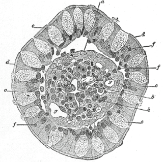
Leydig cells, also known as interstitial cells of the testes and interstitial cells of Leydig, are found adjacent to the seminiferous tubules in the testicle and produce testosterone in the presence of luteinizing hormone (LH). They are polyhedral in shape and have a large, prominent nucleus, an eosinophilic cytoplasm, and numerous lipid-filled vesicles.

Granulocytes are cells in the innate immune system characterized by the presence of specific granules in their cytoplasm. Such granules distinguish them from the various agranulocytes. All myeloblastic granulocytes are polymorphonuclear, that is, they have varying shapes (morphology) of the nucleus ; and are referred to as polymorphonuclear leukocytes. In common terms, polymorphonuclear granulocyte refers specifically to "neutrophil granulocytes", the most abundant of the granulocytes; the other types have varying morphology. Granulocytes are produced via granulopoiesis in the bone marrow.

Parathyroid oxyphil cells are one out of the two types of cells found in the parathyroid gland, the other being parathyroid chief cell. Oxyphil cells are only found in a select few number of species and humans are one of them.
An azurophilic granule is a cellular object readily stainable with a Romanowsky stain. In white blood cells and hyperchromatin, staining imparts a burgundy or merlot coloration. Neutrophils in particular are known for containing azurophils loaded with a wide variety of anti-microbial defensins that fuse with phagocytic vacuoles. Azurophils may contain myeloperoxidase, phospholipase A2, acid hydrolases, elastase, defensins, neutral serine proteases, bactericidal permeability-increasing protein, lysozyme, cathepsin G, proteinase 3, and proteoglycans.
Goblet cells are simple columnar epithelial cells that secrete gel-forming mucins, like mucin 5AC. The goblet cells mainly use the merocrine method of secretion, secreting vesicles into a duct, but may use apocrine methods, budding off their secretions, when under stress. The term goblet refers to the cell's goblet-like shape. The apical portion is shaped like a cup, as it is distended by abundant mucus laden granules; its basal portion lacks these granules and is shaped like a stem.

A proerythroblast is the earliest of four stages in development of the normoblast.

Monoblasts are the committed progenitor cells that differentiated from a committed macrophage or dendritic cell precursor (MDP) in the process of hematopoiesis. They are the first developmental stage in the monocyte series leading to a macrophage. Their myeloid cell fate is induced by the concentration of cytokines they are surrounded by during development. These cytokines induce the activation of transcription factors which push completion of the monoblast's myeloid cell fate. Monoblasts are normally found in bone marrow and do not appear in the normal peripheral blood. They mature into monocytes which, in turn, develop into macrophages. They then are seen as macrophages in the normal peripheral blood and many different tissues of the body. Macrophages can produce a variety of effector molecules that initiate local, systemic inflammatory responses. These monoblast differentiated cells are equipped to fight off foreign invaders using pattern recognition receptors to detect antigen as part of the innate immune response.

A promegakaryocyte is a precursor cell for a megakaryocyte. It arises from a megakaryoblast, into a promegakaryocyte and then into a megakaryocyte, which will eventually break off and become a platelet.

Granular cell tumor is a tumor that can develop on any skin or mucosal surface, but occurs on the tongue 40% of the time.

Toxic granulation refers to dark coarse granules found in granulocytes, particularly neutrophils, in patients with inflammatory conditions.
Micromeritics is the science and technology of small particles pioneered by Joseph M. DallaValle. It is thus the study of the fundamental and derived properties of individual as well as a collection of particles. The knowledge and control of the size of particles has importance in pharmacy and materials science. The size, and hence the surface area of a particle, can be related to the physical, chemical and pharmacological properties of drugs. Clinically, the particle size of a drug can affect its release from dosage forms that are administered orally, parenterally, rectally and topically. The successful formulation of suspensions, emulsions and tablets; both physical stability and pharmacological response also depends on the particle size achieved in the product.
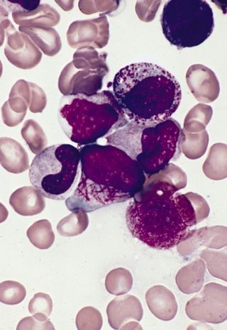
Faggot cells are cells normally found in the hypergranular form of acute promyelocytic leukemia. These promyelocytes have numerous Auer rods in the cytoplasm which gives the appearance of a bundle of sticks, from which the cells are given their name.

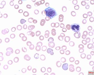
Neutrophil hypersegmentation can be defined as the presence of neutrophils whose nuclei have six or more lobes or the presence of more than 3% of neutrophils with at least five nuclear lobes. This is a clinical laboratory finding. It is visualized by drawing blood from a patient and viewing the blood smeared on a slide under a microscope. Normal neutrophils are uniform in size, with an apparent diameter of about 13 μm in a film. When stained, neutrophils have a segmented nucleus and pink/orange cytoplasm under light microscope. The majority of neutrophils have three nuclear segments (lobes) connected by tapering chromatin strands. A small percentage have four lobes, and occasionally five lobes may be seen. Up to 8% of circulating neutrophils are unsegmented.
Basophilic stippling, also known as punctate basophilia, is the presence of numerous basophilic granules that are dispersed through the cytoplasm of erythrocytes in a peripheral blood smear. They can be demonstrated to be RNA. They are composed of aggregates of ribosomes; degenerating mitochondria and siderosomes may be included in the aggregates. In contrast to Pappenheimer bodies, they are negative with Perls' acid ferrocyanide stain for iron. Basophilic stippling is indicative of disturbed erythropoiesis. It can also be found in some normal individuals.
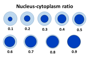
The nuclear-cytoplasmic ratio is a measurement used in cell biology. It is a ratio of the size of the nucleus of a cell to the size of the cytoplasm of that cell.

White blood cells, also called leukocytes or immune cells also called immunocytes, are cells of the immune system that are involved in protecting the body against both infectious disease and foreign invaders. White blood cells include three main subtypes; granulocytes, lymphocytes and monocytes.
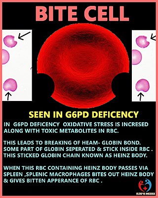
A degmacyte or bite cell is an abnormally shaped mature red blood cell with one or more semicircular portions removed from the cell margin, known as "bites". These "bites" result from the mechanical removal of denatured hemoglobin during splenic filtration as red cells attempt to migrate through endothelial slits from splenic cords into the splenic sinuses. Bite cells are known to be a result from processes of oxidative hemolysis, such as Glucose-6-phosphate dehydrogenase deficiency, in which uncontrolled oxidative stress causes hemoglobin to denature and form Heinz bodies. Bite cells can contain more than one "bite." The "bites" in degmacytes are smaller than the missing red blood cell fragments seen in schistocytes.

The vaginal epithelium is the inner lining of the vagina consisting of multiple layers of (squamous) cells. The basal membrane provides the support for the first layer of the epithelium-the basal layer. The intermediate layers lie upon the basal layer, and the superficial layer is the outermost layer of the epithelium. Anatomists have described the epithelium as consisting of as many as 40 distinct layers. The mucus found on the epithelium is secreted by the cervix and uterus. The rugae of the epithelium create an involuted surface and result in a large surface area that covers 360 cm2. This large surface area allows the trans-epithelial absorption of some medications via the vaginal route.

A white blood cell differential is a medical laboratory test that provides information about the types and amounts of white blood cells in a person's blood. The test, which is usually ordered as part of a complete blood count (CBC), measures the amounts of the five normal white blood cell types – neutrophils, lymphocytes, monocytes, eosinophils and basophils – as well as abnormal cell types if they are present. These results are reported as percentages and absolute values, and compared against reference ranges to determine whether the values are normal, low, or high. Changes in the amounts of white blood cells can aid in the diagnosis of many health conditions, including viral, bacterial, and parasitic infections and blood disorders such as leukemia.

Carcinocythemia, also known as carcinoma cell leukemia, is a condition in which cells from malignant tumours of non-hematopoietic origin are visible on the peripheral blood smear. It is an extremely rare condition, with 33 cases identified in the literature from 1960 to 2018. Carcinocythemia typically occurs secondary to infiltration of the bone marrow by metastatic cancer and carries a very poor prognosis.

