Palatine foramen may refer to:
| This disambiguation page lists articles associated with the title Palatine foramen. If an internal link led you here, you may wish to change the link to point directly to the intended article. |
Palatine foramen may refer to:
| This disambiguation page lists articles associated with the title Palatine foramen. If an internal link led you here, you may wish to change the link to point directly to the intended article. |

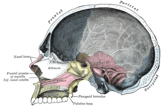
The palatine bones are two irregular bones of the facial skeleton in many animal species. Together with the maxillae they comprise the hard palate.

In anatomy, the orbit is the cavity or socket of the skull in which the eye and its appendages are situated. Anatomical term created by Gerard of Cremona. “Orbit” can refer to the bony socket, or it can also be used to imply the contents. In the adult human, the volume of the orbit is 30 millilitres, of which the eye occupies 6.5 ml. The orbital contents comprise the eye, the orbital and retrobulbar fascia, extraocular muscles, cranial nerves II, III, IV, V, and VI, blood vessels, fat, the lacrimal gland with its sac and nasolacrimal duct, the eyelids, medial and lateral palpebral ligaments, check ligaments, the suspensory ligament, septum, ciliary ganglion and short ciliary nerves.

The maxillary nerve (CN V2) is one of the three branches or divisions of the trigeminal nerve, the fifth (V) cranial nerve. It comprises the principal functions of sensation from the maxillary, nasal cavity, sinuses, the palate and subsequently that of the mid-face, and is intermediate, both in position and size, between the ophthalmic nerve and the mandibular nerve.

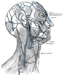
The pterygoid plexus is a venous plexus of considerable size, and is situated between the temporalis muscle and lateral pterygoid muscle, and partly between the two pterygoid muscles.

The descending palatine artery is a branch of the third part of the maxillary artery supplying the hard and soft palate.

In the opening of the incisive foramen, the orifices of two lateral canals are visible; they are named the incisive canals or foramina of Stensen.

In the human mouth, the incisive foramen, also called anterior palatine foramen, or nasopalatine foramen is a funnel-shaped opening in the bone of the oral hard palate immediately behind the incisor teeth where blood vessels and nerves pass. The incisive foramen is continuous with the incisive canal, this foramen or group of foramina is located behind the central incisor teeth in the incisive fossa of the maxilla.

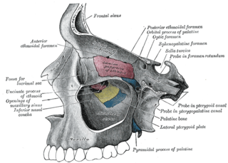
The sphenopalatine foramen is a foramen in the skull that connects the nasal cavity with the pterygopalatine fossa.
The greater palatine artery is a branch of the descending palatine artery and contributes to the blood supply of the hard palate and nasal septum.

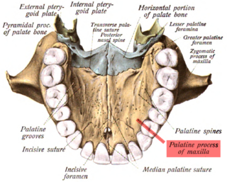
At either posterior angle of the hard palate is the greater palatine foramen, for the transmission of the descending palatine vessels and greater palatine nerve; and running anteriorly (forward) and medially from it is a groove, for the same vessels and nerve.

Behind the greater palatine foramen is the pyramidal process of the palatine bone, perforated by one or more lesser palatine foramina which carry the lesser palatine nerve, and marked by the commencement of a transverse ridge, for the attachment of the tendinous expansion of the tensor veli palatini.

The sphenoidal process of the palatine bone is a thin, compressed plate, much smaller than the orbital, and directed upward and medialward.

The perpendicular plate of palatine bone is the vertical part of the palatine bone, and is thin, of an oblong form, and presents two surfaces and four borders.

In human anatomy of the mouth, the palatine process of maxilla, is a thick, horizontal process of the maxilla. It forms the anterior three quarters of the hard palate, the horizontal plate of the palatine bone making up the rest.

The greater palatine nerve is a branch of the pterygopalatine ganglion that carries both general sensory fibres from the maxillary nerve and parasympathetic fibers from the nerve of the pterygoid canal. It descends through the greater palatine canal, emerges upon the hard palate through the greater palatine foramen, and passes forward in a groove in the hard palate, nearly as far as the incisor teeth.

The greater palatine canal is a passage in the skull that transmits the descending palatine artery, vein, and greater and lesser palatine nerves between the pterygopalatine fossa and the oral cavity.

The lesser palatine nerve (posterior palatine nerve) is one of two palatine nerves that descends through the greater palatine canal, and emerges by the lesser palatine foramen. It is a branch of the maxillary nerve (CN V2) It also has nasal branches that innervate the nasal cavity.

The following outline is provided as an overview of and topical guide to human anatomy:
The sphenopalatine artery passes through the sphenopalatine foramen into the cavity of the nose, at the back part of the superior meatus. Here it gives off its posterior lateral nasal branches which spread forward over the conchæ and meatuses, anastomose with the ethmoidal arteries and the nasal branches of the descending palatine, and assist in supplying the frontal, maxillary, ethmoidal, and sphenoidal sinuses.

Khunnuchelys was a genus of trionychine turtle from the Late Cretaceous of Asia. Three species are known, K. erinhotensis, the type species, K. kizylkumensis, and K. lophorhothon. K. erinhotensis lived in China from the late Turonian until the middle Campanian. K. kizylkumensis existed during the late Turonian of Uzbekistan. The third species, described in 2013 by Danilov et al., persisted from the early to middle Campanian of Kazakhstan.