This page is based on this
Wikipedia article Text is available under the
CC BY-SA 4.0 license; additional terms may apply.
Images, videos and audio are available under their respective licenses.

The vomer is one of the unpaired facial bones of the skull. It is located in the midsagittal line, and articulates with the sphenoid, the ethmoid, the left and right palatine bones, and the left and right maxillary bones. The vomer forms the inferior part of the nasal septum, with the superior part formed by the perpendicular plate of the ethmoid bone. The name is derived from the Latin word for a ploughshare and the shape of the bone.

The raphe nuclei are a moderate-size cluster of nuclei found in the brain stem. They have 5-HT1 receptors which are coupled with Gi/Go protein inhibiting adenyl cyclase. They function as autoreceptors in brain and decreases release of serotonin. The antianxiety drug Buspirone act as partial agonist. Selective serotonin reuptake inhibitor (SSRI) antidepressants are believed to act in these nuclei, as well as at their targets.

The mylohyoid muscle is a paired muscle running from the mandible to the hyoid bone, forming the floor of the oral cavity of the mouth. It is named after its two attachments near the molar teeth. These muscles are mesodermal in embryologic origin. The mylohyoid muscle is derived from the first pharyngeal arch.

The superior pharyngeal constrictor muscle is a muscle in the pharynx. It is the highest located muscle of the three pharyngeal constrictors. The muscle is a quadrilateral muscle, thinner and paler than the inferior pharyngeal constrictor muscle and middle pharyngeal constrictor muscle.
Ferganasaurus was a genus of dinosaur first formally described in 2003 by Alifanov and Averianov. The type species is Ferganasaurus verzilini. It was a sauropod similar to Rhoetosaurus. The fossils were discovered in 1966 in Kyrgyzstan from the Balabansai Formation and date to the Callovian stage of the Middle Jurassic.
The nucleus raphe pallidus receives afferent connections from the periaqueductal gray, the Paraventricular nucleus of hypothalamus, central nucleus of the amygdala, lateral hypothalamic area, and parvocellular reticular nucleus.

The dorsal raphe nucleus is located on the midline of the brainstem and is one of the raphe nuclei. It has rostral and caudal subdivisions.
The median raphe nucleus is composed of polygonal, fusiform and piriform neurons and exists rostral to the nucleus raphes pontis.

The pterygoid hamulus is a hook-like process at the lower extremity of the medial pterygoid plate of the sphenoid bone. Around this the tendon of the tensor veli palatini glides. It is the superior origin of the pterygomandibular raphe.
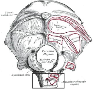
The pharyngeal tubercle is a part of the occipital bone of the head and neck. It is located on the lower surface of the basilar part of occipital bone, about 1 cm. anterior to the foramen magnum. The pharyngeal tubercle gives attachment to the fibrous raphe of the pharynx, also known as the pharyngeal raphe.
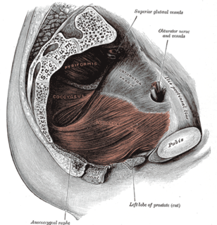
The anococcygeal body is a fibrous median raphe in the floor of the pelvis, which extends between the coccyx and the margin of the anus. It is composed of fibers of the levator ani muscle which unite with the muscle of the opposite side, muscle fibres from external anal sphincter and fibrous connective tissue.
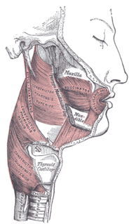
The pterygomandibular raphe is a ligamentous band of the buccopharyngeal fascia, attached superiorly to the pterygoid hamulus of the medial pterygoid plate, and inferiorly to the posterior end of the mylohyoid line of the mandible.

The greater palatine nerve is a branch of the pterygopalatine ganglion that carries both general sensory fibres from the maxillary nerve and parasympathetic fibers from the nerve of the pterygoid canal. It descends through the greater palatine canal, emerges upon the hard palate through the greater palatine foramen, and passes forward in a groove in the hard palate, nearly as far as the incisor teeth.
The median palatal cyst is a rare cyst that may occur anywhere along the median palatal raphe. It may produce swelling because of infection and is treated by excision or surgical removal.
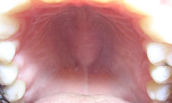
The incisive papilla is a projection on the palate near the incisors.
The term median raphe can refer to one of three different anatomical structures:
Carol Propper, CBE, FBA is Professor of Economics at Imperial College Business School and Professor of Economics of Public Policy at Bristol University. She is also a senior research associate with the Nuffield Trust, and has served on the Economic and Social Research Council Research Grants Board. Her work focuses on economic factors in health care reform, and she has served as an economic advisor to the National Health Service (England).

Gingival cyst is a type of cysts of the jaws that originates from the dental lamina and is found in the mouth parts. It is a superficial cyst in the alveolar mucosa. It can be seen inside the mouth as small and whitish bulge. Depending on the ages in which they develop, the cysts are classified into gingival cyst of newborn and gingival cyst of adult. Structurally, the cyst is lined by thin epithelium and shows a lumen usually filled with desquamated keratin, occasionally containing inflammatory cells. The nodes are formed as a result of cystic degeneration of epithelial rests of the dental lamina.

Timothy Richard Elliott is a Professor at the University of Bristol.

