The facial nerve, also known as the seventh cranial nerve, cranial nerve VII, or simply CN VII, is a cranial nerve that emerges from the pons of the brainstem, controls the muscles of facial expression, and functions in the conveyance of taste sensations from the anterior two-thirds of the tongue. The nerve typically travels from the pons through the facial canal in the temporal bone and exits the skull at the stylomastoid foramen. It arises from the brainstem from an area posterior to the cranial nerve VI and anterior to cranial nerve VIII.

The glossopharyngeal nerve, also known as the ninth cranial nerve, cranial nerve IX, or simply CN IX, is a cranial nerve that exits the brainstem from the sides of the upper medulla, just anterior to the vagus nerve. Being a mixed nerve (sensorimotor), it carries afferent sensory and efferent motor information. The motor division of the glossopharyngeal nerve is derived from the basal plate of the embryonic medulla oblongata, whereas the sensory division originates from the cranial neural crest.

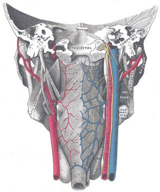
The external carotid artery is a major artery of the head and neck. It arises from the common carotid artery when it splits into the external and internal carotid artery. The external carotid artery supplies blood to the face and neck.

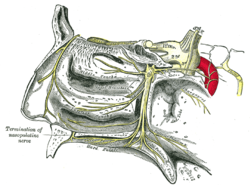
The pterygopalatine ganglion is a parasympathetic ganglion in the pterygopalatine fossa. It is one of four parasympathetic ganglia of the head and neck,.

The facial artery is a branch of the external carotid artery that supplies structures of the superficial face.

The superior pharyngeal constrictor muscle is a quadrilateral muscle of the pharynx. It is the uppermost and thinnest of the three pharyngeal constrictors.
In neuroanatomy, the maxillary nerve (V2) is one of the three branches or divisions of the trigeminal nerve, the fifth (CN V) cranial nerve. It comprises the principal functions of sensation from the maxilla, nasal cavity, sinuses, the palate and subsequently that of the mid-face, and is intermediate, both in position and size, between the ophthalmic nerve and the mandibular nerve.

The pharyngeal arches, also known as visceral arches, are structures seen in the embryonic development of vertebrates that are recognisable precursors for many structures. In fish, the arches are known as the branchial arches, or gill arches.

The aortic arches or pharyngeal arch arteries are a series of six paired embryological vascular structures which give rise to the great arteries of the neck and head. They are ventral to the dorsal aorta and arise from the aortic sac.
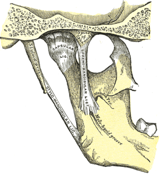
The sphenomandibular ligament is one of the three ligaments of the temporomandibular joint. It is situated medially to - and generally separate from - the articular capsule of the joint. Superiorly, it is attached to the spine of the sphenoid bone; inferiorly, it is attached to the lingula of mandible. The SML acts to limit inferior-ward movement of the mandible.

The pterygoid processes of the sphenoid, one on either side, descend perpendicularly from the regions where the body and the greater wings of the sphenoid bone unite.

The inferior thyroid artery is an artery in the neck. It arises from the thyrocervical trunk and passes upward, in front of the vertebral artery and longus colli muscle. It then turns medially behind the carotid sheath and its contents, and also behind the sympathetic trunk, the middle cervical ganglion resting upon the vessel.

The ascending pharyngeal artery is an artery in the neck that supplies the pharynx, developing from the proximal part of the embryonic second aortic arch.

The maxillary artery supplies deep structures of the face. It branches from the external carotid artery just deep to the neck of the mandible.

The middle cranial fossa is formed by the sphenoid bones, and the temporal bones. It lodges the temporal lobes, and the pituitary gland. It is deeper than the anterior cranial fossa, is narrow medially and widens laterally to the sides of the skull. It is separated from the posterior fossa by the clivus and the petrous crest.

The palatovaginal canal is a canal between the sphenoid bone and the palatine bone that connects the nasopharynx with the pterygopalatine fossa. It transmits the pharyngeal branch of the third part of the maxillary artery. An inconstant vomerovaginal canal may lie between the ala of the vomer and the vaginal process of the sphenoid bone, medial to the palatovaginal canal, and lead into the anterior end of the palatovaginal canal.

Continuous with the dorsal end of the first pharyngeal arch, and growing forward from its cephalic border, is a triangular process, the maxillary prominence, the ventral extremity of which is separated from the mandibular arch by a ">"-shaped notch.
The pharyngeal branch of the vagus nerve is the principal motor nerve of the pharynx. It represents the motor component of the pharyngeal plexus of vagus nerve and ultimately provides motor innervation to most of the muscles of the soft palate, and of the pharynx.
The pharyngeal plexus is a nerve plexus located upon the outer surface of the pharynx. It contains a motor component, a sensory component, and sympathetic component.
The face and neck development of the human embryo refers to the development of the structures from the third to eighth week that give rise to the future head and neck. They consist of three layers, the ectoderm, mesoderm and endoderm, which form the mesenchyme, neural crest and neural placodes. The paraxial mesoderm forms structures named somites and somitomeres that contribute to the development of the floor of the brain and voluntary muscles of the craniofacial region. The lateral plate mesoderm consists of the laryngeal cartilages. The three tissue layers give rise to the pharyngeal apparatus, formed by six pairs of pharyngeal arches, a set of pharyngeal pouches and pharyngeal grooves, which are the most typical feature in development of the head and neck. The formation of each region of the face and neck is due to the migration of the neural crest cells which come from the ectoderm. These cells determine the future structure to develop in each pharyngeal arch. Eventually, they also form the neurectoderm, which forms the forebrain, midbrain and hindbrain, cartilage, bone, dentin, tendon, dermis, pia mater and arachnoid mater, sensory neurons, and glandular stroma.