
The central nervous system (CNS) is the part of the nervous system consisting primarily of the brain and spinal cord. The CNS is so named because the brain integrates the received information and coordinates and influences the activity of all parts of the bodies of bilaterally symmetric and triploblastic animals—that is, all multicellular animals except sponges and diploblasts. It is a structure composed of nervous tissue positioned along the rostral to caudal axis of the body and may have an enlarged section at the rostral end which is a brain. Only arthropods, cephalopods and vertebrates have a true brain, though precursor structures exist in onychophorans, gastropods and lancelets.

In neuroanatomy, the optic chiasm, or optic chiasma, is the part of the brain where the optic nerves cross. It is located at the bottom of the brain immediately inferior to the hypothalamus. The optic chiasm is found in all vertebrates, although in cyclostomes, it is located within the brain.
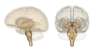
The pineal gland is a small endocrine gland in the brain of most vertebrates. It produces melatonin, a serotonin-derived hormone, which modulates sleep patterns following the diurnal cycles. The shape of the gland resembles a pine cone, which gives it its name. The pineal gland is located in the epithalamus, near the center of the brain, between the two hemispheres, tucked in a groove where the two halves of the thalamus join. It is one of the neuroendocrine secretory circumventricular organs in which capillaries are mostly permeable to solutes in the blood.
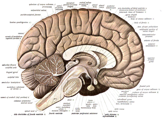
Neuroanatomy is the study of the structure and organization of the nervous system. In contrast to animals with radial symmetry, whose nervous system consists of a distributed network of cells, animals with bilateral symmetry have segregated, defined nervous systems. Their neuroanatomy is therefore better understood. In vertebrates, the nervous system is segregated into the internal structure of the brain and spinal cord and the series of nerves that connect the CNS to the rest of the body. Breaking down and identifying specific parts of the nervous system has been crucial for figuring out how it operates. For example, much of what neuroscientists have learned comes from observing how damage or "lesions" to specific brain areas affects behavior or other neural functions.

Glia, also called glial cells (gliocytes) or neuroglia, are non-neuronal cells in the central nervous system and the peripheral nervous system that do not produce electrical impulses. The neuroglia make up more than one half the volume of neural tissue in the human body. They maintain homeostasis, form myelin in the peripheral nervous system, and provide support and protection for neurons. In the central nervous system, glial cells include oligodendrocytes, astrocytes, ependymal cells and microglia, and in the peripheral nervous system they include Schwann cells and satellite cells.
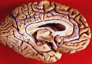
The limbic lobe is an arc-shaped cortical region of the limbic system, on the medial surface of each cerebral hemisphere of the mammalian brain, consisting of parts of the frontal, parietal and temporal lobes. The term is ambiguous, with some authors including the paraterminal gyrus, the subcallosal area, the cingulate gyrus, the parahippocampal gyrus, the dentate gyrus, the hippocampus and the subiculum; while the Terminologia Anatomica includes the cingulate sulcus, the cingulate gyrus, the isthmus of cingulate gyrus, the fasciolar gyrus, the parahippocampal gyrus, the parahippocampal sulcus, the dentate gyrus, the fimbrodentate sulcus, the fimbria of hippocampus, the collateral sulcus, and the rhinal sulcus, and omits the hippocampus.

A cortical column is a group of neurons forming a cylindrical structure through the cerebral cortex of the brain perpendicular to the cortical surface. The structure was first identified by Vernon Benjamin Mountcastle in 1957. He later identified minicolumns as the basic units of the neocortex which were arranged into columns. Each contains the same types of neurons, connectivity, and firing properties. Columns are also called hypercolumn, macrocolumn, functional column or sometimes cortical module. Neurons within a minicolumn (microcolumn) encode similar features, whereas a hypercolumn "denotes a unit containing a full set of values for any given set of receptive field parameters". A cortical module is defined as either synonymous with a hypercolumn (Mountcastle) or as a tissue block of multiple overlapping hypercolumns.

In neuroanatomy, the pretectal area, or pretectum, is a midbrain structure composed of seven nuclei and comprises part of the subcortical visual system. Through reciprocal bilateral projections from the retina, it is involved primarily in mediating behavioral responses to acute changes in ambient light such as the pupillary light reflex, the optokinetic reflex, and temporary changes to the circadian rhythm. In addition to the pretectum's role in the visual system, the anterior pretectal nucleus has been found to mediate somatosensory and nociceptive information.

The supraesophageal ganglion is the first part of the arthropod, especially insect, central nervous system. It receives and processes information from the first, second, and third metameres. The supraesophageal ganglion lies dorsal to the esophagus and consists of three parts, each a pair of ganglia that may be more or less pronounced, reduced, or fused depending on the genus:

Cornelius Ubbo Ariëns Kappers was a Dutch neurologist and anatomist.

Henkjan Honing is a Dutch researcher. He is professor of Music Cognition at both the Faculty of Humanities and the Faculty of Science of the University of Amsterdam. He conducts his research under the auspices of the Institute for Logic, Language and Computation, and the University of Amsterdam's Brain and Cognition center.
Neuromorphology is the study of nervous system form, shape, and structure. The study involves looking at a particular part of the nervous system from a molecular and cellular level and connecting it to a physiological and anatomical point of view. The field also explores the communications and interactions within and between each specialized section of the nervous system. Morphology is distinct from morphogenesis. Morphology is the study of the shape and structure of biological organisms, while morphogenesis is the study of the biological development of the shape and structure of organisms. Therefore, neuromorphology focuses on the specifics of the structure of the nervous system and not the process by which the structure was developed. Neuromorphology and morphogenesis, while two different entities, are nonetheless closely linked.
Constantinus Albertus Josephus Maria "Constantijn" Kortmann was a Dutch professor of constitutional law.

Izak (Ieke) Moerdijk is a Dutch mathematician, currently working at Utrecht University, who in 2012 won the Spinoza prize.
The evolution of nervous systems dates back to the first development of nervous systems in animals. Neurons developed as specialized electrical signaling cells in multicellular animals, adapting the mechanism of action potentials present in motile single-celled and colonial eukaryotes. Primitive systems, like those found in protists, use chemical signalling for movement and sensitivity; data suggests these were precursors to modern neural cell types and their synapses. When some animals started living a mobile lifestyle and eating larger food particles externally, they developed ciliated epithelia, contractile muscles and coordinating & sensitive neurons for it in their outer layer.

Günther K.H. Zupanc is a German-American neurobiologist, researcher, university teacher, book author, journal editor, and educational reformer. He is a Professor in the Department of Biology at Northeastern University in Boston, Massachusetts.

Elizabeth Caroline Crosby was an American neuroanatomist. Crosby received the National Medal of Science from President Jimmy Carter in 1979 "for outstanding contributions to comparative and human neuroanatomy and for the synthesis and transmission of knowledge of the entire nervous system of the vertebrate phylum." Her "careful descriptions" of vertebrate brains - especially reptiles - helped "outline evolutionary history" and her work as a clinical diagnostic assistant to neurosurgeons resulted in "the correlation of anatomy and surgery."
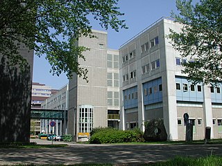
The Netherlands Institute for Neuroscience (NIN) is a research institute of the Royal Netherlands Academy of Arts and Sciences (KNAW) that carries out neuroscience research with special emphasis on the brain and visual system. Although the institute's focus is on understanding the fundamental mechanisms underlying brain function, its research spans the development, plasticity and aging of the brain and is often linked to clinical research questions. The research program is carried out in 17 research groups. In addition, the NIN includes the Netherlands Brain Bank and the Netherlands Sleep Registry.

In anatomy a chiasm is the spot where two structures cross, forming an X-shape. Examples of chiasms are: