Sphenoid sinus

Last updated
Sphenoid sinus
Gray856.png
Lateral wall of nasal cavity; the three nasal conchæ have been removed. (Sphenoidal sinus visible at upper right, in dark circle.)
Illu nose nasal cavities.jpg
Nose and nasal cavities. (Sphenoid sinus labeled at upper right.)
Details
Nerve posterior ethmoidal nerves, and orbital branches of the pterygopalatine ganglion
Identifiers
Latin sinus sphenoidalis
MeSH D013101
TA98 A06.1.03.003
TA2 3178
FMA 54683
Anatomical terms of bone

The sphenoid sinus is a paired paranasal sinus occurring within the body of the sphenoid bone. It represents one pair of the four paired paranasal sinuses. [1] The pair of sphenoid sinuses are separated in the middle by a septum of sphenoid sinuses. Each sphenoid sinus communicates with the nasal cavity via the opening of sphenoidal sinus. [2] :500 The two sphenoid sinuses vary in size and shape, and are usually asymmetrical. [3]

Contents

Anatomy

Paranasal sinuses Paranasal Sinuses lat.jpg
Paranasal sinuses

On average, a sphenoid sinus measures 2.2 cm vertical height, 2 cm in transverse breadth; and 2.2 cm antero-posterior depth. [3]

Each spehoid sinus is contained within the body of sphenoid bone, being situated just inferior to the sella turcica. The two sphenoid sinuses are separated medially by the septum of sphenoidal sinuses (which is usually asymmetrical). [2] :500

An opening of sphenoidal sinus forms a passage between each sphenoidal sinus, [2] :500 and the nasal cavity. Posteriorly, an opening of sphenoidal sinus opens into the sphenoidal sinus by an aperture high on the anterior wall the sinus; anteriorly, an opening of sphenoidal sinus opens into the roof of the nasal cavity via an aperture on the posterior wall of the sphenoethmoidal recess (occurring just superior the choana). [4]

Innervation

The mucous membrane receives sensory[ citation needed ] innervation from the posterior ethmoidal nerve (branch of the ophthalmic nerve (CN V1)), and branches of the maxillary nerve (CN V2). [5]

Postganglionic parasympathetic fibers of the facial nerve that synapsed at the pterygopalatine ganglion control mucus secretion.[ citation needed ]

Anatomical relations

Proximal structures include: the optic canal and optic nerve, internal carotid artery, cavernous sinus, trigeminal nerve, pituitary gland, and the anterior ethmoidal cells. [2] :500 One study found that carotid canal protrudation into the sphenoid sinus wall was present 23.9-32.1% in males and 35.5-36.2% in females, dehiscence in carotid canal was detected more in females (34%) compared to males (22%), optic canal protrudation was 33.3 and 30.5% in males and females, and optic canal dehiscence was detected 11.3 and 9.9% in males and females. [6]

Anatomical variation

The sphenoid sinuses vary in size and shape, and, owing to the lateral displacement of the intervening septum of sphenoid sinuses, are rarely symmetrical. [3]

When exceptionally large, the sphenoid sinuses may extend into the roots of the pterygoid processes or greater wings of sphenoid bone, and may invade the basilar part of the occipital bone. [3]

The septum of the sphenoidal sinuses may be partially or completely absent. Additional incomplete septa may also be present. [2] :500

Development

The sphenoidal sinuses are minute at birth; [3] [4] their main development takes place after puberty. [4]

Clinical significance

The sphenoid sinuses cannot be palpated on physical examination. [1] However, patients with isolated sphenoid sinusitis may complain of occipital or vertex headache, retro-orbital pain, otalgia, drowsiness, or meningitis-like symptoms. [7]

A potential complication of sphenoidal sinusitis is cavernous sinus thrombosis. [7]

If a fast-growing tumor erodes the floor of the sphenoidal sinus, the vidian nerve could be in danger.[ citation needed ] If the tumor spreads laterally, the cavernous sinus and all its constituent nerves could be in danger. [8]

An endonasal surgical procedure called a sphenoidotomy may be carried out to enlarge the sphenoid sinus, usually in order to drain it. [8]

Of note, the sphenoid sinus should be distinguished from an Onodi cell, an anatomic variant that is the posterior-most ethmoidal air cell. Onodi cells typically extend posteriorly to lie superolateral to the sphenoid sinus and thus in close proximity to the optic nerve and internal carotid artery. [9] Failure to recognize an Onodi cell on CT scan prior to surgery may put these structures at risk. One study found that an Onodi cell was present 26.6% and 19.1% in males and females, respectively. [6]

Transsphenoidal surgery

Because only thin shelves of bone separate the sphenoidal sinuses from the nasal cavities below and hypophyseal fossa above, the pituitary gland can be surgically approached through the roof of the nasal cavities by first passing through the anterioinferior aspect of the sphenoid bone and into the sinuses, followed by entry through the top of the sphenoid bone into the hypophyseal fossa.[ citation needed ]

Related Research Articles

<span class="mw-page-title-main">Paranasal sinuses</span> Air-filled spaces surrounding the nasal cavity

Paranasal sinuses are a group of four paired air-filled spaces that surround the nasal cavity. The maxillary sinuses are located under the eyes; the frontal sinuses are above the eyes; the ethmoidal sinuses are between the eyes and the sphenoidal sinuses are behind the eyes. The sinuses are named for the facial bones and sphenoid bone in which they are located. Their role is disputed and no function has been confirmed.

Articles related to anatomy include:

<span class="mw-page-title-main">Ethmoid bone</span> Bone in the skull

The ethmoid bone is an unpaired bone in the skull that separates the nasal cavity from the brain. It is located at the roof of the nose, between the two orbits. The cubical bone is lightweight due to a spongy construction. The ethmoid bone is one of the bones that make up the orbit of the eye.

<span class="mw-page-title-main">Sphenoid bone</span> Bone at the front of the skull

The sphenoid bone is an unpaired bone of the neurocranium. It is situated in the middle of the skull towards the front, in front of the basilar part of the occipital bone. The sphenoid bone is one of the seven bones that articulate to form the orbit. Its shape somewhat resembles that of a butterfly or bat with its wings extended.

<span class="mw-page-title-main">Nasal cavity</span> Large, air-filled space above and behind the nose in the middle of the face

The nasal cavity is a large, air-filled space above and behind the nose in the middle of the face. The nasal septum divides the cavity into two cavities, also known as fossae. Each cavity is the continuation of one of the two nostrils. The nasal cavity is the uppermost part of the respiratory system and provides the nasal passage for inhaled air from the nostrils to the nasopharynx and rest of the respiratory tract.

<span class="mw-page-title-main">Internal carotid artery</span> Artery of the human brain

The internal carotid artery is an artery in the neck which supplies the anterior circulation of the brain.

<span class="mw-page-title-main">Orbit (anatomy)</span> Cavity or socket of the skull in which the eye and its appendages are situated

In anatomy, the orbit is the cavity or socket/hole of the skull in which the eye and its appendages are situated. "Orbit" can refer to the bony socket, or it can also be used to imply the contents. In the adult human, the volume of the orbit is 30 millilitres, of which the eye occupies 6.5 ml. The orbital contents comprise the eye, the orbital and retrobulbar fascia, extraocular muscles, cranial nerves II, III, IV, V, and VI, blood vessels, fat, the lacrimal gland with its sac and duct, the eyelids, medial and lateral palpebral ligaments, cheek ligaments, the suspensory ligament, septum, ciliary ganglion and short ciliary nerves.

<span class="mw-page-title-main">Maxillary sinus</span> Largest of the paranasal sinuses, and drains into the middle meatus of the nose

The pyramid-shaped maxillary sinus is the largest of the paranasal sinuses, located in the maxilla. It drains into the middle meatus of the nose through the semilunar hiatus. It is located to the side of the nasal cavity, and below the orbit.

<span class="mw-page-title-main">Orbital part of frontal bone</span>

The orbital or horizontal part of the frontal bone consists of two thin triangular plates, the orbital plates, which form the vaults of the orbits, and are separated from one another by a median gap, the ethmoidal notch.

<span class="mw-page-title-main">Ethmoid sinus</span> Air-filled space near the nasal cavity

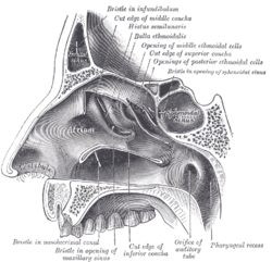
The ethmoid sinuses or ethmoid air cells of the ethmoid bone are one of the four paired paranasal sinuses. Unlike the other three pairs of paranasal sinuses which consist of one or two large cavities, the ethmoidal sinuses entail a number of small air-filled cavities. The cells are located within the lateral mass (labyrinth) of each ethmoid bone and are variable in both size and number. The cells are grouped into anterior, middle, and posterior groups; the groups differ in their drainage modalities, though all ultimately drain into either the superior or the middle nasal meatus of the lateral wall of the nasal cavity.

<span class="mw-page-title-main">Cavernous sinus</span> Sinus in the human head

The cavernous sinus within the human head is one of the dural venous sinuses creating a cavity called the lateral sellar compartment bordered by the temporal bone of the skull and the sphenoid bone, lateral to the sella turcica.

<span class="mw-page-title-main">Ethmoidal labyrinth</span>

The ethmoidal labyrinth or lateral mass of the ethmoid bone consists of a number of thin-walled cellular cavities, the ethmoid air cells, arranged in three groups, anterior, middle, and posterior, and interposed between two vertical plates of bone; the lateral plate forms part of the orbit, the medial plate forms part of the nasal cavity. In the disarticulated bone many of these cells are opened into, but when the bones are articulated, they are closed in at every part, except where they open into the nasal cavity.

<span class="mw-page-title-main">Anterior ethmoidal artery</span>

The anterior ethmoidal artery is a branch of the ophthalmic artery in the orbit. It exits the orbit through the anterior ethmoidal foramen alongside the anterior ethmoidal nerve. It contributes blood supply to the ethmoid sinuses, frontal sinuses, the dura mater, lateral nasal wall, and nasal septum. It issues a meningeal branch, and nasal branches.

<span class="mw-page-title-main">Sphenoethmoidal recess</span>

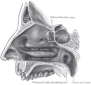
The sphenoethmoidal recess is a small triangular space of the superior nasal meatus of the nasal cavity into which the sphenoidal sinus and the posterior ethmoidal air cells open. The sphenoethmoidal recess is situated superoposterior to the superior nasal concha, between the superior nasal concha and the anterior aspect of the body of the sphenoid bone.

<span class="mw-page-title-main">Middle cranial fossa</span>

The middle cranial fossa is formed by the sphenoid bones, and the temporal bones. It lodges the temporal lobes, and the pituitary gland. It is deeper than the anterior cranial fossa, is narrow medially and widens laterally to the sides of the skull. It is separated from the posterior cranial fossa by the clivus and the petrous crest.

<span class="mw-page-title-main">Anterior cranial fossa</span>

The anterior cranial fossa is a depression in the floor of the cranial base which houses the projecting frontal lobes of the brain. It is formed by the orbital plates of the frontal, the cribriform plate of the ethmoid, and the small wings and front part of the body of the sphenoid; it is limited behind by the posterior borders of the small wings of the sphenoid and by the anterior margin of the chiasmatic groove. The lesser wings of the sphenoid separate the anterior and middle fossae.

<span class="mw-page-title-main">Body of sphenoid bone</span>

The body of the sphenoid bone, more or less cubical in shape, is hollowed out in its interior to form two large cavities, the sphenoidal sinuses, which are separated from each other by a septum.

<span class="mw-page-title-main">Human nose</span> Feature of the human face

The human nose is the most protruding part of the human face. It bears the nostrils and is the first organ of the respiratory system. It is also the principal organ in the olfactory system. The shape of the nose is determined by the nasal bones and the nasal cartilages, including the nasal septum which separates the nostrils and divides the nasal cavity into two.

<span class="mw-page-title-main">Base of skull</span> Inferior area of the skull, composed of the endocranium and lower parts of the skull roof

The base of skull, also known as the cranial base or the cranial floor, is the most inferior area of the skull. It is composed of the endocranium and the lower parts of the calvaria.

<span class="mw-page-title-main">Outline of human anatomy</span> Overview of and topical guide to human anatomy

The following outline is provided as an overview of and topical guide to human anatomy:

References

PD-icon.svgThis article incorporates text in the public domain from page 998 of the 20th edition of Gray's Anatomy (1918)

  1. 1 2 Illustrated Anatomy of the Head and Neck, Fehrenbach and Herring, Elsevier, 2012, page 64
  2. 1 2 3 4 5 Sobotta anatomy textbook. Friedrich Paulsen, Tobias M. Böckers, J. Waschke, Stephan Winkler, Katja Dalkowski, Jörg Mair. Amsterdam. 2018. ISBN   978-0-7206-7617-4. OCLC   1082911887.{{cite book}}: CS1 maint: location missing publisher (link) CS1 maint: others (link)
  3. 1 2 3 4 5 Gray, Henry (1918). Gray's Anatomy (20th ed.). pp. 998–999.
  4. 1 2 3 Human Anatomy, Jacob, Elsevier, 2008, page 211
  5. Morton, David A. (2019). The Big Picture: Gross Anatomy. K. Bo Foreman, Kurt H. Albertine (2nd ed.). New York. p. 246. ISBN   978-1-259-86264-9. OCLC   1044772257.{{cite book}}: CS1 maint: location missing publisher (link)
  6. 1 2 Asal, Neşe; Bayar Muluk, Nuray; Inal, Mikail; Şahan, Mehmet Hamdi; Doğan, Adil; Arıkan, Osman Kürşat (June 2019). "Carotid canal and optic canal at sphenoid sinus". Neurosurgical Review. 42 (2): 519–529. doi:10.1007/s10143-018-0995-4. ISSN   1437-2320. PMID   29926302. S2CID   49331277.
  7. 1 2 Ishak, NL; Subha, ST; Abu Bakar, S (2019-04-30). "Isolated sphenoid sinusitis: A big headache". Malaysian Family Physician. 14 (1): 29–30. ISSN   1985-207X. PMC   6612268 . PMID   31289629.
  8. 1 2 Kozłowski, Z; Mazerant, M; Skóra, W; Dabrowska, K (2008). "[Sphenoidotomy--the treatment of patients with isolated sphenoid sinus diseases]". Otolaryngologia Polska = the Polish Otolaryngology. 62 (5): 582–6. doi:10.1016/S0030-6657(08)70319-6. PMID   19004262.
  9. Gaillard, Frank. "Sphenoethmoidal air cell | Radiology Reference Article | Radiopaedia.org". Radiopaedia. Retrieved 2023-11-12.