In the human skull, the zygomatic bone, also called cheekbone or malar bone, is a paired irregular bone which articulates with the maxilla, the temporal bone, the sphenoid bone and the frontal bone. It is situated at the upper and lateral part of the face and forms the prominence of the cheek, part of the lateral wall and floor of the orbit, and parts of the temporal fossa and the infratemporal fossa. It presents a malar and a temporal surface; four processes, and four borders.

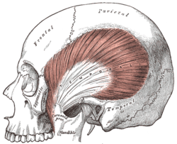
In anatomy, the temporalis muscle, also known as the temporal muscle, is one of the muscles of mastication (chewing). It is a broad, fan-shaped convergent muscle on each side of the head that fills the temporal fossa, superior to the zygomatic arch so it covers much of the temporal bone.Temporal refers to the head's temples.

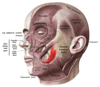
In human anatomy, the masseter is one of the muscles of mastication. Found only in mammals, it is particularly powerful in herbivores to facilitate chewing of plant matter. The most obvious muscle of mastication is the masseter muscle, since it is the most superficial and one of the strongest.

The auriculotemporal nerve is a sensory branch of the mandibular nerve (CN V3) that runs with the superficial temporal artery and vein, and provides sensory innervation to parts of the external ear, scalp, and temporomandibular joint. The nerve also conveys post-ganglionic parasympathetic fibres from the otic ganglion to the parotid gland.
The risorius muscle is a highly variable muscle of facial expression. It has numerous and very variable origins, and inserts into the angle of the mouth. It receives motor innervation from branches of facial nerve. It may be absent or asymmetrical in some people. It pulls the angle of the mouth sidewise, such as during smiling.

The temporal fossa is a fossa on the side of the skull bounded by the temporal lines and terminating below the level of the zygomatic arch.

In human anatomy, the superficial temporal artery is a major artery of the head. It arises from the external carotid artery when it splits into the superficial temporal artery and maxillary artery.

The zygomaticotemporal nerve (zygomaticotemporal branch, temporal branch) is a small nerve of the face. It is derived from the zygomatic nerve, a branch of the maxillary nerve (CN V2). It is distributed to the skin of the side of the forehead. It communicates with the facial nerve and with the auriculotemporal branch of the mandibular nerve.

The maxillary artery supplies deep structures of the face. It branches from the external carotid artery just deep to the neck of the mandible.

The squamous part of temporal bone, or temporal squama, forms the front and upper part of the temporal bone, and is scale-like, thin, and translucent.

The squamous part of the frontal bone is the superior portion when viewed in standard anatomical orientation. There are two surfaces of the squamous part of the frontal bone: the external surface, and the internal surface.

The infratemporal fossa is an irregularly shaped cavity that is a part of the skull. It is situated below and medial to the zygomatic arch. It is not fully enclosed by bone in all directions. It contains superficial muscles, including the lower part of the temporalis muscle, the lateral pterygoid muscle, and the medial pterygoid muscle. It also contains important blood vessels such as the middle meningeal artery, the pterygoid plexus, and the retromandibular vein, and nerves such as the mandibular nerve (CN V3) and its branches.

The deep cervical fascia lies under cover of the platysma, and invests the muscles of the neck; it also forms sheaths for the carotid vessels, and for the structures situated in front of the vertebral column. Its attachment to the hyoid bone prevents the formation of a dewlap.

The masseteric fascia and parotideomasseteric fascia are fascias of the head varyingly described depending upon the source consulted. They may or may not be described as one and the same structure.

In anatomy, the middle temporal artery is a major artery which arises immediately above the zygomatic arch, and, perforating the temporal fascia, gives branches to the temporalis, anastomosing with the deep temporal branches of the internal maxillary.

The temporal branches of the facial nerve crosses the zygomatic arch to the temporal region, supplying the auriculares anterior and superior, and joining with the zygomaticotemporal branch of the maxillary nerve, and with the auriculotemporal branch of the mandibular nerve.
The middle temporal artery occasionally gives off a zygomatico-orbital branch, which runs along the upper border of the zygomatic arch, between the two layers of the temporal fascia, to the lateral angle of the orbit.
The investing layer of deep cervical fascia is the most superficial part of the deep cervical fascia, and encloses the whole neck.

The following outline is provided as an overview of and topical guide to human anatomy:
The parotid fascia is a tough fascia enclosing the parotid gland. It has a superficial layer and a deep layer.