Thoracic aperture may refer to:
| This disambiguation page lists articles associated with the title Thoracic aperture. If an internal link led you here, you may wish to change the link to point directly to the intended article. |
Thoracic aperture may refer to:
| This disambiguation page lists articles associated with the title Thoracic aperture. If an internal link led you here, you may wish to change the link to point directly to the intended article. |
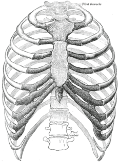
In vertebrate anatomy, ribs are the long curved bones which form the rib cage, part of the axial skeleton. In most tetrapods, ribs surround the chest, enabling the lungs to expand and thus facilitate breathing by expanding the chest cavity. They serve to protect the lungs, heart, and other internal organs of the thorax. In some animals, especially snakes, ribs may provide support and protection for the entire body.

The thoracic cavity is the chamber of the body of vertebrates that is protected by the thoracic wall. The central compartment of the thoracic cavity is the mediastinum. There are two openings of the thoracic cavity, a superior thoracic aperture known as the thoracic inlet and a lower inferior thoracic aperture known as the thoracic outlet.

The thorax or chest is a part of the anatomy of humans and various other animals located between the neck and the abdomen. The thorax includes the thoracic cavity and the thoracic wall. It contains organs including the heart, lungs, and thymus gland, as well as muscles and various other internal structures. Many diseases may affect the chest, and one of the most common symptoms is chest pain.

In human anatomy, the thoracic duct is the larger of the two lymph ducts of the lymphatic system. It is also known as the left lymphatic duct, alimentary duct, chyliferous duct, and Van Hoorne's canal. The other duct is the right lymphatic duct. It carries chyle, a liquid containing both lymph and emulsified fats, rather than pure lymph. Thus when it ruptures, the resulting flood of liquid into the pleural cavity is known as chylothorax.

The thoracic diaphragm, or simply the diaphragm, is a sheet of internal skeletal muscle in humans and other mammals that extends across the bottom of the thoracic cavity. The diaphragm separates the thoracic cavity, containing the heart and lungs, from the abdominal cavity and performs an important function in respiration: as the diaphragm contracts, the volume of the thoracic cavity increases, a negative vacuum is created which draws air into the lungs.
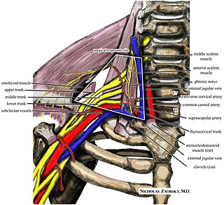
Thoracic outlet syndrome (TOS) is a condition in which there is compression of the nerves, arteries, or veins in the passageway from the lower neck to the armpit. There are three main types: neurogenic, venous, and arterial. The neurogenic type is the most common and presents with pain, weakness, and occasionally loss of muscle at the base of the thumb. The venous type results in swelling, pain, and possibly a bluish coloration of the arm. The arterial type results in pain, coldness, and paleness of the arm.

A thoracic aortic aneurysm is an aortic aneurysm that presents primarily in the thorax.
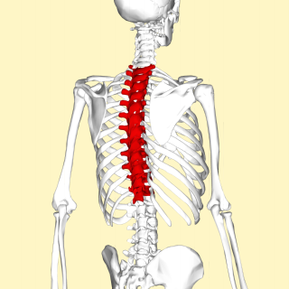
In vertebrates, thoracic vertebrae compose the middle segment of the vertebral column, between the cervical vertebrae and the lumbar vertebrae. In humans, there are twelve thoracic vertebrae and they are intermediate in size between the cervical and lumbar vertebrae; they increase in size going towards the lumbar vertebrae, with the lower ones being a lot larger than the upper. They are distinguished by the presence of facets on the sides of the bodies for articulation with the heads of the ribs, as well as facets on the transverse processes of all, except the eleventh and twelfth, for articulation with the tubercles of the ribs. By convention, the human thoracic vertebrae are numbered T1–T12, with the first one (T1) located closest to the skull and the others going down the spine toward the lumbar region.
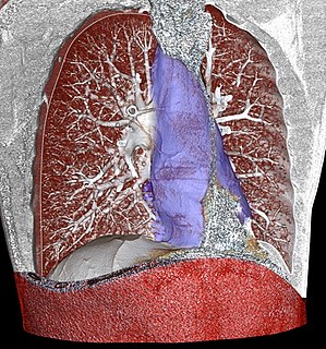
The mediastinum is the central compartment of the thoracic cavity surrounded by loose connective tissue, as an undelineated region that contains a group of structures within the thorax. The mediastinum contains the heart and its vessels, the esophagus, the trachea, the phrenic and cardiac nerves, the thoracic duct, the thymus and the lymph nodes of the central chest.

In human anatomy, the internal thoracic artery (ITA), previously known as the internal mammary artery, is an artery that supplies the anterior chest wall and the breasts. It is a paired artery, with one running along each side of the sternum, to continue after its bifurcation as the superior epigastric and musculophrenic arteries.
The thoracic inlet, also known as the superior thoracic aperture, refers to the opening at the top of the thoracic cavity. It is also clinically referred to as the thoracic outlet, in the case of thoracic outlet syndrome; this refers to the superior thoracic aperture, and not to the lower, larger opening, the inferior thoracic aperture.

The descending thoracic aorta is a part of the aorta located in the thorax. It is a continuation of the descending aorta and contained in the posterior mediastinal cavity. The descending thoracic aorta begins at the lower border of the fourth thoracic vertebra where it is continuous with the aortic arch, and ends in front of the lower border of the twelfth thoracic vertebra, at the aortic hiatus in the diaphragm where it becomes the abdominal aorta.

The intercostal nerves are part of the somatic nervous system, and arise from the anterior rami of the thoracic spinal nerves from T1 to T11. The intercostal nerves are distributed chiefly to the thoracic pleura and abdominal peritoneum and differ from the anterior rami of the other spinal nerves in that each pursues an independent course without plexus formation.

The posterior thoracic nucleus, is a group of interneurons found in the medial part of lamina VII, also known as the intermediate zone, of the spinal cord. It is mainly located from the cervical vertebra C8 to lumbar L3-L4 levels and is an important structure for proprioception of the lower limb.

The thoracic wall or chest wall is the boundary of the thoracic cavity.

The intercostal arteries are a group of arteries that supply the area between the ribs ("costae"), called the intercostal space. The highest intercostal artery is an artery in the human body that usually gives rise to the first and second posterior intercostal arteries, which supply blood to their corresponding intercostal space. It usually arises from the costocervical trunk, which is a branch of the subclavian artery. Some anatomists may contend that there is no supreme intercostal artery, only a supreme intercostal vein.

The thoracic ganglia are paravertebral ganglia. The thoracic portion of the sympathetic trunk typically has 12 thoracic ganglia. Emerging from the ganglia are thoracic splanchnic nerves that help provide sympathetic innervation to abdominal structures. The thoracic part of sympathetic trunk lies posterior to the costovertebral pleura and is hence not a content of the posterior mediastinum
Omentopexy is a surgical procedure whereby the greater omentum is sutured to a nearby organ. Suture to the abdominal wall is used to induce circulation away from portal circulation into caval circulation. It may also be sutured to another organ to increase arterial circulation.

The vertebral column, also known as the backbone or spine, is part of the axial skeleton. The vertebral column is the defining characteristic of a vertebrate in which the notochord found in all chordates has been replaced by a segmented series of bone: vertebrae separated by intervertebral discs. The vertebral column houses the spinal canal, a cavity that encloses and protects the spinal cord.

In the vertebrate spinal column, each vertebra is an irregular bone with a complex structure composed of bone and some hyaline cartilage, the proportions of which vary according to the segment of the backbone and the species of vertebrate.