Related Research Articles
Articles related to anatomy include:

The brainstem is the posterior stalk-like part of the brain that connects the cerebrum with the spinal cord. In the human brain the brainstem is composed of the midbrain, the pons, and the medulla oblongata. The midbrain is continuous with the thalamus of the diencephalon through the tentorial notch, and sometimes the diencephalon is included in the brainstem.

The oculomotor nerve, also known as the third cranial nerve, cranial nerve III, or simply CN III, is a cranial nerve that enters the orbit through the superior orbital fissure and innervates extraocular muscles that enable most movements of the eye and that raise the eyelid. The nerve also contains fibers that innervate the intrinsic eye muscles that enable pupillary constriction and accommodation. The oculomotor nerve is derived from the basal plate of the embryonic midbrain. Cranial nerves IV and VI also participate in control of eye movement.

In neuroanatomy, the trigeminal nerve (lit. triplet nerve), also known as the fifth cranial nerve, cranial nerve V, or simply CN V, is a cranial nerve responsible for sensation in the face and motor functions such as biting and chewing; it is the most complex of the cranial nerves. Its name (trigeminal, from Latin tri- 'three' and -geminus 'twin') derives from each of the two nerves (one on each side of the pons) having three major branches: the ophthalmic nerve (V1), the maxillary nerve (V2), and the mandibular nerve (V3). The ophthalmic and maxillary nerves are purely sensory, whereas the mandibular nerve supplies motor as well as sensory (or "cutaneous") functions. Adding to the complexity of this nerve is that autonomic nerve fibers as well as special sensory fibers (taste) are contained within it.

The fibers of the oculomotor nerve arise from a nucleus in the midbrain, which lies in the gray substance of the floor of the cerebral aqueduct and extends in front of the aqueduct for a short distance into the floor of the third ventricle. From this nucleus the fibers pass forward through the tegmentum, the red nucleus, and the medial part of the substantia nigra, forming a series of curves with a lateral convexity, and emerge from the oculomotor sulcus on the medial side of the cerebral peduncle.

The solitary nucleus(SN) (nucleus of the solitary tract, nucleus solitarius, or nucleus tractus solitarii) is a series of neurons whose cell bodies form a roughly vertical column of grey matter in the medulla oblongata of the brainstem. Their axons form the bulk of the enclosed solitary tract. The solitary nucleus can be divided into different parts including dorsomedial, dorsolateral, and ventrolateral subnuclei.

The nucleus ambiguus is a group of large motor neurons, situated deep in the medullary part of the reticular formation named by Jacob Clarke. The nucleus ambiguus contains the cell bodies of neurons that innervate the muscles of the soft palate, pharynx, and larynx which are associated with speech and swallowing. As well as motor neurons, the nucleus ambiguus contains preganglionic parasympathetic neurons which innervate postganglionic parasympathetic neurons in the heart.

The medial longitudinal fasciculus (MLF) is a prominent bundle of nerve fibres which pass within the ventral/anterior portion of periaqueductal gray of the mesencephalon (midbrain). It contains the interstitial nucleus of Cajal, responsible for oculomotor control, head posture, and vertical eye movement.
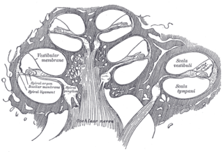
The cochlear nerve is one of two parts of the vestibulocochlear nerve, a cranial nerve present in amniotes, the other part being the vestibular nerve. The cochlear nerve carries auditory sensory information from the cochlea of the inner ear directly to the brain. The other portion of the vestibulocochlear nerve is the vestibular nerve, which carries spatial orientation information to the brain from the semicircular canals, also known as semicircular ducts.

A cranial nerve nucleus is a collection of neurons in the brain stem that is associated with one or more of the cranial nerves. Axons carrying information to and from the cranial nerves form a synapse first at these nuclei. Lesions occurring at these nuclei can lead to effects resembling those seen by the severing of nerve(s) they are associated with. All the nuclei except that of the trochlear nerve supply nerves of the same side of the body.

The paramedian pontine reticular formation (PPRF) is a subset of neurons of the oral and caudal pontine reticular nuclei mediating horizontal gaze. It is situated in the pons adjacent to the abducens nucleus. It projects to the ipsilateral abducens nucleus, and contralateral oculomotor nucleus to mediate conjugate horizontal eye movements and saccades.

The facial motor nucleus is a collection of neurons in the brainstem that belong to the facial nerve. These lower motor neurons innervate the muscles of facial expression and the stapedius.

The nucleus of the trochlear nerve is a motor nucleus in the medial midbrain giving rise to the trochlear nerve.

The cochlear nucleus (CN) or cochlear nuclear complex comprises two cranial nerve nuclei in the human brainstem, the ventral cochlear nucleus (VCN) and the dorsal cochlear nucleus (DCN). The ventral cochlear nucleus is unlayered whereas the dorsal cochlear nucleus is layered. Auditory nerve fibers, fibers that travel through the auditory nerve carry information from the inner ear, the cochlea, on the same side of the head, to the nerve root in the ventral cochlear nucleus. At the nerve root the fibers branch to innervate the ventral cochlear nucleus and the deep layer of the dorsal cochlear nucleus. All acoustic information thus enters the brain through the cochlear nuclei, where the processing of acoustic information begins. The outputs from the cochlear nuclei are received in higher regions of the auditory brainstem.
The vestibular nuclei (VN) are the cranial nuclei for the vestibular nerve located in the brainstem.

The dorsal nucleus of vagus nerve is a cranial nerve nucleus of the vagus nerve situated in the medulla oblongata of the brainstem ventral to the floor of the fourth ventricle. It contains nerve cell bodies of parasympathetic neurons of CN X that provide parasympathetic innervation to the gastrointestinal tract and lungs as well as other thoracic and abdominal organs. These functions include, among others, bronchoconstriction and gland secretion.
The salivatory nuclei are pre-ganglionic parasympathetic neurons in the caudal pons representing the general visceral efferent (GVE) cranial nerve nuclei giving rise to axons which join the facial nerve and glossopharyngeal nerve to reach and innervate the salivary as well as lacrimal glands. The nuclei may also be involved in parasympathetic control of head vasculature.

The nucleus prepositus or nucleus prepositus hypoglossi is one of the largest of the three perihypoglossal nuclei. It is situated in the caudal pons and rostral medulla oblongata. It contributes to several aspects of gaze control including the horizontal gaze holding system.
Perihypoglossal nuclei are three prominent groups of neurons in the caudal medulla oblongata near the hypoglossal nucleus: the nucleus prepositus hypoglossi, intercalated nucleus, and sublingual nucleus. They are involved in controlling eye movements: they send their principal projections to the three cranial nerve nuclei controlling extrinsic eye muscles via the medial longitudinal fasciculus.
References
John Alan Kiernan, Murray Llewellyn Barr. Barr's The Human Nervous System: An Anatomical Viewpoint. 2008