Ovarian torsion

Last updated
Ovarian torsion
Other namesAdnexal torsion [1]
Gray1170.png
Arteries of the female reproductive tract: uterine artery, ovarian artery and vaginal arteries. (Ovary and ovarian artery visible in upper right.)
Specialty Gynecology
Symptoms Pelvic pain [2]
Complications Infertility [2]
Usual onsetClassically sudden [2]
Risk factors Ovarian cysts, ovarian enlargement, ovarian tumors, pregnancy, tubal ligation [3] [2]
Diagnostic method Based on symptoms, ultrasound, CT scan [1] [2]
Differential diagnosis Appendicitis, kidney infection, kidney stones, ectopic pregnancy [2]
TreatmentSurgery [1]
Frequency6 per 100,000 women per year [2]

Ovarian torsion (OT) or adnexal torsion is an abnormal condition where an ovary twists on its attachment to other structures, such that blood flow is decreased. [3] [4] Symptoms typically include pelvic pain on one side. [2] [5] While classically the pain is sudden in onset, this is not always the case. [2] Other symptoms may include nausea. [2] Complications may include infection, bleeding, or infertility. [2] [5]

Contents

Risk factors include ovarian cysts, ovarian enlargement, ovarian tumors, pregnancy, fertility treatment, and prior tubal ligation. [3] [2] [5] The diagnosis may be supported by an ultrasound done via the vagina or CT scan, but these do not completely rule out the diagnosis. [2] Surgery is the most accurate method of diagnosis. [2]

Treatment is by surgery to either untwist and fix the ovary in place or to remove it. [2] [1] The ovary will often recover, even if the condition has been present for some time. [5] In those who have had a prior ovarian torsion, there is a 10% chance the other will also be affected. [4] The diagnosis is relatively rare, affecting about 6 per 100,000 women per year. [2] While it most commonly occurs in those of reproductive age, it can occur at any age. [2]

Signs and symptoms

Patients with ovarian torsion often present with sudden onset of sharp and usually unilateral lower abdominal pain, in 70% of cases accompanied by nausea and vomiting. [6]

Pathophysiology

The development of an ovarian mass is related to the development of torsion. In the reproductive years, regular growth of large corpus luteal cysts are a risk factor for rotation. The mass effect of ovarian tumors is also a common cause of torsion. Torsion of the ovary usually occurs with torsion of the fallopian tube as well on their shared vascular pedicle around the broad ligament, although in rare cases the ovary rotates around the mesovarium or the fallopian tube rotates around the mesosalpinx. In 80%, torsion happens unilaterally, with slight predominance on the right. In ovarian torsion, the ovary rotates around both the infundibulopelvic ligament (ie, suspensory ligament) and the utero-ovarian ligament (i.e. ovarian ligament), disrupting blood flow to the ovary.[ citation needed ]

Diagnosis

Ovarian torsion is difficult to diagnose accurately, and operation is often performed before certain diagnosis is made. A study at an obstetrics and gynaecology department found that preoperative diagnosis of ovarian torsion was confirmed in only 46% of people. [7]

Ultrasound

Gynecologic ultrasonography is the imaging modality of choice. [8] Use of doppler ultrasound in the diagnosis has been suggested. [9] [10] However, doppler flow is not always absent in torsion – the definitive diagnosis is often made in the operating room. [11]

Lack of ovarian blood flow on doppler sonography seems to be a good predictor of ovarian torsion. Women with pathologically low flow are more likely to have torsion. [7] The sensitivity and specificity of abnormal ovarian flow are 44% and 92%, respectively, with a positive and negative predictive value of 78% and 71%, respectively. [7] Specific flow features on Doppler sonography include: [8]

Normal vascularity does not exclude intermittent torsion. There may occasionally be normal Doppler flow because of the ovary's dual blood supply from both the ovarian arteries and uterine arteries.[ citation needed ]

Other ultrasonographic features include: [8]

Treatment

Surgical treatment of ovarian torsion includes laparoscopy to uncoil the torsed ovary and possibly oophoropexy to fixate the ovary which is likely to twist again. [12] In severe cases, where blood flow is cut off to the ovary for an extended period of time, necrosis of the ovary can occur. In these cases the ovary must be surgically removed. [13] [14]

Epidemiology

Ovarian torsion accounts for about 3% of gynecologic emergencies. The incidence of ovarian torsion among women of all ages is 5.9 per 100,000 women, and the incidence among women of reproductive age (15–45 years) is 9.9 per 100,000 women. [15] In 70% of cases, it is diagnosed in women between 20 and 39 years of age. The risk is greater during pregnancy and in menopause. Risk factors include increased length of the ovarian ligaments, pathologically enlarged ovaries (more than 6 cm), ovarian masses or cysts, and enlarged corpus luteum in pregnancy.[ citation needed ]

See also

Related Research Articles

<span class="mw-page-title-main">Polycystic ovary syndrome</span> Set of symptoms caused by abnormal hormones in females

Polycystic ovary syndrome, or polycystic ovarian syndrome (PCOS), is the most common endocrine disorder in women of reproductive age. The syndrome is named after cysts which form on the ovaries of some people with this condition, though this is not a universal symptom, and not the underlying cause of the disorder.

<span class="mw-page-title-main">Ectopic pregnancy</span> Female reproductive system health issue

Ectopic pregnancy is a complication of pregnancy in which the embryo attaches outside the uterus. Signs and symptoms classically include abdominal pain and vaginal bleeding, but fewer than 50 percent of affected women have both of these symptoms. The pain may be described as sharp, dull, or crampy. Pain may also spread to the shoulder if bleeding into the abdomen has occurred. Severe bleeding may result in a fast heart rate, fainting, or shock. With very rare exceptions, the fetus is unable to survive.

<span class="mw-page-title-main">Ovarian cyst</span> Fluid-filled sac in the ovary

An ovarian cyst is a fluid-filled sac within the ovary. Often they cause no symptoms. Occasionally they may produce bloating, lower abdominal pain, or lower back pain. The majority of cysts are harmless. If the cyst either breaks open or causes twisting of the ovary, it may cause severe pain. This may result in vomiting or feeling faint, and even cause head aches.

<span class="mw-page-title-main">Hysterectomy</span> Surgical removal of the uterus

Hysterectomy is the surgical removal of the uterus and cervix. Supracervical hysterectomy refers to removal of the uterus while the cervix is spared. These procedures may also involve removal of the ovaries (oophorectomy), fallopian tubes (salpingectomy), and other surrounding structures. The term “partial” or “total” hysterectomy are lay-terms that incorrectly describe the addition or omission of oophorectomy at the time of hysterectomy. These procedures are usually performed by a gynecologist. Removal of the uterus renders the patient unable to bear children and has surgical risks as well as long-term effects, so the surgery is normally recommended only when other treatment options are not available or have failed. It is the second most commonly performed gynecological surgical procedure, after cesarean section, in the United States. Nearly 68 percent were performed for conditions such as endometriosis, irregular bleeding, and uterine fibroids. It is expected that the frequency of hysterectomies for non-malignant indications will continue to fall given the development of alternative treatment options.

<span class="mw-page-title-main">Ovarian cancer</span> Cancer originating in or on the ovary

Ovarian cancer is a cancerous tumor of an ovary. It may originate from the ovary itself or more commonly from communicating nearby structures such as fallopian tubes or the inner lining of the abdomen. The ovary is made up of three different cell types including epithelial cells, germ cells, and stromal cells. When these cells become abnormal, they have the ability to divide and form tumors. These cells can also invade or spread to other parts of the body. When this process begins, there may be no or only vague symptoms. Symptoms become more noticeable as the cancer progresses. These symptoms may include bloating, vaginal bleeding, pelvic pain, abdominal swelling, constipation, and loss of appetite, among others. Common areas to which the cancer may spread include the lining of the abdomen, lymph nodes, lungs, and liver.

<span class="mw-page-title-main">Gynecologic ultrasonography</span> Application of medical ultrasonography to the female pelvic organs

Gynecologic ultrasonography or gynecologic sonography refers to the application of medical ultrasonography to the female pelvic organs as well as the bladder, the adnexa, and the recto-uterine pouch. The procedure may lead to other medically relevant findings in the pelvis.This technique is useful to detect myomas or mullerian malformations.

<span class="mw-page-title-main">Abdominal pregnancy</span> Medical condition

An abdominal pregnancy is a rare type of ectopic pregnancy where the embryo or fetus is growing and developing outside the uterus, in the abdomen, and not in a fallopian tube, an ovary, or the broad ligament.

<span class="mw-page-title-main">Bicornuate uterus</span> Medical condition

A bicornuate uterus or bicornate uterus, is a type of müllerian anomaly in the human uterus, where there is a deep indentation at the fundus (top) of the uterus.

An adnexal mass is a lump in the tissue of the adnexa of the uterus. Adnexal masses can be benign (noncancerous) or malignant (cancerous), and they can be categorized as simple or complex.

<span class="mw-page-title-main">Velamentous cord insertion</span> Velamentous placenta

Velamentous cord insertion is a complication of pregnancy where the umbilical cord is inserted in the fetal membranes. It is a major cause of antepartum hemorrhage that leads to loss of fetal blood and associated with high perinatal mortality. In normal pregnancies, the umbilical cord inserts into the middle of the placental mass and is completely encased by the amniotic sac. The vessels are hence normally protected by Wharton's jelly, which prevents rupture during pregnancy and labor. In velamentous cord insertion, the vessels of the umbilical cord are improperly inserted in the chorioamniotic membrane, and hence the vessels traverse between the amnion and the chorion towards the placenta. Without Wharton's jelly protecting the vessels, the exposed vessels are susceptible to compression and rupture.

<span class="mw-page-title-main">Corpus luteum cyst</span> Medical condition

A corpus luteum cyst is a type of ovarian cyst which may rupture about the time of menstruation, and take up to three months to disappear entirely. A corpus luteum cyst rarely occurs in women over the age of 50, because eggs are no longer being released after menopause. Corpus luteum cysts may contain blood and other fluids. The physical shape of a corpus luteum cyst may appear as an enlargement of the ovary itself, rather than a distinct mass -like growth on the surface of the ovary.

Reproductive surgery is surgery in the field of reproductive medicine. It can be used for contraception, e.g. in vasectomy, wherein the vasa deferentia of a male are severed, but is also used plentifully in assisted reproductive technology. Reproductive surgery is generally divided into three categories: surgery for infertility, in vitro fertilization, and fertility preservation.

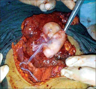
Ovarian pregnancy refers to an ectopic pregnancy that is located in the ovary. Typically the egg cell is not released or picked up at ovulation, but fertilized within the ovary where the pregnancy implants. Such a pregnancy usually does not proceed past the first four weeks of pregnancy. An untreated ovarian pregnancy causes potentially fatal intra-abdominal bleeding and thus may become a medical emergency.

Theca lutein cyst is a type of bilateral functional ovarian cyst filled with clear, straw-colored fluid. These cysts result from exaggerated physiological stimulation due to elevated levels of beta-human chorionic gonadotropin (beta-hCG) or hypersensitivity to beta-hCG. On ultrasound and MRI, theca lutein cysts appear in multiples on ovaries that are enlarged.

Ovarian apoplexy is a sudden rupture in the ovary, commonly at the site of a cyst, accompanied by hemorrhage in the ovarian tissue and/or intraperitoneal bleeding.

<span class="mw-page-title-main">Paraovarian cyst</span> Medical condition

Paraovarian cysts or paratubal cysts are epithelium-lined fluid-filled cysts in the adnexa adjacent to the fallopian tube and ovary. The terms are used interchangeably, and depend on the location of the cyst.

<span class="mw-page-title-main">Tubo-ovarian abscess</span> One of the late complications of pelvic inflammatory disease

A tubo-ovarian abscess (TOA) is one of the late complications of pelvic inflammatory disease (PID) and can be life-threatening if the abscess ruptures and results in sepsis. It consists of an encapsulated or confined pocket of pus with defined boundaries that forms during an infection of a fallopian tube and ovary. These abscesses are found most commonly in reproductive age women and typically result from upper genital tract infection. It is an inflammatory mass involving the fallopian tube, ovary and, occasionally, other adjacent pelvic organs. A TOA can also develop as a complication of a hysterectomy.

<span class="mw-page-title-main">Prophylactic salpingectomy</span> Surgical technique

Prophylactic salpingectomy is a preventative surgical technique performed on patients who are at higher risk of having ovarian cancer, such as individuals who may have pathogenic variants of the BRCA1 or BRCA2 gene. Originally salpingectomy was used in cases of ectopic pregnancies. As a preventative surgery however, it involves the removal of the fallopian tubes. By not removing the ovaries this procedure is advantageous to individuals who are still of child bearing age. It also reduces risks such as cardiovascular disease and osteoporosis which are associated with removal of the ovaries.

Ovarian germ cell tumors (OGCTs) are heterogeneous tumors that are derived from the primitive germ cells of the embryonic gonad, which accounts for about 2.6% of all ovarian malignancies. There are four main types of OGCTs, namely dysgerminomas, yolk sac tumor, teratoma, and choriocarcinoma.

<span class="mw-page-title-main">Ovarian squamous cell carcinoma</span> Medical condition

Ovarian squamous cell carcinoma (oSCC) or squamous ovarian carcinoma (SOC) is a rare tumor that accounts for 1% of ovarian cancers. Included in the World Health Organization's classification of ovarian cancer, it mainly affects women above 45 years of age. Survival depends on how advanced the disease is and how different or similar the individual cancer cells are.

References

  1. 1 2 3 4 "Adnexal Torsion". Merck Manuals Professional Edition. Retrieved 12 September 2018.
  2. 1 2 3 4 5 6 7 8 9 10 11 12 13 14 15 16 17 Robertson JJ, Long B, Koyfman A (April 2017). "Myths in the Evaluation and Management of Ovarian Torsion". The Journal of Emergency Medicine. 52 (4): 449–456. doi:10.1016/j.jemermed.2016.11.012. PMID   27988260.
  3. 1 2 3 Asfour V, Varma R, Menon P (2015). "Clinical risk factors for ovarian torsion". Journal of Obstetrics and Gynaecology. 35 (7): 721–5. doi:10.3109/01443615.2015.1004524 (inactive 31 January 2024). PMID   26212687.{{cite journal}}: CS1 maint: DOI inactive as of January 2024 (link)
  4. 1 2 Ros PR, Mortele KJ (2007). CT and MRI of the Abdomen and Pelvis: A Teaching File. Lippincott Williams & Wilkins. p. 395. ISBN   9780781772372.
  5. 1 2 3 4 Wall R (2017). Rosen's Emergency Medicine: Concepts and Clinical Practice (9 ed.). Elsevier. p. 1232. ISBN   978-0323354790.
  6. Ovarian Torsion at eMedicine
  7. 1 2 3 Bar-On S, Mashiach R, Stockheim D, Soriano D, Goldenberg M, Schiff E, Seidman DS (April 2010). "Emergency laparoscopy for suspected ovarian torsion: are we too hasty to operate?". Fertility and Sterility. 93 (6): 2012–5. doi:10.1016/j.fertnstert.2008.12.022. PMID   19159873.
  8. 1 2 3 Weerakkody Y, Dixon A. "Ovarian torsion". Radiopaedia .
  9. Peña JE, Ufberg D, Cooney N, Denis AL (May 2000). "Usefulness of Doppler sonography in the diagnosis of ovarian torsion". Fertility and Sterility. 73 (5): 1047–50. doi: 10.1016/S0015-0282(00)00487-8 . PMID   10785237.
  10. Zanforlin Filho SM, Araujo Júnior E, Serafini P, Guimarães Filho HA, Pires CR, Nardozza LM, Moron AF (April 2008). "Diagnosis of ovarian torsion by three-dimensional power Doppler in first trimester of pregnancy". The Journal of Obstetrics and Gynaecology Research. 34 (2): 266–70. doi:10.1111/j.1447-0756.2008.00768.x. PMID   18412795. S2CID   25469572.
  11. Tintinalli J (2004). Emergency Medicine. McGraw Hill Professional. p. 904. ISBN   978-0-07-138875-7.
  12. "oophoropexy". Repropedia.
  13. Crouch NS, Gyampoh B, Cutner AS, Creighton SM (December 2003). "Ovarian torsion: to pex or not to pex? Case report and review of the literature". Journal of Pediatric and Adolescent Gynecology. 16 (6): 381–4. doi:10.1016/j.jpag.2003.09.017. PMID   14642961.
  14. Eckler K, Laufer MR, Perlman SE (August 2000). "Conservative management of bilateral asynchronous adnexal torsion with necrosis in a prepubescent girl". Journal of Pediatric Surgery. 35 (8): 1248–51. doi:10.1053/jpsu.2000.8764. PMID   10945705.
  15. Yuk JS, Kim LY, Shin JY, Choi DY, Kim TY, Lee JH (May 2015). "A national population-based study of the incidence of adnexal torsion in the Republic of Korea". International Journal of Gynaecology and Obstetrics. 129 (2): 169–70. doi:10.1016/j.ijgo.2014.11.027. PMID   25721499. S2CID   12427870.