Related Research Articles

The cervix or cervix uteri is the lower part of the uterus (womb) in the human female reproductive system. The cervix is usually 2 to 3 cm long and roughly cylindrical in shape, which changes during pregnancy. The narrow, central cervical canal runs along its entire length, connecting the uterine cavity and the lumen of the vagina. The opening into the uterus is called the internal os, and the opening into the vagina is called the external os. The lower part of the cervix, known as the vaginal portion of the cervix, bulges into the top of the vagina. The cervix has been documented anatomically since at least the time of Hippocrates, over 2,000 years ago.

The endometrium is the inner epithelial layer, along with its mucous membrane, of the mammalian uterus. It has a basal layer and a functional layer: the basal layer contains stem cells which regenerate the functional layer. The functional layer thickens and then is shed during menstruation in humans and some other mammals, including apes, Old World monkeys, some species of bat, the elephant shrew and the Cairo spiny mouse. In most other mammals, the endometrium is reabsorbed in the estrous cycle. During pregnancy, the glands and blood vessels in the endometrium further increase in size and number. Vascular spaces fuse and become interconnected, forming the placenta, which supplies oxygen and nutrition to the embryo and fetus. The speculated presence of an endometrial microbiota has been argued against.

The uterus or womb is the organ in the reproductive system of most female mammals, including humans, that accommodates the embryonic and fetal development of one or more embryos until birth. The uterus is a hormone-responsive sex organ that contains glands in its lining that secrete uterine milk for embryonic nourishment.

The female reproductive system is made up of the internal and external sex organs that function in the reproduction of new offspring. In humans, the female reproductive system is immature at birth and develops to maturity at puberty to be able to produce gametes, and to carry a fetus to full term. The internal sex organs are the vagina, uterus, fallopian tubes, and ovaries. The female reproductive tract includes the vagina, uterus, and fallopian tubes and is prone to infections. The vagina allows for sexual intercourse and childbirth, and is connected to the uterus at the cervix. The uterus or womb accommodates the embryo which develops into the fetus. The uterus also produces secretions which help the transit of sperm to the fallopian tubes, where sperm fertilize ova produced by the ovaries. The external sex organs are also known as the genitals and these are the organs of the vulva including the labia, clitoris, and vaginal opening.

The round ligament of the uterus is a ligament that connects the uterus to the labia majora. It originates at the junction of the uterus and uterine tube. It passes through the inguinal canal to insert at the labium majus.

The uterine artery is an artery that supplies blood to the uterus in females.

The greater omentum is a large apron-like fold of visceral peritoneum that hangs down from the stomach. It extends from the greater curvature of the stomach, passing in front of the small intestines and doubles back to ascend to the transverse colon before reaching to the posterior abdominal wall. The greater omentum is larger than the lesser omentum, which hangs down from the liver to the lesser curvature. The common anatomical term "epiploic" derives from "epiploon", from the Greek epipleein, meaning to float or sail on, since the greater omentum appears to float on the surface of the intestines. It is the first structure observed when the abdominal cavity is opened anteriorly.

The broad ligament of the uterus is the wide fold of peritoneum that connects the sides of the uterus to the walls and floor of the pelvis.

The cervical canal is the spindle-shaped, flattened canal of the cervix, the neck of the uterus.
The Uterovaginal plexus is a division of the inferior hypogastric plexus. In older texts, it is referred to as two structures, the "vaginal plexus" and "uterine plexus".
The ovarian ligament is a fibrous ligament that connects the ovary to the lateral surface of the uterus.
The perimetrium is the outer serosal layer of the uterus, derived from the peritoneum overlying the uterine fundus, and can be considered a visceral peritoneum. It consists of a superficial layer of mesothelium, and a thin layer of loose connective tissue beneath it.

The uterosacral ligaments are major ligaments of uterus that extend posterior-ward from the cervix to attach onto the sacrum.

The cardinal ligament is a major ligament of the uterus formed as a thickening of connective tissue of the base of the broad ligament of the uterus. It extends laterally from the cervix and vaginal fornix to attach onto the lateral wall of the pelvis. The female ureter, uterine artery, and inferior hypogastric (nervous) plexus course within the cardinal ligament. The cardinal ligament supports the uterus.
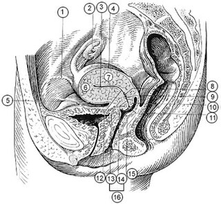
The supravaginal portion of the cervix is separated in front from the bladder by fibrous tissue (parametrium), which extends also on to its sides and lateralward between the layers of the broad ligaments.

Parametritis is an infection of the parametrium. It is considered a form of pelvic inflammatory disease.
The development of the reproductive system is the part of embryonic growth that results in the sex organs and contributes to sexual differentiation. Due to its large overlap with development of the urinary system, the two systems are typically described together as the urogenital or genitourinary system.
The fallopian tubes, also known as uterine tubes, oviducts or salpinges, are paired tubes in the human female body that stretch from the uterus to the ovaries. The fallopian tubes are part of the female reproductive system. In other mammals, they are only called oviducts.

The vaginal support structures are those muscles, bones, ligaments, tendons, membranes and fascia, of the pelvic floor that maintain the position of the vagina within the pelvic cavity and allow the normal functioning of the vagina and other reproductive structures in the female. Defects or injuries to these support structures in the pelvic floor leads to pelvic organ prolapse. Anatomical and congenital variations of vaginal support structures can predispose a woman to further dysfunction and prolapse later in life. The urethra is part of the anterior wall of the vagina and damage to the support structures there can lead to incontinence and urinary retention.
References
 This article incorporates text in the public domain from page 1259 of the 20th edition of Gray's Anatomy (1918)
- 1 2 "NCI Dictionary of Cancer Terms". National Cancer Institute. 2 February 2011.