
The cingulate cortex is a part of the brain situated in the medial aspect of the cerebral cortex. The cingulate cortex includes the entire cingulate gyrus, which lies immediately above the corpus callosum, and the continuation of this in the cingulate sulcus. The cingulate cortex is usually considered part of the limbic lobe.

Brodmann area 23 (BA23) is a region in the brain that lies inside the posterior cingulate cortex. It lies between Brodmann area 30 and Brodmann area 31 and is located on the medial wall of the cingulate gyrus between the callosal sulcus and the cingulate sulcus.
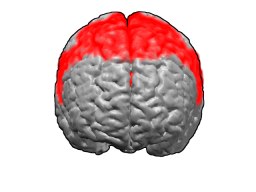
Brodmann area 6 (BA6) part of the frontal cortex in the human brain. Situated just anterior to the primary motor cortex (BA4), it is composed of the premotor cortex and, medially, the supplementary motor area, or SMA. This large area of the frontal cortex is believed to play a role in the planning of complex, coordinated movements.

Brodmann area 44, or BA44, is part of the frontal cortex in the human brain. Situated just anterior to premotor cortex (BA6) and on the lateral surface, inferior to BA9.

Brodmann area 20, or BA20, is part of the temporal cortex in the human brain. The region encompasses most of the ventral temporal cortex, a region believed to play a part in high-level visual processing and recognition memory.

Brodmann area 11 is one of Brodmann's cytologically defined regions of the brain. It is in the orbitofrontal cortex which is above the eye sockets (orbitae). It is involved in decision making and processing rewards, planning, encoding new information into long-term memory, and reasoning.

Brodmann area 4 refers to the primary motor cortex of the human brain. It is located in the posterior portion of the frontal lobe.

The primary somatosensory cortex is located in the postcentral gyrus, and is part of the somatosensory system. It was initially defined from surface stimulation studies of Wilder Penfield, and parallel surface potential studies of Bard, Woolsey, and Marshall. Although initially defined to be roughly the same as Brodmann areas 3, 1 and 2, more recent work by Kaas has suggested that for homogeny with other sensory fields only area 3 should be referred to as "primary somatosensory cortex", as it receives the bulk of the thalamocortical projections from the sensory input fields.

The Brodmann area 32, also known in the human brain as the dorsal anterior cingulate area 32, refers to a subdivision of the cytoarchitecturally defined cingulate cortex. In the human it forms an outer arc around the anterior cingulate gyrus. The cingulate sulcus defines approximately its inner boundary and the superior rostral sulcus (H) its ventral boundary; rostrally it extends almost to the margin of the frontal lobe. Cytoarchitecturally it is bounded internally by the ventral anterior cingulate area 24, externally by medial margins of the agranular frontal area 6, intermediate frontal area 8, granular frontal area 9, frontopolar area 10, and prefrontal area 11-1909. (Brodmann19-09).
Brodmann area 16 is a subdivision of the cerebral cortex of the guenon defined on the basis of cytoarchitecture. It is a relatively undifferentiated cortical area that Brodmann regarded as part of the insula because of the relation of its innermost multiform layer (VI) with the claustrum (VICl). The laminar organization of cortex is almost totally lacking. The molecular layer (I) is wide as in area 15 of Brodmann-1905. The space between layer I and layer VI is composed of a mixture of pyramidal cells and spindle cells with no significant number of granule cells. Pyramidal cells clump in the outer part to form glomeruli similar to those seen in some of the primary olfactory areas (Brodmann-1905).

Brodmann area 26 is the name for a small part of the brain.

Brodmann area 29, also known as granular retrolimbic area 29 or granular retrosplenial cortex, is a cytoarchitecturally defined portion of the retrosplenial region of the cerebral cortex. In the human it is a narrow band located in the isthmus of cingulate gyrus. Cytoarchitecturally it is bounded internally by the ectosplenial area 26 and externally by the agranular retrolimbic area 30 (Brodmann-1909).

Brodmann area 43, the subcentral area, is a structurally distinct area of the cerebral cortex defined on the basis of cytoarchitecture. Along with Brodmann Area 1, 2, and 3, Brodmann area 43 is a subdivision of the postcentral region of the brain, suggesting a somatosensory function. The histological structure of Area 43 was initially described by Korbinian Brodmann, but it was not labeled on his map of cortical areas.
The external granular layer of the cerebral cortex is commonly known as layer II. It is different from the internal granular layer of the cerebral cortex.
Cortical patterning is a field of developmental neuroscience which aims to determine how the various functional areas of the cerebral cortex are generated, what size and shape they will be, and how their spatial pattern across the surface of the cortex is specified. Early brain lesion studies indicated that different parts of the cortex served different cognitive functions, such as visual, somatosensory, and motor functions, beautifully assimilated by Brodmann in 1909. Today the field supports the idea of a 'protomap', which is a molecular pre-pattern of the cortical areas during early embryonic stages. The protomap is a feature of the cortical ventricular zone, which contains the primary stem cells of the cortex known as radial glial cells. A system of signaling centers, positioned strategically at the midline and edges of the cortex, produce secreted signaling proteins that establish concentration gradients in the cortical primordium. This provides positional information for each stem cell, and regulates proliferation, neurogenesis, and areal identity. After the initial establishment of areal identity, axons from the developing thalamus arrive at their correct cortical areal destination through the process of axon guidance and begin to form synapses. Many activity-dependent processes are then thought to play important roles in the maturation of each area.

