In chemistry, biochemistry, and pharmacology, a dissociation constant is a specific type of equilibrium constant that measures the propensity of a larger object to separate (dissociate) reversibly into smaller components, as when a complex falls apart into its component molecules, or when a salt splits up into its component ions. The dissociation constant is the inverse of the association constant. In the special case of salts, the dissociation constant can also be called an ionization constant.
A radioligand is a radioactive biochemical substance that is used for diagnosis or for research-oriented study of the receptor systems of the body.

In biochemistry and pharmacology, a receptor is a protein molecule that receives chemical signals from outside a cell. When such chemical signals bind to a receptor, they cause some form of cellular/tissue response, e.g. a change in the electrical activity of a cell. There are three main ways the action of the receptor can be classified: relay of signal, amplification, or integration. Relaying sends the signal onward, amplification increases the effect of a single ligand, and integration allows the signal to be incorporated into another biochemical pathway. In this sense, a receptor is a protein-molecule that recognizes and responds to endogenous chemical signals, e.g. an acetylcholine receptor recognizes and responds to its endogenous ligand, acetylcholine. However, sometimes in pharmacology, the term is also used to include other proteins that are drug targets, such as enzymes, transporters, and ion channels.
Pharmacodynamics (PD) is the study of the biochemical and physiologic effects of drugs. The effects can include those manifested within animals, microorganisms, or combinations of organisms. Pharmacodynamics is the study of how a drug affects an organism, whereas pharmacokinetics is the study of how the organism affects the drug. Both together influence dosing, benefit, and adverse effects. Pharmacodynamics is sometimes abbreviated as PD and pharmacokinetics as PK, especially in combined reference.
![Host–guest chemistry [[Supramolecular chemistry|Supramolecular structures]] held together other than by covalent bonds](https://upload.wikimedia.org/wikipedia/commons/thumb/9/98/Cucurbit-6-uril_ActaCrystallB-Stru_1984_382.jpg/320px-Cucurbit-6-uril_ActaCrystallB-Stru_1984_382.jpg)
In supramolecular chemistry, host–guest chemistry describes complexes that are composed of two or more molecules or ions that are held together in unique structural relationships by forces other than those of full covalent bonds. Host–guest chemistry encompasses the idea of molecular recognition and interactions through non-covalent bonding. Non-covalent bonding is critical in maintaining the 3D structure of large molecules, such as proteins and is involved in many biological processes in which large molecules bind specifically but transiently to one another. There are four commonly mentioned types of non-covalent interactions: hydrogen bonds, ionic bonds, van der Waals forces, and hydrophobic interactions.
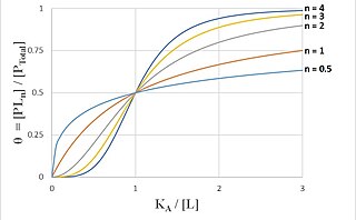
[[File:Hill Curves for Increasing Hill Coefficients.jpg|thumb|370x370px|Biochemical binding curves showing the characteristically sigmoidal curves generated by using the Hill equation to model cooperative binding. Each curve corresponds to a different Hill coefficient, labeled to the curve's right. The vertical axis displays the fraction of occupied ligand-binding sites on a protein receptor, equal to ratio of the concentration of ligand-bound protein to the total concentration of protein receptor. The horizontal axis is the ratio of the ligand concentration producing half occupation to the free ligand concentration .]]
The Scatchard equation is an equation used in molecular biology for calculating the affinity constant of a ligand with a protein.
A Patlak plot is a graphical analysis technique based on the compartment model that uses linear regression to identify and analyze pharmacokinetics of tracers involving irreversible uptake, such as in the case of deoxyglucose. It is used for the evaluation of nuclear medicine imaging data after the injection of a radioopaque or radioactive tracer.
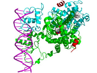
In the field of molecular biology, nuclear receptors are a class of proteins found within cells that are responsible for sensing steroid and thyroid hormones and certain other molecules. In response, these receptors work with other proteins to regulate the expression of specific genes, thereby controlling the development, homeostasis, and metabolism of the organism.
The binding constant, or association constant, is a special case of the equilibrium constant K, and is the inverse of the dissociation constant. It is associated with the binding and unbinding reaction of receptor (R) and ligand (L) molecules, which is formalized as:
In biochemistry, receptor–ligand kinetics is a branch of chemical kinetics in which the kinetic species are defined by different non-covalent bindings and/or conformations of the molecules involved, which are denoted as receptor(s) and ligand(s). Receptor–ligand binding kinetics also involves the on- and off-rates of binding.
Schild regression analysis, named for Heinz Otto Schild, is a useful tool for studying the effects of agonists and antagonists on the cellular response caused by the receptor or on ligand-receptor binding.

Translocator protein (TSPO) is an 18 kDa protein mainly found on the outer mitochondrial membrane. It was first described as peripheral benzodiazepine receptor (PBR), a secondary binding site for diazepam, but subsequent research has found the receptor to be expressed throughout the body and brain. In humans, the translocator protein is encoded by the TSPO gene. It belongs to family of tryptophan-rich sensory proteins. Regarding intramitochondrial cholesterol transport, TSPO has been proposed to interact with StAR to transport cholesterol into mitochondria, though evidence is mixed.
Copper-64 (64Cu) is a positron emitting isotope of copper, with applications for molecular radiotherapy and positron emission tomography.

Altanserin is a compound that binds to the 5-HT2A receptor. Labeled with the isotope fluorine-18 it is used as a radioligand in positron emission tomography (PET) studies of the brain, i.e., studies of the 5-HT2A neuroreceptors. Besides human neuroimaging studies altanserin has also been used in the study of rats.

Nisoxetine, originally synthesized in the Lilly research laboratories during the early 1970s, is a potent and selective inhibitor for the reuptake of norepinephrine (noradrenaline) into synapses. It currently has no clinical applications in humans, although it was originally researched as an antidepressant. Nisoxetine is now widely used in scientific research as a standard selective norepinephrine reuptake inhibitor. It has been used to research obesity and energy balance, and exerts some local analgesia effects.
Binding selectivity is defined with respect to the binding of ligands to a substrate forming a complex. A selectivity coefficient is the equilibrium constant for the reaction of displacement by one ligand of another ligand in a complex with the substrate. Binding selectivity is of major importance in biochemistry and in chemical separation processes.
A Logan plot is a graphical analysis technique based on the compartment model that uses linear regression to analyze pharmacokinetics of tracers involving reversible uptake. It is mainly used for the evaluation of nuclear medicine imaging data after the injection of a labeled ligand that binds reversibly to specific receptor or enzyme.
Ligand binding assays (LBA) is an assay, or an analytic procedure, whose procedure or method relies on the binding of ligand molecules to receptors, antibodies or other macromolecules. A detection method is used to determine the presence and extent of the ligand-receptor complexes formed, and this is usually determined electrochemically or through a fluorescence detection method. This type of analytic test can be used to test for the presence of target molecules in a sample that are known to bind to the receptor.