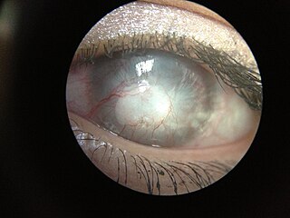
Contact lenses, or simply contacts, are thin lenses placed directly on the surface of the eyes. Contact lenses are ocular prosthetic devices used by over 150 million people worldwide, and they can be worn to correct vision or for cosmetic or therapeutic reasons. In 2010, the worldwide market for contact lenses was estimated at $6.1 billion, while the US soft lens market was estimated at $2.1 billion. Multiple analysts estimated that the global market for contact lenses would reach $11.7 billion by 2015. As of 2010, the average age of contact lens wearers globally was 31 years old, and two-thirds of wearers were female.

Keratoconus (KC) is a disorder of the eye that results in progressive thinning of the cornea. This may result in blurry vision, double vision, nearsightedness, irregular astigmatism, and light sensitivity leading to poor quality-of-life. Usually both eyes are affected. In more severe cases a scarring or a circle may be seen within the cornea.

The cornea is the transparent front part of the eye that covers the iris, pupil, and anterior chamber. Along with the anterior chamber and lens, the cornea refracts light, accounting for approximately two-thirds of the eye's total optical power. In humans, the refractive power of the cornea is approximately 43 dioptres. The cornea can be reshaped by surgical procedures such as LASIK.

LASIK or Lasik, commonly referred to as laser eye surgery or laser vision correction, is a type of refractive surgery for the correction of myopia, hyperopia, and an actual cure for astigmatism, since it is in the cornea. LASIK surgery is performed by an ophthalmologist who uses a laser or microkeratome to reshape the eye's cornea in order to improve visual acuity.

Keratitis is a condition in which the eye's cornea, the clear dome on the front surface of the eye, becomes inflamed. The condition is often marked by moderate to intense pain and usually involves any of the following symptoms: pain, impaired eyesight, photophobia, red eye and a 'gritty' sensation. Diagnosis of infectious keratitis is usually made clinically based on the signs and symptoms as well as eye examination, but corneal scrapings may be obtained and evaluated using microbiological culture or other testing to identify the causative pathogen.

Orthokeratology, also referred to as Night lenses, Ortho-K, OK, Overnight Vision Correction, Corneal Refractive Therapy (CRT), Accelerated Orthokeretology, Cornea Corrective Contacts, Eccentricity Zero Molding, and Gentle Vision Shaping System (GVSS), is the use of gas-permeable contact lenses that temporarily reshape the cornea to reduce refractive errors such as myopia, hyperopia, and astigmatism.

Corneal abrasion is a scratch to the surface of the cornea of the eye. Symptoms include pain, redness, light sensitivity, and a feeling like a foreign body is in the eye. Most people recover completely within three days.

Corneal cross-linking (CXL) with riboflavin (vitamin B2) and UV-A light is a surgical treatment for corneal ectasia such as keratoconus, PMD, and post-LASIK ectasia.

A scleral lens, also known as a scleral contact lens, is a large contact lens that rests on the sclera and creates a tear-filled vault over the cornea. Scleral lenses are designed to treat a variety of eye conditions, many of which do not respond to other forms of treatment.

An intrastromal corneal ring segment (ICRS) (also known as intrastromal corneal ring, corneal implant or corneal insert) is a small device surgically implanted in the cornea of the eye to correct vision. Two crescent or semi-circular shaped ring segments are inserted between the layers of the corneal stroma, one on each side of the pupil, This is intended to flatten the cornea and change the refraction of light passing through the cornea on its way into the eye.
George Jessen (1916–1987) was an optometrist who was an early pioneer of the contact lens. He is credited with being one of the first to employ the concept of orthokeratology, a direct attempt to reduce refractive error with the use of a contact lens, under the term orthofocus.

Astigmatism is a type of refractive error due to rotational asymmetry in the eye's refractive power. This results in distorted or blurred vision at any distance. Other symptoms can include eyestrain, headaches, and trouble driving at night. Astigmatism often occurs at birth and can change or develop later in life. If it occurs in early life and is left untreated, it may result in amblyopia.

Acanthamoeba keratitis (AK) is a rare disease in which amoebae of the genus Acanthamoeba invade the clear portion of the front (cornea) of the eye. It affects roughly 100 people in the United States each year. Acanthamoeba are protozoa found nearly ubiquitously in soil and water and can cause infections of the skin, eyes, and central nervous system.

Corneal neovascularization (CNV) is the in-growth of new blood vessels from the pericorneal plexus into avascular corneal tissue as a result of oxygen deprivation. Maintaining avascularity of the corneal stroma is an important aspect of healthy corneal physiology as it is required for corneal transparency and optimal vision. A decrease in corneal transparency causes visual acuity deterioration. Corneal tissue is avascular in nature and the presence of vascularization, which can be deep or superficial, is always pathologically related.

Corneal ulcer, also called keratitis, is an inflammatory or, more seriously, infective condition of the cornea involving disruption of its epithelial layer with involvement of the corneal stroma. It is a common condition in humans particularly in the tropics and in farming. In developing countries, children afflicted by vitamin A deficiency are at high risk for corneal ulcer and may become blind in both eyes persisting throughout life. In ophthalmology, a corneal ulcer usually refers to having an infection, while the term corneal abrasion refers more to a scratch injury.

Pellucid marginal degeneration (PMD) is a degenerative corneal condition, often confused with keratoconus. It typically presents with painless vision loss affecting both eyes. Rarely, it may cause acute vision loss with severe pain due to perforation of the cornea. It is typically characterized by a clear, bilateral thinning (ectasia) in the inferior and peripheral region of the cornea, although some cases affect only one eye. The cause of the disease remains unclear.

Long-term contact lens use can lead to alterations in corneal thickness, stromal thickness, curvature, corneal sensitivity, cell density, and epithelial oxygen uptake, etc. Other changes may include the formation of epithelial vacuoles and microcysts as well as the emergence of polymegethism in the corneal endothelium. Decreased corneal sensitivity, vision loss, and photophobia have also been observed in patients who have worn contact lenses for an extended period of time. Many contact lens-induced changes in corneal structure are reversible if contact lenses are removed for an extended period of time.
Post-LASIK ectasia is a condition similar to keratoconus where the cornea starts to bulge forwards at a variable time after LASIK, PRK, or SMILE corneal laser eye surgery. However, the physiological processes of post-LASIK ectasia seem to be different from keratoconus. The visible changes in the basal epithelial cell and anterior and posterior keratocytes linked with keratoconus were not observed in post-LASIK ectasia.
Corneal ectatic disorders or corneal ectasia are a group of uncommon, noninflammatory, eye disorders characterised by bilateral thinning of the central, paracentral, or peripheral cornea.
Exposure keratopathy is medical condition affecting the cornea of eyes. It can lead to corneal ulceration and permanent loss of vision due to corneal opacity.