
The epiglottis is a flap in the throat that keeps food from entering the windpipe and the lungs. The flap is made of elastic cartilage covered with a mucous membrane, attached to the entrance of the larynx. It projects obliquely upwards behind the tongue and the hyoid bone, pointing dorsally. It stands open during breathing, allowing air into the larynx. During swallowing, it closes to prevent aspiration and forcing the swallowed liquids or food to go along the esophagus instead. It is thus the valve that diverts passage to either the trachea or the esophagus.

The nictitating membrane is a transparent or translucent third eyelid present in some animals that can be drawn across the eye from the medial canthus for protection and to moisten it while maintaining vision. Some reptiles, birds, and sharks have full nictitating membranes; in many mammals, a small, vestigial portion of the membrane remains in the corner of the eye. Some mammals, such as camels, polar bears, seals and aardvarks, have full nictitating membranes. Often called a third eyelid or haw, it may be referred to in scientific terminology as the plica semilunaris, membrana nictitans, or palpebra tertia.

The nasolacrimal duct carries tears from the lacrimal sac of the eye into the nasal cavity. The duct begins in the eye socket between the maxillary and lacrimal bones, from where it passes downwards and backwards. The opening of the nasolacrimal duct into the inferior nasal meatus of the nasal cavity is partially covered by a mucosal fold. Excess tears flow through nasolacrimal duct which drains into the inferior nasal meatus.

The paired sublingual glands are major salivary glands in the mouth. They are the smallest, most diffuse, and the only unencapsulated major salivary glands. They provide only 3-5% of the total salivary volume. There are also two other types of salivary glands; they are submandibular and Parotid glands.

The recto-uterine pouch, also known by various other names, is the extension of the peritoneal cavity between the rectum and the posterior wall of the uterus in the female human body.

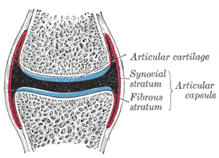
In anatomy, a joint capsule or articular capsule is an envelope surrounding a synovial joint. Each joint capsule has two parts: an outer fibrous layer or membrane, and an inner synovial layer or membrane.

The base of the cartilaginous portion of the auditory tube lies directly under the mucous membrane of the nasal part of the pharynx, where it forms an elevation, the torus tubarius, the torus of the auditory tube, or cushion, behind the pharyngeal orifice of the tube. The torus tubarius is very close to the tubal tonsil, which is sometimes also called the tonsil of (the) torus tubarius. Equating the torus with its tonsil however might be seen as incorrect or imprecise.
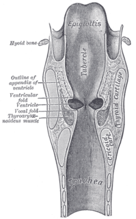
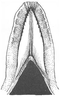
The portion of the cavity of the larynx above the vocal folds is called the laryngeal vestibule; it is wide and triangular in shape, its base or anterior wall presenting, however, about its center the backward projection of the tubercle of the epiglottis. It contains the vestibular folds, and between these and the vocal folds are the laryngeal ventricles.

The fimbriated fold of tongue, also plica fimbriata is a slight fold of the mucous membrane on the underside of the tongue which runs laterally on either side of the frenulum. The free edge of the fimbriated fold occasionally exhibits a series of fringe-like processes..

The plica semilunaris is a small fold of bulbar conjunctiva on the medial canthus of the eye. It functions during movement of the eye, to help maintain tear drainage via the lacrimal lake, and to permit greater rotation of the globe, for without the plica the conjunctiva would attach directly to the eyeball, restricting movement. It is the vestigial remnant of the nictitating membrane which is drawn across the eye for protection, and is present in other animals such as birds, reptiles, and fish, but is rare in mammals, mainly found in monotremes and marsupials. Its associated muscles are also vestigial. It is loose, thus eye movements are not restricted by it. Only one species of primate, the Calabar angwantibo, is known to have a functioning nictitating membrane.

The peritoneum of the anterior pelvic wall covers the superior surface of the bladder, and on either side of this viscus forms a depression, termed the paravesical fossa, which is limited laterally by the fold of peritoneum covering the ductus deferens.
The excretory ducts of the sublingual gland are from eight to twenty in number. Of the smaller sublingual ducts, some join the submandibular duct; others open separately into the mouth, on the elevated crest of mucous membrane, caused by the projection of the gland, on either side of the frenulum linguae. One or more join to form the major sublingual duct, which opens into the submandibular duct.
Plica syndrome is a condition that occurs when a plica becomes irritated, enlarged, or inflamed.
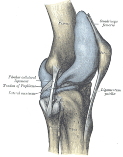
The articular capsule of the knee joint is wide and lax; thin in front and at the side; and contains the patella, ligaments, menisci, and bursae. The capsule consists of a synovial and a fibrous membrane separated by fatty deposits anteriorly and posteriorly.

The tonsillar fossa is a space delineated by the triangular fold of the palatoglossal and palatopharyngeal arches within the lateral wall of the oral cavity. In this space lie the palatine tonsils.
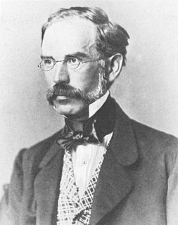
Václav Treitz was a Czech pathologist who was a native of Hostomice, Bohemia.
Skin folds or skinfolds are areas of skin where it folds. Many skin folds are distinct, heritable anatomical features, and may be used for identification of animal species, while others are non-specific and may be produced either by individual development of an organism or by arbitrary application of force to skin, either by the actions of the muscles of the body or by external force, e.g., gravity. Anatomical folds can also be found in other structures and tissues besides the skin, such as the ileocecal fold beneath the terminal ileum of the cecum.
In anatomy, capsulitis is inflammation of a capsule.
The sublingua ("under-tongue") is a muscular secondary tongue found below the primary tongue in tarsiers and living strepsirrhine primates, which includes lemurs and lorisoids. Although it is most fully developed in these primates, similar structures can be found in some other mammals, such as marsupials, treeshrews, and colugos. This "second tongue" lacks taste buds, and in lemuriforms, it is thought to be used to remove hair and other debris from the toothcomb, a specialized dental structure used to comb the fur during oral grooming.

