
Apoptosis is a form of programmed cell death that occurs in multicellular organisms and in some eukaryotic, single-celled microorganisms such as yeast. Biochemical events lead to characteristic cell changes (morphology) and death. These changes include blebbing, cell shrinkage, nuclear fragmentation, chromatin condensation, DNA fragmentation, and mRNA decay. The average adult human loses between 50 and 70 billion cells each day due to apoptosis. For an average human child between eight and fourteen years old, each day the approximate lost is 20 to 30 billion cells.

Caspases are a family of protease enzymes playing essential roles in programmed cell death. They are named caspases due to their specific cysteine protease activity – a cysteine in its active site nucleophilically attacks and cleaves a target protein only after an aspartic acid residue. As of 2009, there are 12 confirmed caspases in humans and 10 in mice, carrying out a variety of cellular functions.
Programmed cell death is the death of a cell as a result of events inside of a cell, such as apoptosis or autophagy. PCD is carried out in a biological process, which usually confers advantage during an organism's lifecycle. For example, the differentiation of fingers and toes in a developing human embryo occurs because cells between the fingers apoptose; the result is that the digits are separate. PCD serves fundamental functions during both plant and animal tissue development.

Howard Robert Horvitz ForMemRS NAS AAA&S APS NAM is an American biologist best known for his research on the nematode worm Caenorhabditis elegans, for which he was awarded the 2002 Nobel Prize in Physiology or Medicine, together with Sydney Brenner and John E. Sulston, whose "seminal discoveries concerning the genetic regulation of organ development and programmed cell death" were "important for medical research and have shed new light on the pathogenesis of many diseases".

Cell death is the event of a biological cell ceasing to carry out its functions. This may be the result of the natural process of old cells dying and being replaced by new ones, as in programmed cell death, or may result from factors such as diseases, localized injury, or the death of the organism of which the cells are part. Apoptosis or Type I cell-death, and autophagy or Type II cell-death are both forms of programmed cell death, while necrosis is a non-physiological process that occurs as a result of infection or injury.

Nerve growth factor (NGF) is a neurotrophic factor and neuropeptide primarily involved in the regulation of growth, maintenance, proliferation, and survival of certain target neurons. It is perhaps the prototypical growth factor, in that it was one of the first to be described. Since it was first isolated by Nobel Laureates Rita Levi-Montalcini and Stanley Cohen in 1956, numerous biological processes involving NGF have been identified, two of them being the survival of pancreatic beta cells and the regulation of the immune system.

The death-effector domain (DED) is a protein interaction domain found only in eukaryotes that regulates a variety of cellular signalling pathways. The DED domain is found in inactive procaspases and proteins that regulate caspase activation in the apoptosis cascade such as FAS-associating death domain-containing protein (FADD). FADD recruits procaspase 8 and procaspase 10 into a death induced signaling complex (DISC). This recruitment is mediated by a homotypic interaction between the procaspase DED and a second DED that is death effector domain in an adaptor protein that is directly associated with activated TNF receptors. Complex formation allows proteolytic activation of procaspase into the active caspase form which results in the initiation of apoptosis. Structurally the DED domain are a subclass of protein motif known as the death fold and contains 6 alpha helices, that closely resemble the structure of the Death domain (DD).
Guy Salvesen is a South African-born biochemist, best known for his work in the field of apoptosis. His research focuses on proteases and their inhibitors in humans, with particular emphasis on the caspases of the apoptotic cell death pathway.
Phenoptosis is a conception of the self-programmed death of an organism proposed by Vladimir Skulachev in 1999.
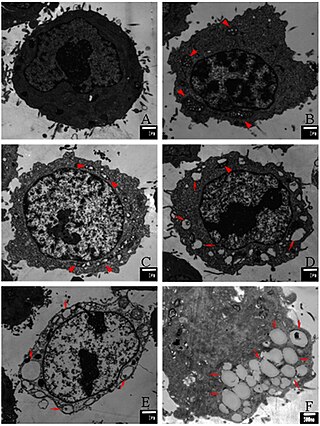
UV-induced apoptosis UV-induced apoptosis is an adequate (physiological) reaction of a cell damaged by UV radiation (UVR) in a sufficiently large (lethal) dose and it prevents the disordered destruction of UV damaged cells by help necrosis. Cell elimination by apoptosis occurs when UV-induced cell damage which cannot be repaired by the intracellular repair system exceeds at it certain limit. Through apoptosis, the cells are self-disassembled into compartments with their subsequent utilization. The first time sign of the beginning of the apoptosis system is working in a UV damaged cell is the activation of restriction enzymes, which divide cell DNA into fragments convenient for utilization. But too large a dose of UVR can lead to breakdown (inactivation) of the energy-dependent mechanism of apoptosis. In this case, cell destruction occurs randomly, not orderly, and during a significantly longer time interval. UV-irradiated cells do not change their appearance for a long time [1, 6], as a result of which the researchers may make the erroneous conclusion that “revealed an unexpected response to a dose at which a higher dose of UV increased the viability of keratinocytes” [2]. The fact that UV-induced apoptosis at high doses of UVR begins to be replaced by necrosis was established in 2000 [3]. For keratinocytes, the proportion of cells that have elimination by help apoptosis, with an increase in UVR dose can reach to achieve 45%, but with a further increase in the dose of UVR, destruction of damaged cells by help necrosis and the part of cells that eliminated by apoptosis begins to decrease [4, 11]. In the dose range of UVR from “lethal” to “super-lethal”, “pro-inflammatory” apoptosis can be manifested, which was experimentally discovered in 2003 [5]. This may be the result of partial damage to the apoptosis mechanism by UV radiation [1]. If at moderate doses “pure” apoptosis does not cause an inflammatory reaction, then at sufficiently large doses, an inflammatory reaction arises due to pro-inflammatory apoptosis, which leads to the appearance of “fast” erythema for UV irradiated skin keratinocytes. Kinetic of “fast” erythema is much faster by the time of development of UV erythema caused by necrosis of UV damaged keratinocytes [6]. The most erythemogenic is UVB the spectral range of UVR, since radiation in this range is less absorbed by the outer layers of the skin, which allows UVB radiation, in contrast to UVC, to reach more deep layers skin and act on keratinocytes of the deep-lying basal layer of the epidermis of the skin. The ability to induce apoptosis for UVB and UVC radiation is due to the fact that the DNA of the nucleus [7] and / or mitochondria [8] of the cell absorbs UVR well in the UVC and UVB spectral range. Keratinocytes of the skin are in a state of programmed apoptosis, during which the keratinocytes of the basal layer are removed from it and during the transition through all layers of the epidermis within 28 days turn into flakes of the outer stratum corneum, which are subsequently desquamated. It is clear that the keratinocyte response to UV exposure will depend on what phase of programmed apoptosis the keratinocyte experienced UV exposure, and this is the main reason for the difference of the UV effect for UVC and UVB on the skin. There are also differences in the initiation of mitochondrial (internal) and caspase-dependent (external) apoptosis for the UVC and UVB spectral ranges [9]. Sunburn cells (SBS) are the keratinocytes in the process of UV-induced apoptosis. The appearance of SBC may be not associated with an inflammatory reaction, but the role of UV-induced apoptosis of skin keratinocytes in the development of UV erythema of the skin has been established, which allowed the development of a patent-protected METHOD FOR QUANTITATIVE ASSESSMENT OF APOPTOSIS SYSTEM [10], in which “the brightest lamp of skin display "(photoerythema) is used to diagnose the state of the body systems involved in the elimination of UV-induced damage. Such systems include the immune system, the intracellular repair system, the microcirculation system and not only.

Survivin, also called baculoviral inhibitor of apoptosis repeat-containing 5 or BIRC5, is a protein that, in humans, is encoded by the BIRC5 gene.
Apoptosis is the process of programmed cell death. From its early conceptual beginnings in the 1950s, it has exploded as an area of research within the life sciences community. As well as its implication in many diseases, it is an integral part of biological development.

The BCL2 associated agonist of cell death (BAD) protein is a pro-apoptotic member of the Bcl-2 gene family which is involved in initiating apoptosis. BAD is a member of the BH3-only family, a subfamily of the Bcl-2 family. It does not contain a C-terminal transmembrane domain for outer mitochondrial membrane and nuclear envelope targeting, unlike most other members of the Bcl-2 family. After activation, it is able to form a heterodimer with anti-apoptotic proteins and prevent them from stopping apoptosis.

Death-associated protein 6 also known as Daxx is a protein that in humans is encoded by the DAXX gene.

Caspase-3 is a caspase protein that interacts with caspase-8 and caspase-9. It is encoded by the CASP3 gene. CASP3 orthologs have been identified in numerous mammals for which complete genome data are available. Unique orthologs are also present in birds, lizards, lissamphibians, and teleosts.
In cellular biology, dependence receptors are proteins that mediate programmed cell death by monitoring the absence of certain trophic factors that otherwise serve as ligands (interactors) for the dependence receptors. A trophic ligand is a molecule whose protein binding stimulates cell growth, differentiation, and/or survival. Cells depend for their survival on stimulation that is mediated by various receptors and sensors, and integrated via signaling within the cell and between cells. The withdrawal of such trophic support leads to a form of cellular suicide.
Paraptosis is a type of programmed cell death, morphologically distinct from apoptosis and necrosis. The defining features of paraptosis are cytoplasmic vacuolation, independent of caspase activation and inhibition, and lack of apoptotic morphology. Paraptosis lacks several of the hallmark characteristics of apoptosis, such as membrane blebbing, chromatin condensation, and nuclear fragmentation. Like apoptosis and other types of programmed cell death, the cell is involved in causing its own death, and gene expression is required. This is in contrast to necrosis, which is non-programmed cell death that results from injury to the cell.
Emad Saleem Alnemri is a professor in Biochemistry & Molecular Biology at the Sidney Kimmel Cancer Center, Thomas Jefferson University, researching apoptosis and the inflammasome.

Death regulator Nedd2-like caspase was firstly identified and characterised in Drosophila in 1999 as a cysteine protease containing an amino-terminal caspase recruitment domain. At first, it was thought of as an effector caspase involved in apoptosis, but subsequent findings have proved that it is, in fact, an initiator caspase with a crucial role in said type of programmed cell death.
Alfred Lewis Goldberg was an American cell biologist-biochemist and professor at Harvard University. His major discoveries have concerned the mechanisms and physiological importance of protein degradation in cells. Of wide impact have been his lab's demonstration that all cells contain a pathway for selectively eliminating misfolded proteins, his discoveries about the role of proteasomes in this process and of the enzyme systems catalyzing protein breakdown in bacteria, his elucidating the mechanisms for muscle atrophy and the role of proteasomes in antigen presentation to the immune system, and his introduction of proteasome inhibitors now widely used as research tools and in the treatment of blood cancers.
