
Perception is the organization, identification, and interpretation of sensory information in order to represent and understand the presented information or environment.
The receptive field, or sensory space, is a delimited medium where some physiological stimuli can evoke a sensory neuronal response in specific organisms.
Meridian is used in perimetry and in specifying visual fields. According to IPS Perimetry Standards 1978 (2002): "Perimetry is the measurement of [an observer's] visual functions ... at topographically defined loci in the visual field. The visual field is that portion of the external environment of the observer [in which when he or she is] steadily fixating ...[he or she] can detect visual stimuli."

Eye movement includes the voluntary or involuntary movement of the eyes, helping in acquiring, fixating and tracking visual stimuli. A special type of eye movement, rapid eye movement, occurs during REM sleep.
Closed-eye hallucinations and closed-eye visualizations (CEV) are a distinct class of hallucination. These types of hallucinations generally only occur when one's eyes are closed or when one is in a darkened room. They can be a form of phosphene. Some people report closed-eye hallucinations under the influence of psychedelics. These are reportedly of a different nature than the "open-eye" hallucinations of the same compounds. Similar hallucinations that occur due to loss of vision are called visual release hallucinations.
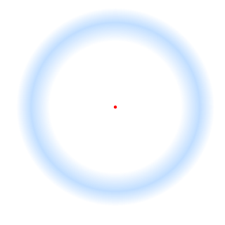
Neural adaptation or sensory adaptation is a gradual decrease over time in the responsiveness of the sensory system to a constant stimulus. It is usually experienced as a change in the stimulus. For example, if a hand is rested on a table, the table's surface is immediately felt against the skin. Subsequently, however, the sensation of the table surface against the skin gradually diminishes until it is virtually unnoticeable. The sensory neurons that initially respond are no longer stimulated to respond; this is an example of neural adaptation.
In vision, filling-in phenomena are those responsible for the completion of missing information across the physiological blind spot, and across natural and artificial scotomata. There is also evidence for similar mechanisms of completion in normal visual analysis. Classical demonstrations of perceptual filling-in involve filling in at the blind spot in monocular vision, and images stabilized on the retina either by means of special lenses, or under certain conditions of steady fixation. For example, naturally in monocular vision at the physiological blind spot, the percept is not a hole in the visual field, but the content is “filled-in” based on information from the surrounding visual field. When a textured stimulus is presented centered on but extending beyond the region of the blind spot, a continuous texture is perceived. This partially inferred percept is paradoxically considered more reliable than a percept based on external input..

Fixation or visual fixation is the maintaining of the visual gaze on a single location. An animal can exhibit visual fixation if they possess a fovea in the anatomy of their eye. The fovea is typically located at the center of the retina and is the point of clearest vision. The species in which fixational eye movement has been found thus far include humans, primates, cats, rabbits, turtles, salamanders, and owls. Regular eye movement alternates between saccades and visual fixations, the notable exception being in smooth pursuit, controlled by a different neural substrate that appears to have developed for hunting prey. The term "fixation" can either be used to refer to the point in time and space of focus or the act of fixating. Fixation, in the act of fixating, is the point between any two saccades, during which the eyes are relatively stationary and virtually all visual input occurs. In the absence of retinal jitter, a laboratory condition known as retinal stabilization, perceptions tend to rapidly fade away. To maintain visibility, the nervous system carries out a mechanism called fixational eye movement, which continuously stimulates neurons in the early visual areas of the brain responding to transient stimuli. There are three categories of fixational eye movements: microsaccades, ocular drifts, and ocular microtremor. Although the existence of these movements has been known since the 1950s, only recently their functions have started to become clear.
Binocular disparity refers to the difference in image location of an object seen by the left and right eyes, resulting from the eyes’ horizontal separation (parallax). The brain uses binocular disparity to extract depth information from the two-dimensional retinal images in stereopsis. In computer vision, binocular disparity refers to the difference in coordinates of similar features within two stereo images.
Flash suppression is a phenomenon of visual perception in which an image presented to one eye is suppressed by a flash of another image presented to the other eye.

The neural basis of prey detection, recognition, and orientation was studied in depth by Jörg-Peter Ewert in a series of experiments that made the toad visual system a model system in neuroethology. He began by observing the natural prey catching behavior of the common European toad.
Stabilized Images are images that remain immobile on the retina. Under natural viewing conditions, the eyes are always in motion. Small eye movements continually occur even when attempting to maintain steady gaze on a single point. Experiments by Riggs and Ratliff in the early 1950s established the remarkable finding that stabilized images result in the fading and disappearance of the visual percept. Some think that this demonstrates retinal adaptation to a stationary field, but it may have a more profound involvement in the functioning of neural cliques, cell assemblies and patterns for memory. More recently, work from Michele Rucci's laboratory at Boston University has indicated that stabilizing vision between saccades selectively impairs vision of fine spatial detail.
Feature detection is a process by which the nervous system sorts or filters complex natural stimuli in order to extract behaviorally relevant cues that have a high probability of being associated with important objects or organisms in their environment, as opposed to irrelevant background or noise.

Impossible colors are colors that do not appear in ordinary visual functioning. Different color theories suggest different hypothetical colors that humans are incapable of seeing for one reason or another, and fictional colors are routinely created in popular culture. While some such colors have no basis in reality, phenomena such as cone cell fatigue enable colors to be perceived in certain circumstances that would not be otherwise.
Transsaccadic memory is the neural process that allows humans to perceive their surroundings as a seamless, unified image despite rapid changes in fixation points. Transsaccadic memory is a relatively new topic of interest in the field of psychology. Conflicting views and theories have spurred several types of experiments intended to explain transsaccadic memory and the neural mechanisms involved.
Binocular neurons are neurons in the visual system that assist in the creation of stereopsis from binocular disparity. They have been found in the primary visual cortex where the initial stage of binocular convergence begins. Binocular neurons receive inputs from both the right and left eyes and integrate the signals together to create a perception of depth.
Surround suppression is where the relative firing rate of a neuron may under certain conditions decrease when a particular stimulus is enlarged. It has been observed in electrophysiology studies of the brain and has been noted in many sensory neurons, most notably in the early visual system. Surround suppression is defined as a reduction in the activity of a neuron in response to a stimulus outside its classical receptive field.
Cerebral diplopia or polyopia describes seeing two or more images arranged in ordered rows, columns, or diagonals after fixation on a stimulus. The polyopic images occur monocular bilaterally and binocularly, differentiating it from ocular diplopia or polyopia. The number of duplicated images can range from one to hundreds. Some patients report difficulty in distinguishing the replicated images from the real images, while others report that the false images differ in size, intensity, or color. Cerebral polyopia is sometimes confused with palinopsia, in which multiple images appear while watching an object. However, in cerebral polyopia, the duplicated images are of a stationary object which are perceived even after the object is removed from the visual field. Movement of the original object causes all of the duplicated images to move, or the polyopic images disappear during motion. In palinoptic polyopia, movement causes each polyopic image to leave an image in its wake, creating hundreds of persistent images (entomopia).
Microperimetry, sometimes called fundus-controlled perimetry, is a type of visual field test which uses one of several technologies to create a "retinal sensitivity map" of the quantity of light perceived in specific parts of the retina in people who have lost the ability to fixate on an object or light source. The main difference with traditional perimetry instruments is that, microperimetry includes a system to image the retina and an eye tracker to compensate eye movements during visual field testing.
Binocular switch suppression (BSS) is a technique to suppress usually salient images from an individual's awareness, a type of experimental manipulation used in visual perception and cognitive neuroscience. In BSS, two images of differing signal strengths are repetitively switched between the left and right eye at a constant rate of 1 Hertz. During this process of switching, the image of lower contrast and signal strength is perceptually suppressed for a period of time.
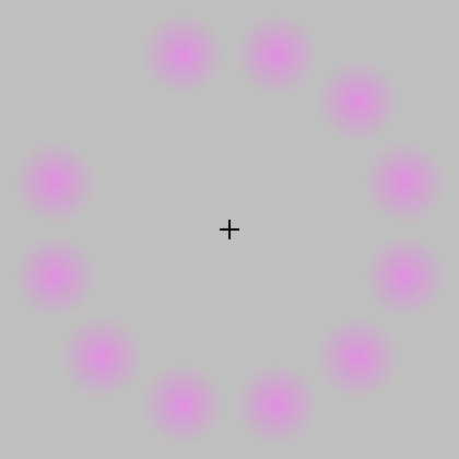
![The neural adaptation effect of Troxler's fading can be experienced by looking at the cross from a short distance without moving the eyes. After a few seconds, the colors seem to vanish. [Click on image to enlarge] Troxler-Effekt.jpg](http://upload.wikimedia.org/wikipedia/commons/thumb/1/1b/Troxler-Effekt.jpg/420px-Troxler-Effekt.jpg)