Related Research Articles

Radiation therapy or radiotherapy, often abbreviated RT, RTx, or XRT, is a therapy using ionizing radiation, generally as part of cancer treatment to control or kill malignant cells and normally delivered by a linear accelerator. Radiation therapy may be curative in a number of types of cancer if they are localized to one area of the body. It may also be used as part of adjuvant therapy, to prevent tumor recurrence after surgery to remove a primary malignant tumor. Radiation therapy is synergistic with chemotherapy, and has been used before, during, and after chemotherapy in susceptible cancers. The subspecialty of oncology concerned with radiotherapy is called radiation oncologist.

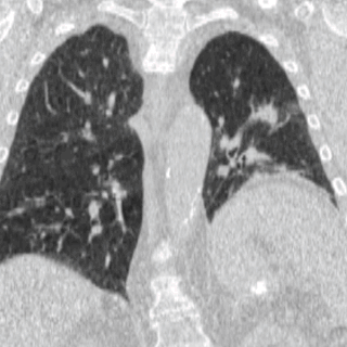
A CT scan or computed tomography scan is a medical imaging technique that uses computer-processed combinations of multiple X-ray measurements taken from different angles to produce tomographic (cross-sectional) images of a body, allowing the user to see inside the body without cutting. The personnel that perform CT scans are called radiographers or radiologic technologists.

Radiography is an imaging technique using X-rays, gamma rays, or similar ionizing radiation and non-ionizing radiation to view the internal form of an object. Applications of radiography include medical radiography and industrial radiography. Similar techniques are used in airport security. To create an image in conventional radiography, a beam of X-rays is produced by an X-ray generator and is projected toward the object. A certain amount of the X-rays or other radiation is absorbed by the object, dependent on the object's density and structural composition. The X-rays that pass through the object are captured behind the object by a detector. The generation of flat two dimensional images by this technique is called projectional radiography. In computed tomography an X-ray source and its associated detectors rotate around the subject which itself moves through the conical X-ray beam produced. Any given point within the subject is crossed from many directions by many different beams at different times. Information regarding attenuation of these beams is collated and subjected to computation to generate two dimensional images in three planes which can be further processed to produce a three dimensional image.

Radiology is the medical discipline that uses medical imaging to diagnose and treat diseases within the bodies of animals, including humans.

Medical imaging is the technique and process of creating visual representations of the interior of a body for clinical analysis and medical intervention, as well as visual representation of the function of some organs or tissues (physiology). Medical imaging seeks to reveal internal structures hidden by the skin and bones, as well as to diagnose and treat disease. Medical imaging also establishes a database of normal anatomy and physiology to make it possible to identify abnormalities. Although imaging of removed organs and tissues can be performed for medical reasons, such procedures are usually considered part of pathology instead of medical imaging.
External beam radiotherapy (EBRT) is the most common form of radiotherapy. The patient sits or lies on a couch and an external source of ionizing radiation is pointed at a particular part of the body. In contrast to brachytherapy and unsealed source radiotherapy, in which the radiation source is inside the body, external beam radiotherapy directs the radiation at the tumour from outside the body. Orthovoltage ("superficial") X-rays are used for treating skin cancer and superficial structures. Megavoltage X-rays are used to treat deep-seated tumours, whereas megavoltage electron beams are typically used to treat superficial lesions extending to a depth of approximately 5 cm. X-rays and electron beams are by far the most widely used sources for external beam radiotherapy. A small number of centers operate experimental and pilot programs employing beams of heavier particles, particularly protons, owing to the rapid dropoff in absorbed dose beneath the depth of the target.
A radiation oncologist is a specialist physician who uses ionizing radiation in the treatment of cancer. Radiation oncology is one of the three primary specialties, the other two being surgical and medical oncology, involved in the treatment of cancer. Radiation can be given as a curative modality, either alone or in combination with surgery and/or chemotherapy. It may also be used palliatively, to relieve symptoms in patients with incurable cancers. A radiation oncologist may also use radiation to treat some benign diseases, including benign tumors. In some countries, radiotherapy and chemotherapy are controlled by a single oncologist who is a "clinical oncologist". Radiation oncologists work closely with other physicians such as surgical oncologists, interventional radiologists, internal medicine subspecialists, and medical oncologists, as well as medical physicists and technicians as part of the multi-disciplinary cancer team.. Radiation oncologists undergo four years of oncology-specific training whereas oncologists who deliver chemotherapy have two years of additional training in cancer care during fellowship after internal medicine residency in the United States.

Radiosurgery is surgery using radiation, that is, the destruction of precisely selected areas of tissue using ionizing radiation rather than excision with a blade. Like other forms of radiation therapy, it is usually used to treat cancer. Radiosurgery was originally defined by the Swedish neurosurgeon Lars Leksell as "a single high dose fraction of radiation, stereotactically directed to an intracranial region of interest".
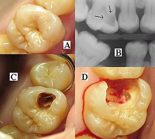
Dental radiographs are commonly called X-rays. Dentists use radiographs for many reasons: to find hidden dental structures, malignant or benign masses, bone loss, and cavities.

In radiotherapy, radiation treatment planning (RTP) is the process in which a team consisting of radiation oncologists, radiation therapist, medical physicists and medical dosimetrists plan the appropriate external beam radiotherapy or internal brachytherapy treatment technique for a patient with cancer.
Isocenter in aerial photography: it is a point where a line cuts an angle of 90 degree of tier/2. It is the point on the aerial photo platform that directly falls on a line half-way between the Principal point and the Nadir point. In imaging physics and radiation oncology, the isocenter is termed as the point in space through which the central rays of the radiation beams pass.

Tomotherapy is a radiation therapy modality, in which the patient is scanned across a modulated strip-beam, so that only one “slice” of the target is exposed at any one time by the linear accelerator (linac) beam. The three components distinctive to this modality are: (1) a collimator pair that defines the length of the strip, (2) a binary multileaf collimator whose leaves open and close during treatment to modulate the strip’s intensity, and (3) a couch that scans the patient across the beam at a fixed speed the during treatment delivery.

Projectional radiography, also known as conventional radiography, is a form of radiography and medical imaging that produces two-dimensional images by x-ray radiation. The image acquisition is generally performed by radiographers, and the images are often examined by radiologists. Both the procedure and any resultant images are often simply called "X-ray". Plain radiography or roentgenography generally refers to projectional radiography. Plain radiography can also refer to radiography without a radiocontrast agent or radiography that generates single static images, as contrasted to fluoroscopy, which are technically also projectional.
Image-guided radiation therapy is the process of frequent two and three-dimensional imaging, during a course of radiation treatment, used to direct radiation therapy utilizing the imaging coordinates of the actual radiation treatment plan. The patient is localized in the treatment room in the same position as planned from the reference imaging dataset. An example of IGRT would include localization of a cone beam computed tomography (CBCT) dataset with the planning computed tomography (CT) dataset from planning. IGRT would also include matching planar kilovoltage (kV) radiographs or megavoltage (MV) images with digital reconstructed radiographs (DRRs) from the planning CT. These two methods comprise the bulk of IGRT strategies currently employed circa 2013.
A dose-volume histogram (DVH) is a histogram relating radiation dose to tissue volume in radiation therapy planning. DVHs are most commonly used as a plan evaluation tool and to compare doses from different plans or to structures. DVHs were introduced by Michael Goitein and Verhey in 1979. DVH summarizes 3D dose distributions in a graphical 2D format. In modern radiation therapy, 3D dose distributions are typically created in a computerized treatment planning system (TPS) based on a 3D reconstruction of a CT scan. The "volume" referred to in DVH analysis is a target of radiation treatment, a healthy organ nearby a target, or an arbitrary structure.

Automatic Exposure Control (AEC) is an X-ray exposure termination device. A medical radiography x-ray exposure is always initiated by a human operator but an AEC detector system may be used to terminate the exposure when a predetermined amount of radiation has been received. The intention of AEC is to provide consistent x-ray image exposure, whether to film, a digital detector or a CT scanner. AEC systems may also automatically set exposure factors such as the X-ray tube current and voltage.

Cone beam computed tomography is a medical imaging technique consisting of X-ray computed tomography where the X-rays are divergent, forming a cone.
Four-dimensional computed tomography (4DCT) is a type of CT scanning which records multiple images over time. It allows playback of the scan as a video, so that physiological processes can be observed and internal movement can be tracked. The name is derived from the addition of time to traditional 3D computed tomography. Alternatively, the phase of a particular process, such as respiration, may be considered the fourth dimension.
Proton Computed Tomography (pCT), or Proton CT, is an imaging modality first proposed by Cormack in 1963 and initial experiment explorations identified several advantages over conventional x-ray CT (xCT). However, particle interactions such as multiple Coulomb scattering (MCS) and (in)elastic nuclear scattering events deflect the proton trajectory, resulting in nonlinear paths which can only be approximated via statistical assumptions, leading to lower spatial resolution than X-ray tomography. Further experiments were largely abandoned until the advent of proton radiation therapy in the 1990s which renewed interest in the topic due to the potential benefits of imaging and treating patients with the same particle.
A central nervous system tumor is an abnormal growth of cells from the tissues of the brain or spinal cord. CNS tumor is a generic term encompassing over 120 distinct tumor types. Common symptoms of CNS tumors include vomiting, headache, changes in vision, nausea, and seizures. A CNS tumor can be detected and classified via neurological examination, medical imaging, such as x-ray imaging, magnetic resonance imaging (MRI) or computed tomography (CT), or after analysis of a biopsy.
References
- Faiz Kahn and Roger Potish (Eds.) (1998).Treatment Planning in Radiation Oncology. Williams & Wilkins. ISBN 0-683-04607-1.
- Jacob Van Dyk (Ed.) (1999). The Modern Technology of Radiation Oncology. Medical Physics Publishing. ISBN 0-944838-38-3.
- Ross I. Berbeco (Ed.) (2018). Beam's Eye View Imaging in Radiation Oncology. CRC Press. ISBN 978-1-4987-3634-3.
- Louis Lemieux, Roger Jagoe, David R. Fish, Neil D. Kitchen, David G. Thomas, A patient-to-computed-tomography image registration method based on digitally reconstructed radiographs. Med. Phys. 21(11), 1749–1760 (1994). https://doi.org/10.1118/1.597276