Keratoconus (KC) is a disorder of the eye that results in progressive thinning of the cornea. This may result in blurry vision, double vision, nearsightedness, irregular astigmatism, and light sensitivity leading to poor quality-of-life. Usually both eyes are affected. In more severe cases a scarring or a circle may be seen within the cornea.

Sir Charles Bell was a Scottish surgeon, anatomist, physiologist, neurologist, artist, and philosophical theologian. He is noted for discovering the difference between sensory nerves and motor nerves in the spinal cord. He is also noted for describing Bell's palsy.
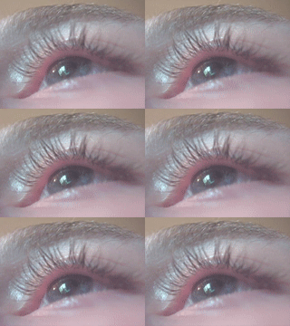
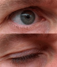
Blinking is a bodily function; it is a semi-autonomic rapid closing of the eyelid. A single blink is determined by the forceful closing of the eyelid or inactivation of the levator palpebrae superioris and the activation of the palpebral portion of the orbicularis oculi, not the full open and close. It is an essential function of the eye that helps spread tears across and remove irritants from the surface of the cornea and conjunctiva.

Dry eye syndrome (DES), also known as keratoconjunctivitis sicca (KCS), is the condition of having dry eyes. Other associated symptoms include irritation, redness, discharge, and easily fatigued eyes. Blurred vision may also occur. Symptoms range from mild and occasional to severe and continuous. Scarring of the cornea may occur in untreated cases.

The human eye is a sensory organ, part of the sensory nervous system, that reacts to visible light and allows us to use visual information for various purposes including seeing things, keeping our balance, and maintaining circadian rhythm.

An eye examination is a series of tests performed to assess vision and ability to focus on and discern objects. It also includes other tests and examinations pertaining to the eyes. Eye examinations are primarily performed by an optometrist, ophthalmologist, or an orthoptist. Health care professionals often recommend that all people should have periodic and thorough eye examinations as part of routine primary care, especially since many eye diseases are asymptomatic.

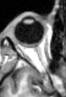
The extraocular muscles, are the seven extrinsic muscles of the human eye. Six of the extraocular muscles, the four recti muscles, and the superior and inferior oblique muscles, control movement of the eye and the other muscle, the levator palpebrae superioris, controls eyelid elevation. The actions of the six muscles responsible for eye movement depend on the position of the eye at the time of muscle contraction.

Arcus senilis (AS), also known as gerontoxon, arcus lipoides, arcus cornae, corneal arcus, arcus adiposus, or arcus cornealis, are rings in the peripheral cornea. It‘s usually caused by cholesterol deposits, so it may be a sign of high cholesterol. It is the most common peripheral corneal opacity, and is usually found in the elderly where it is considered a benign condition. When AS is found in patients less than 50 years old it is termed arcus juvenilis. The finding of arcus juvenilis in combination with hyperlipidemia in younger men represents an increased risk for cardiovascular disease.

Corneal abrasion is a scratch to the surface of the cornea of the eye. Symptoms include pain, redness, light sensitivity, and a feeling like a foreign body is in the eye. Most people recover completely within three days.

The orbicularis oculi is a muscle in the face that closes the eyelids. It arises from the nasal part of the frontal bone, from the frontal process of the maxilla in front of the lacrimal groove, and from the anterior surface and borders of a short fibrous band, the medial palpebral ligament.

Blepharospasm is any abnormal contraction of the orbicularis oculi muscle. The condition should be distinguished from the more common, and milder, involuntary quivering of an eyelid, known as myokymia, or fasciculation. In most cases, blepharospasm symptoms last for a few days and then disappear without treatment, but in some cases the twitching is chronic and persistent, causing life-long challenges. In these cases, the symptoms are often severe enough to result in functional blindness. The person's eyelids feel like they are clamping shut and will not open without great effort. People have normal eyes, but for periods of time are effectively blind due to their inability to open their eyelids. In contrast, the reflex blepharospasm is due to any pain in and around the eye.

The corneal reflex, also known as the blink reflex or eyelid reflex, is an involuntary blinking of the eyelids elicited by stimulation of the cornea, though it could result from any peripheral stimulus. Stimulation should elicit both a direct and consensual response. The reflex occurs at a rapid rate of 0.1 seconds. The purpose of this reflex is to protect the eyes from foreign bodies and bright lights. The blink reflex also occurs when sounds greater than 40–60 dB are made.

The nasociliary nerve is a branch of the ophthalmic nerve, itself a branch of the trigeminal nerve. It is intermediate in size between the other two branches of the ophthalmic nerve, the frontal nerve and lacrimal nerve.

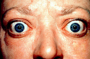
Graves’ ophthalmopathy, also known as thyroid eye disease (TED), is an autoimmune inflammatory disorder of the orbit and periorbital tissues, characterized by upper eyelid retraction, lid lag, swelling, redness (erythema), conjunctivitis, and bulging eyes (exophthalmos). It occurs most commonly in individuals with Graves' disease, and less commonly in individuals with Hashimoto's thyroiditis, or in those who are euthyroid.
The red reflex refers to the reddish-orange reflection of light from the back of the eye, or fundus, observed when using an ophthalmoscope or retinoscope. The reflex relies on the transparency of optical media and reflects off the fundus back through media into the aperture of the ophthalmoscope. The red reflex is considered abnormal if there is any asymmetry between the eyes, dark spots, or white reflex (Leukocoria).

Marcus Gunn phenomenon is an autosomal dominant condition with incomplete penetrance, in which nursing infants will have rhythmic upward jerking of their upper eyelid. This condition is characterized as a synkinesis: when two or more muscles that are independently innervated have either simultaneous or coordinated movements.

Keratoprosthesis is a surgical procedure where a diseased cornea is replaced with an artificial cornea. Traditionally, keratoprosthesis is recommended after a person has had a failure of one or more donor corneal transplants. More recently, a less invasive, non-penetrating artificial cornea has been developed which can be used in more routine cases of corneal blindness. While conventional cornea transplant uses donor tissue for transplant, an artificial cornea is used in the Keratoprosthesis procedure. The surgery is performed to restore vision in patients with severely damaged cornea due to congenital birth defects, infections, injuries and burns.

Conjunctivochalasis, also known as Mechanical Dry Eye (MDE), is a common eye surface condition characterized by the presence of excess folds of the conjunctiva located between the globe of the eye and the eyelid margin.
Eye injuries during general anaesthesia are reasonably common if care is not taken to prevent them.
Exposure keratopathy is medical condition affecting the cornea of eyes. It can lead to corneal ulceration and permanent loss of vision due to corneal opacity.


