Related Research Articles

Albinism is a congenital condition characterized in humans by the partial or complete absence of pigment in the skin, hair and eyes. Albinism is associated with a number of vision defects, such as photophobia, nystagmus, and amblyopia. Lack of skin pigmentation makes for more susceptibility to sunburn and skin cancers. In rare cases such as Chédiak–Higashi syndrome, albinism may be associated with deficiencies in the transportation of melanin granules. This also affects essential granules present in immune cells leading to increased susceptibility to infection.

Melanin is a broad term for a group of natural pigments found in most organisms. Melanin is produced through a multistage chemical process known as melanogenesis, where the oxidation of the amino acid tyrosine is followed by polymerization. The melanin pigments are produced in a specialized group of cells known as melanocytes.

Vitiligo is a long-term skin condition characterized by patches of the skin losing their pigment. The patches of skin affected become white and usually have sharp margins. The hair from the skin may also become white. The inside of the mouth and nose may also be involved. Typically both sides of the body are affected. Often the patches begin on areas of skin that are exposed to the sun. It is more noticeable in people with dark skin. Vitiligo may result in psychological stress and those affected are sometimes stigmatized.

Hair color is the pigmentation of hair follicles due to two types of melanin: eumelanin and pheomelanin. Generally, if more melanin is present, the color of the hair is darker; if less melanin is present, the hair is lighter. The tone of the hair is dependent on the ratio of black or brown eumelanin to yellow or red pheomelanin. Levels of melanin can vary over time causing a person's hair color to change, and it is possible to have hair follicles of more than one color on the same person. Some hair colors are associated with some ethnic groups due to observed higher frequency of particular hair color within their geographical region, e.g. straight dark hair amongst East Asians, a large variety of dark, fair, curly, wavy and bushy hair amongst Europeans, curly, dark, and uniquely helical hair with Africans, whilst gray, white hair or "silver", is often associated with age and wisdom.

Cyanosis is the change of body tissue color to a bluish-purple hue as a result of having decreased amounts of oxygen bound to the hemoglobin in the red blood cells of the capillary bed. Body tissues that reflect cyanosis are usually in locations where the skin is thinner, including the mucous membranes, lips, nail beds, and ear lobes. It is important to note that medications containing amiodarone or silver, Mongolian spots, large birth marks, and the consumption of food products with blue or purple dyes can also result in the bluish skin tissue discoloration and may be mistaken for cyanosis.

Teething is the process by which an infant's first teeth sequentially appear by emerging through the gums, typically arriving in pairs. The mandibular central incisors are the first primary teeth to erupt, usually between 6 and 10 months of age. It can take several years for all 20 teeth to complete the tooth eruption. Though the process of teething is sometimes referred to as "cutting teeth", when teeth emerge through the gums they do not cut through the flesh. Instead, hormones are released within the body that cause some cells in the gums to die and separate, allowing the teeth to come through.

The gums or gingiva consist of the mucosal tissue that lies over the mandible and maxilla inside the mouth. Gum health and disease can have an effect on general health.
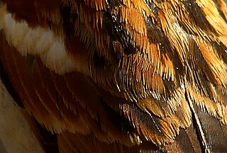
Plumage is a layer of feathers that cover a bird and the pattern, colour, and arrangement of those feathers. The pattern and colours of plumage differ between species and subspecies and may vary with age classes. Within species, there can be different colour morphs. The placement of feathers on a bird is not haphazard, but rather emerge in organized, overlapping rows and groups, and these feather tracts are known by standardized names.

Ectodermal dysplasia (ED) is a group of genetic syndromes all deriving from abnormalities of the ectodermal structures. More than 150 different syndromes have been identified.
Black hairy tongue syndrome (BHT) is a condition of the tongue in which the small bumps on the tongue elongate with black or brown discoloration, giving a black and hairy appearance. The appearance may be alarming, but it is a harmless condition. Predisposing factors include smoking, xerostomia, soft diet, poor oral hygiene and certain medications. Management is facilitated by improving oral hygiene, especially scraping or brushing the tongue.
Chrysiasis is a dermatological condition induced by the parenteral administration of gold salts, usually for the treatment of rheumatoid arthritis. Such treatment has been superseded as the best practice for treating the disease because of "numerous side effects and monitoring requirements, their limited efficacy, and very slow onset of action".

Piebaldism refers to the absence of mature melanin-forming cells (melanocytes) in certain areas of the skin and hair. It is a rare autosomal dominant disorder of melanocyte development. Common characteristics include a congenital white forelock, scattered normal pigmented and hypopigmented macules and a triangular shaped depigmented patch on the forehead. There is nevertheless great variation in the degree and pattern of presentation, even within affected families. In some cases, piebaldism occurs together with severe developmental problems, as in Waardenburg syndrome and Hirschsprung's disease.

A white horse is born predominantly white and stays white throughout its life. A white horse has mostly pink skin under its hair coat, and may have brown, blue, or hazel eyes. "True white" horses, especially those that carry one of the dominant white (W) genes, are rare. Most horses that are commonly referred to as "white" are actually "gray" horses whose hair coats are completely white. Gray horses may be born of any color and their hairs gradually turn white as time goes by and take on a white appearance. Nearly all gray horses have dark skin, except under any white markings present at birth. Skin color is the most common method for an observer to distinguish between mature white and gray horses.
Gum depigmentation, also known as gum bleaching, is a procedure used in cosmetic dentistry to lighten or remove black spots or patches on the gums consisting of melanin. Melanin in skin is very common in inhabitants in many parts of the world due to genetic factors. Melanin pigmentation in skin, oral mucosa, inner ear and other organs is a detoxification mechanism. Some toxic agents bind to melanin and will move out of the tissue with the ageing cells and are expelled to the tissue surfaces. Also in the gums and oral mucosa a visible pigmentation is most often caused by genetic factors, but also by tobacco smoking or in a few cases by long-term use of certain medications. If stopping smoking or change of medication do not solve the problem with a disfigurating melanin pigmentation, a surgical operation may be performed. The procedure itself can involve laser ablation techniques.
Oculocutaneous albinism type I or type 1A is an autosomal recessive skin disease. This subtype of oculocutaneous albinism is caused when the gene for tyrosinase does not function properly.
Burton's line, also known as the Burton line or Burtonian line, is a clinical sign found in patients with chronic lead poisoning. It is a very thin, black-blue line visible along the margin of the gums, at the base of the teeth.
Oral pigmentation is asymptomatic and does not usually cause any alteration to the texture or thickness of the affected area. The colour can be uniform or speckled and can appear solitary or as multiple lesions. Depending on the site, depth, and quantity of pigment, the appearance can vary considerably.

Amalgam tattoo is a grey, blue or black area of discoloration on the mucous membranes of the mouth, typically on the gums of the lower jaw. It is a healthcare caused lesion, due to entry of dental amalgam into the soft tissues. It is common, painless, and benign, but it can be mistaken for melanoma.
Hereditary gingival fibromatosis (HGF), also known as idiopathic gingival hyperplasia, is a rare condition of gingival overgrowth. HGF is characterized as a benign, slowly progressive, nonhemorrhagic, fibrous enlargement of keratinized gingiva. It can cover teeth in various degrees, and can lead to aesthetic disfigurement. Fibrous enlargement is most common in areas of maxillary and mandibular tissues of both arches in the mouth. Phenotype and genotype frequency of HGF is 1:175,000 where males and females are equally affected but the cause is not entirely known. It mainly exists as an isolated abnormality but can also be associated with a multi-system syndrome.

Albinism is the congenital absence of any pigmentation or colouration in an animal, plant, or person, resulting in white hair, feathers, scales and skin and pink eyes in mammals, birds, reptiles, amphibians and fish and invertebrates as well. Individuals with the condition are referred to as albino.