Brown–Vialetto–Van Laere syndrome

Last updated
Brown–Vialetto–Van Laere syndrome
Other namesBVVLS1 [1]

Brown-Vialetto-Van-Laere syndrome (BVVL), sometimes known as Brown's syndrome, is a rare degenerative disorder often initially characterized by progressive sensorineural deafness. [2]

Contents

The syndrome most often affects children, adolescents, and young adults. As knowledge of BVVL grows some adult patients have now been diagnosed. With prompt treatment the prognosis may be positive with some patients stabilizing and even major improvements noted in certain cases.

Symptoms and signs

BVVL is marked by a number of cranial nerve palsies, including those of the motor components involving the 7th and 9th-12th cranial nerves, spinal motor nerves, and upper motor neurons. [2] Major features of BVVL include facial and neck weakness, fasciculation of the tongue, and neurological disorders from the cranial nerves. [3] The neurological manifestations develop insidiously: they usually begin with sensorineural deafness, progress inexorably to paralysis, and often culminate in respiratory failure. Most mortality in patients has been from either respiratory infections or respiratory muscle paralysis. Pathological descriptions of BVVL include injury and depletion of 3rd-7th cranial nerves, loss of the spinal anterior horn cells, degeneration of Purkinje cells, as well as degeneration of the spinocerebellar and pyramidal tracts. [3] The first symptoms in nearly all cases of BVVL is progressive vision loss and deafness, and the first initial symptoms are seen anywhere from one to three years. [4]

Most cases of deafness are followed by a latent period that can extend anywhere from weeks to years, and this time is usually marked by cranial nerve degeneration. Neurological symptoms of BVVL include optic atrophy, cerebellar ataxia, retinitis pigmentosa, epilepsy and autonomic dysfunction. [4] Non-neurological symptoms can include diabetes, auditory hallucinations, respiratory difficulties, color blindness, and hypertension.[ citation needed ]

Genetics

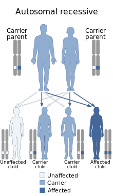
BVVL has autosomal recessive pattern of inheritance Autosomal recessive - en.svg
BVVL has autosomal recessive pattern of inheritance

The disorder has been associated with various mutations in the SLC52A2 and SLC52A3 genes. [1] [5] [6] This gene is thought to be involved in transport of riboflavin. [7]

BVVL is allelic and phenotypically similar to Fazio–Londe disease and likewise is inherited in an autosomal recessive manner.[ citation needed ]

Diagnosis

Diagnosis requires a neurological examination and neuroimaging can be helpful. [8]

BVVL can be differentially diagnosed from similar conditions like Fazio-Londe syndrome and amyotrophic lateral sclerosis, in that those two conditions don't involve sensorineural hearing loss, while BVVL, Madras motor neuron disease, Nathalie syndrome, and Boltshauser syndrome do. Nathalie syndrome does not involve lower cranial nerve symptoms, so it can be excluded if those are present. If there is evidence of lower motor neuron involvement, Boltshauser syndrome can be excluded. Finally, if there is a family history of the condition, then BVVL is more likely than MMND, as MMND tends to be sporadic. [8]

Genetic testing is able to identify genetic mutations underlying BVVL. [6]

Treatment

As of 2017, there were case reports that treatment with high doses of riboflavin 5 phosphate can halt the progress of Brown–Vialetto–Van Laere syndrome and in some cases can improve symptoms. [9] [10]

The first report of BVVL syndrome in Japanese literature was of a woman that had BVVL and showed improvement after such treatments. The patient was a sixty-year-old woman who had symptoms such as sensorineural deafness, weakness, and atrophy since she was 15 years old. Around the age of 49 the patient was officially diagnosed with BVVL, intubated, and then attached to a respirator to improve her CO₂ narcosis. After the treatments, the patient still required respiratory assistance during sleep; however, the patient no longer needed assistance by a respirator during the daytime. [11]

Prognosis

The clinical course of BVVL can vary from one patient to another. There have been cases with progressive deterioration, deterioration followed by periods of stabilization, and deterioration with abrupt periods of increasing severity. [3]

The syndrome has previously been considered to have a high mortality rate but the initial response of most patients to the Riboflavin protocol are very encouraging and seem to indicate a significantly improved life expectancy could be achievable. There are three documented cases of BVVL where the patient died within the first five years of the disease. On the contrary, most patients have survived more than 10 years after the onset of their first symptom, and several cases have survived 20–30 years after the onset of their first symptom. [4]

Families with multiple cases of BVVL and, more generally, multiple cases of infantile progressive bulbar palsy can show variability in age of disease onset and survival. Dipti and Childs described such a situation in which a family had five children that had Infantile PBP. In this family, three siblings showed sensorineural deafness and other symptoms of BVVL at an older age. The other two siblings showed symptoms of Fazio-Londe disease and died before the age of two. [12]

Epidemiology

As of 2015, there are approximately 70 known cases of Brown-Vialetto-Van-Laere syndrome worldwide. [2] BVVL was first described in a Portuguese family, and has since been described in a number of ethnic groups. Reports have shown that BVVL infects females more than males at a rate of 5:1 respectively. However, males usually exhibit more severe symptoms, an earlier onset of deafness and a tendency to die earlier in life. [2]

History

The syndrome was first described by Charles Brown in 1894; [13] further accounts by Vialetto [14] and Van Laere [15] followed in 1936 and 1966, respectively. There were very few cases reported in the medical literature over the 100 odd years since its first description however with the specific genetic testing available in recent years more cases are being identified and treated.[ citation needed ]

See also

Related Research Articles

<span class="mw-page-title-main">Bell's palsy</span> Facial paralysis resulting from dysfunction in the cranial nerve VII (facial nerve)

Bell's palsy is a type of facial paralysis that results in a temporary inability to control the facial muscles on the affected side of the face. In most cases, the weakness is temporary and significantly improves over weeks. Symptoms can vary from mild to severe. They may include muscle twitching, weakness, or total loss of the ability to move one or, in rare cases, both sides of the face. Other symptoms include drooping of the eyebrow, a change in taste, and pain around the ear. Typically symptoms come on over 48 hours. Bell's palsy can trigger an increased sensitivity to sound known as hyperacusis.

<span class="mw-page-title-main">Medulla oblongata</span> Structure of the brain stem

The medulla oblongata or simply medulla is a long stem-like structure which makes up the lower part of the brainstem. It is anterior and partially inferior to the cerebellum. It is a cone-shaped neuronal mass responsible for autonomic (involuntary) functions, ranging from vomiting to sneezing. The medulla contains the cardiac, respiratory, vomiting and vasomotor centers, and therefore deals with the autonomic functions of breathing, heart rate and blood pressure as well as the sleep–wake cycle.

<span class="mw-page-title-main">Alexander disease</span> Rare genetic disorder of the white matter of the brain

Alexander disease is a very rare autosomal dominant leukodystrophy, which are neurological conditions caused by anomalies in the myelin which protects nerve fibers in the brain. The most common type is the infantile form that usually begins during the first two years of life. Symptoms include mental and physical developmental delays, followed by the loss of developmental milestones, an abnormal increase in head size and seizures. The juvenile form of Alexander disease has an onset between the ages of 2 and 13 years. These children may have excessive vomiting, difficulty swallowing and speaking, poor coordination, and loss of motor control. Adult-onset forms of Alexander disease are less common. The symptoms sometimes mimic those of Parkinson’s disease or multiple sclerosis, or may present primarily as a psychiatric disorder.

<span class="mw-page-title-main">Facial nerve paralysis</span> Medical condition

Facial nerve paralysis is a common problem that involves the paralysis of any structures innervated by the facial nerve. The pathway of the facial nerve is long and relatively convoluted, so there are a number of causes that may result in facial nerve paralysis. The most common is Bell's palsy, a disease of unknown cause that may only be diagnosed by exclusion of identifiable serious causes.

<span class="mw-page-title-main">Progressive supranuclear palsy</span> Medical condition

Progressive supranuclear palsy (PSP) is a late-onset neurodegenerative disease involving the gradual deterioration and death of specific volumes of the brain. The condition leads to symptoms including loss of balance, slowing of movement, difficulty moving the eyes, and cognitive impairment. PSP may be mistaken for other types of neurodegeneration such as Parkinson's disease, frontotemporal dementia and Alzheimer's disease. The cause of the condition is uncertain, but involves the accumulation of tau protein within the brain. Medications such as levodopa and amantadine may be useful in some cases.

<span class="mw-page-title-main">Krabbe disease</span> Medical condition

Krabbe disease (KD) is a rare and often fatal lysosomal storage disease that results in progressive damage to the nervous system. KD involves dysfunctional metabolism of sphingolipids and is inherited in an autosomal recessive pattern. The disease is named after the Danish neurologist Knud Krabbe (1885–1961).

<span class="mw-page-title-main">Fazio–Londe disease</span> Medical condition

Fazio–Londe disease (FLD), also called progressive bulbar palsy of childhood, is a very rare inherited motor neuron disease of children and young adults and is characterized by progressive paralysis of muscles innervated by cranial nerves. FLD, along with Brown–Vialetto–Van Laere syndrome (BVVL), are the two forms of infantile progressive bulbar palsy, a type of progressive bulbar palsy in children.

Progressive bulbar palsy (PBP) is a medical condition. It belongs to a group of disorders known as motor neuron diseases. PBP is a disease that attacks the nerves supplying the bulbar muscles. These disorders are characterized by the degeneration of motor neurons in the cerebral cortex, spinal cord, brain stem, and pyramidal tracts. This specifically involves the glossopharyngeal nerve (IX), vagus nerve (X), and hypoglossal nerve (XII).

Pseudobulbar palsy is a medical condition characterized by the inability to control facial movements and caused by a variety of neurological disorders. Patients experience difficulty chewing and swallowing, have increased reflexes and spasticity in tongue and the bulbar region, and demonstrate slurred speech, sometimes also demonstrating uncontrolled emotional outbursts.

<span class="mw-page-title-main">Foix–Chavany–Marie syndrome</span> Medical condition

Foix–Chavany–Marie syndrome (FCMS), also known as bilateral opercular syndrome, is a neuropathological disorder characterized by paralysis of the facial, tongue, pharynx, and masticatory muscles of the mouth that aid in chewing. The disorder is primarily caused by thrombotic and embolic strokes, which cause a deficiency of oxygen in the brain. As a result, bilateral lesions may form in the junctions between the frontal lobe and temporal lobe, the parietal lobe and cortical lobe, or the subcortical region of the brain. FCMS may also arise from defects existing at birth that may be inherited or nonhereditary. Symptoms of FCMS can be present in a person of any age and it is diagnosed using automatic-voluntary dissociation assessment, psycholinguistic testing, neuropsychological testing, and brain scanning. Treatment for FCMS depends on the onset, as well as on the severity of symptoms, and it involves a multidisciplinary approach.

Bulbar palsy refers to a range of different signs and symptoms linked to impairment of function of the glossopharyngeal nerve, the vagus nerve, the accessory nerve, and the hypoglossal nerve. It is caused by a lower motor neuron lesion in the medulla oblongata, or from lesions to these nerves outside the brainstem, and also botulism. This may be caused by any of a number of genetic, vascular, degenerative, inflammatory, and other underlying conditions. It can be differentiated from pseudobulbar palsy. When there is airway obstruction, intubation is used.

Conjugate gaze palsies are neurological disorders affecting the ability to move both eyes in the same direction. These palsies can affect gaze in a horizontal, upward, or downward direction. These entities overlap with ophthalmoparesis and ophthalmoplegia.

Infantile progressive bulbar palsy is a rare type of progressive bulbar palsy that occurs in children. The disease exists in both rapid and slow onsets, and involves inflammation of the gray matter of the bulb. Infantile PBP is a disease that manifests itself in two forms: Fazio–Londe syndrome (FL) and Brown–Vialetto–Van Laere syndrome (BVVL).

Madras motor neuron disease is a rare motor neuron disease originating in South India. Two other forms of the disease have been found, Familial Madras Motor Neuron Disease (FMMND) and the variant Madras Motor Neuron Disease (MMNDV). The symptoms of MMND include weakness in the arms and legs, loss of vision, and deafness. Most affected individuals are diagnosed by the age of 15 and occurs at the same frequency in males and females. While the cause of the disease and its origins are not yet known, supportive care is available to individuals affected by the disease.

<span class="mw-page-title-main">ALS</span> Rare neurodegenerative disease

Amyotrophic lateral sclerosis (ALS), also known as motor neurone disease (MND) or Lou Gehrig's disease, is a rare and terminal neurodegenerative disease that results in the progressive loss of motor neurons that control voluntary muscles. ALS is the most common form of the motor neuron diseases. Early symptoms of ALS include stiff muscles, muscle twitches, gradual increasing weakness, and muscle wasting. Limb-onset ALS begins with weakness in the arms or legs, while bulbar-onset ALS begins with difficulty in speaking or swallowing. Around half of people with ALS develop at least mild difficulties with thinking and behavior, and about 15% develop frontotemporal dementia. Motor neuron loss continues until the abilities to eat, speak, move, or, lastly, breathe are lost.

<span class="mw-page-title-main">Distal spinal muscular atrophy type 1</span> Medical condition

Distal spinal muscular atrophy type 1 (DSMA1), also known as spinal muscular atrophy with respiratory distress type 1 (SMARD1), is a rare neuromuscular disorder involving death of motor neurons in the spinal cord which leads to a generalised progressive atrophy of body muscles.

<span class="mw-page-title-main">SLC52A3</span> Protein-coding gene in the species Homo sapiens

Solute carrier family 52, member 3, formerly known as chromosome 20 open reading frame 54 and riboflavin transporter 2, is a protein that in humans is encoded by the SLC52A3 gene.

Alternating hemiplegia is a form of hemiplegia that has an ipsilateral cranial nerve palsies and contralateral hemiplegia or hemiparesis of extremities of the body. The disorder is characterized by recurrent episodes of paralysis on one side of the body. There are multiple forms of alternating hemiplegia, Weber's syndrome, middle alternating hemiplegia, and inferior alternating hemiplegia. This type of syndrome can result from a unilateral lesion in the brainstem affecting both upper motor neurons and lower motor neurons. The muscles that would receive signals from these damaged upper motor neurons result in spastic paralysis. With a lesion in the brainstem, this affects the majority of limb and trunk muscles on the contralateral side due to the upper motor neurons decussation after the brainstem. The cranial nerves and cranial nerve nuclei are also located in the brainstem making them susceptible to damage from a brainstem lesion. Cranial nerves III (Oculomotor), VI (Abducens), and XII (Hypoglossal) are most often associated with this syndrome given their close proximity with the pyramidal tract, the location which upper motor neurons are in on their way to the spinal cord. Damages to these structures produce the ipsilateral presentation of paralysis or palsy due to the lack of cranial nerve decussation before innervating their target muscles. The paralysis may be brief or it may last for several days, many times the episodes will resolve after sleep. Some common symptoms of alternating hemiplegia are mental impairment, gait and balance difficulties, excessive sweating and changes in body temperature.

<span class="mw-page-title-main">Arts syndrome</span> Medical condition

Arts syndrome is a rare metabolic disorder that causes serious neurological problems in males due to a malfunction of the PRPP synthetase 1 enzyme. Arts Syndrome is part of a spectrum of PRPS-1 related disorders with reduced activity of the enzyme that includes Charcot–Marie–Tooth disease and X-linked non-syndromic sensorineural deafness.

CAPOS syndrome is a rare genetic neurological disorder which is characterized by abnormalities of the feet, eyes and brain which affect their normal function. These symptoms occur episodically when a fever-related infection is present within the body. The name is an acronym for "cerebellar ataxia, areflexia, pes cavus, optic atrophy, and sensorineural hearing loss".

References

  1. 1 2 Online Mendelian Inheritance in Man (OMIM): 211530
  2. 1 2 3 4 Voudris, KA; Skardoutsou, A; Vagiakou, EA (2002). "Infantile progressive bulbar palsy with deafness". Brain & Development. 24 (7): 732–735. doi:10.1016/S0387-7604(02)00085-2. PMID   12427524. S2CID   7690110.
  3. 1 2 3 Prabhu, HV; Brown, MJ (June 2005). "Brown-Vialetto-Van Laere syndrome: a rare syndrome in otology". The Journal of Laryngology & Otology. 119 (6): 470–2. doi:10.1258/0022215054273179. PMID   15992475. S2CID   28865108.
  4. 1 2 3 Sathasivam, S; O’Sullivan, S; Nicolson, A; Tilley, PJB; Shaw, PJ (2000). "Brown-Vialetto-Van Laere syndrome: Case report and literature review". Amyotrophic Lateral Sclerosis and Other Motor Neuron Disorders. 1 (4): 277–281. doi:10.1080/14660820050515106. PMID   11465021. S2CID   42001695.
  5. Green, P; Wiseman, M; Crow, YJ; et al. (March 2010). "Brown-Vialetto-Van Laere syndrome, a ponto-bulbar palsy with deafness, is caused by mutations in c20orf54". American Journal of Human Genetics. 86 (3): 485–9. doi:10.1016/j.ajhg.2010.02.006. PMC   2833371 . PMID   20206331.
  6. 1 2 Johnson, JO; Gibbs, JR; Van Maldergem, L; Houlden, H; Singleton, AB (October 2010). "Exome sequencing in Brown-Vialetto-van Laere syndrome". American Journal of Human Genetics. 87 (4): 567–9, author reply 569–70. doi:10.1016/j.ajhg.2010.05.021. PMC   2948797 . PMID   20920669.
  7. Yamamoto, S; Inoue, K; Ohta, K; Fukatsu, R; Maeda, J; Yoshida, Y; Yuasa, H (2009). "Identification and Functional Characterization of Rat Riboflavin Transporter 2". Journal of Biochemistry. 145 (4): 437–443. doi:10.1093/jb/mvn181. PMID   19122205.
  8. 1 2 Sathasivam, S (17 April 2008). "Brown-Vialetto-Van Laere syndrome". Orphanet Journal of Rare Diseases. 3: 9. doi: 10.1186/1750-1172-3-9 . PMC   2346457 . PMID   18416855.
  9. Allison, T; Roncero, I; Forsyth, R; Coffman, K; Pichon, JL (May 2017). "Brown-Vialetto-Van Laere Syndrome as a Mimic of Neuroimmune Disorders: 3 Cases From the Clinic and Review of the Literature". Journal of Child Neurology. 32 (6): 528–532. doi:10.1177/0883073816689517. PMID   28116953. S2CID   31769793.
  10. Bosch, Annet M; Stroek, Kevin; Abeling, Nico G; Waterham, Hans R; IJlst, Lodewijk; Wanders, Ronald JA (2012). "The Brown-Vialetto-Van Laere and Fazio Londe syndrome revisited: natural history, genetics, treatment and future perspectives". Orphanet Journal of Rare Diseases. 7 (1): 83. doi: 10.1186/1750-1172-7-83 . PMC   3517535 . PMID   23107375.
  11. Nemoto, H; Konno, S; Nomoto, N; Wakata, N; Kurihara, T (May 2005). "[A case of Brown-Vialetto-van Laere (BVVL) syndrome in Japan]". Rinsho Shinkeigaku. 45 (5): 357–61. PMID   15960172.
  12. Dipti, S; Childs, AM; Livingston, JH; Aggarwal, AK; Miller, M; Williams, C; Crow, YJ (September 2005). "Brown-Vialetto-Van Laere syndrome; variability in age at onset and disease progression highlighting the phenotypic overlap with Fazio-Londe disease". Brain & Development. 27 (6): 443–6. doi:10.1016/j.braindev.2004.10.003. PMID   16122634. S2CID   32223440.
  13. Brown CH (1894). "Infantile amyotrophic lateral sclerosis of the family type". Journal of Nervous and Mental Disease. 21 (2): 707–716. doi:10.1097/00005053-189411000-00003. S2CID   143353104.
  14. Vialetto E (1936). "Contributo alla forma ereditaria della paralisi bulbare progressive". Rivista sperimentale di freniatria e medicina legale delle alienazioni mentali. 40: 1–24.
  15. Van Laere, J (August 1966). "Paralysie bulbo-pontine chronique progressive familiale avec surdité. Un cas de syndrome de Klippel-Trenaunay dans la même fratrie – problèmes diagnostiques et génétiques [Familial progressive chronic bulbo-pontine paralysis with deafness. A case of Klippel-Trenaunay syndrome in siblings of the same family. Diagnostic and genetic problems]". Revue neurologique. 115 (2): 289–95. PMID   5969547.

Further reading