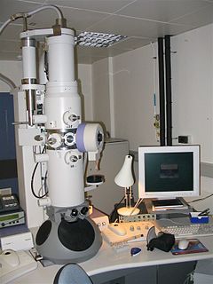
An electron microscope is a microscope that uses a beam of accelerated electrons as a source of illumination. As the wavelength of an electron can be up to 100,000 times shorter than that of visible light photons, electron microscopes have a higher resolving power than light microscopes and can reveal the structure of smaller objects. A scanning transmission electron microscope has achieved better than 50 pm resolution in annular dark-field imaging mode and magnifications of up to about 10,000,000x whereas most light microscopes are limited by diffraction to about 200 nm resolution and useful magnifications below 2000x.
The Protein Data Bank (PDB) is a database for the three-dimensional structural data of large biological molecules, such as proteins and nucleic acids. The data, typically obtained by X-ray crystallography, NMR spectroscopy, or, increasingly, cryo-electron microscopy, and submitted by biologists and biochemists from around the world, are freely accessible on the Internet via the websites of its member organisations. The PDB is overseen by an organization called the Worldwide Protein Data Bank, wwPDB.
XSD, a recommendation of the World Wide Web Consortium (W3C), specifies how to formally describe the elements in an Extensible Markup Language (XML) document. It can be used by programmers to verify each piece of item content in a document. They can check if it adheres to the description of the element it is placed in.

Tomography is imaging by sections or sectioning, through the use of any kind of penetrating wave. The method is used in radiology, archaeology, biology, atmospheric science, geophysics, oceanography, plasma physics, materials science, astrophysics, quantum information, and other areas of science. The word tomography is derived from Ancient Greek τόμος tomos, "slice, section" and γράφω graphō, "to write". A device used in tomography is called a tomograph, while the image produced is a tomogram.

Transmission electron cryomicroscopy (CryoTEM), commonly known as cryo-EM, is a form of cryogenic electron microscopy, more specifically a type of transmission electron microscopy (TEM) where the sample is studied at cryogenic temperatures. Cryo-EM is gaining popularity in structural biology.

Electron cryotomography (CryoET) is an imaging technique used to produce high-resolution (~1-4 nm) three-dimensional views of samples, typically biological macromolecules and cells. CryoET is a specialized application of transmission electron cryomicroscopy (CryoTEM) in which samples are imaged as they are tilted, resulting in a series of 2D images that can be combined to produce a 3D reconstruction, similar to a CT scan of the human body. In contrast to other electron tomography techniques, samples are immobilized in non-crystalline ("vitreous") ice and imaged under cryogenic conditions, allowing them to be imaged without dehydration or chemical fixation, which could otherwise disrupt or distort biological structures.

The Oxygen XML Editor is a multi-platform XML editor, XSLT/XQuery debugger and profiler with Unicode support. It is a Java application, so it can run in Windows, Mac OS X, and Linux. It also has a version that can run as an Eclipse plugin.

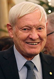
Richard Henderson, CH, FRS, FMedSci, HonFRSC is a Scottish molecular biologist and biophysicist and pioneer in the field of electron microscopy of biological molecules. Henderson shared the Nobel Prize in Chemistry in 2017 with Jacques Dubochet and Joachim Frank.
The EM Data Bank or Electron Microscopy Data Bank (EMDB) collects 3D EM maps and associated experimental data determined using electron microscopy of biological specimens. It was established in 2002 at the MSD/PDBe group of the European Bioinformatics Institute (EBI), where the European site of the EMDataBank.org consortium is located. As of 2015, the resource contained over 2,600 entries with a mean resolution of 15Å.
Electron tomography (ET) is a tomography technique for obtaining detailed 3D structures of sub-cellular macro-molecular objects. Electron tomography is an extension of traditional transmission electron microscopy and uses a transmission electron microscope to collect the data. In the process, a beam of electrons is passed through the sample at incremental degrees of rotation around the center of the target sample. This information is collected and used to assemble a three-dimensional image of the target. For biological applications, the typical resolution of ET systems are in the 5–20 nm range, suitable for examining supra-molecular multi-protein structures, although not the secondary and tertiary structure of an individual protein or polypeptide.

EMSL, or the William R. Wiley Environmental Molecular Sciences Laboratory, is a national scientific user facility located at Pacific Northwest National Laboratory in Richland, Washington. EMSL is funded by the Department of Energy’s Office of Biological and Environmental Research. EMSL provides experimental and computational resources to address the environmental molecular science challenges facing DOE and the nation.
Biology data visualization is a branch of bioinformatics concerned with the application of computer graphics, scientific visualization, and information visualization to different areas of the life sciences. This includes visualization of sequences, genomes, alignments, phylogenies, macromolecular structures, systems biology, microscopy, and magnetic resonance imaging data. Software tools used for visualizing biological data range from simple, standalone programs to complex, integrated systems.

Single particle analysis is a group of related computerized image processing techniques used to analyze images from transmission electron microscopy (TEM). These methods were developed to improve and extend the information obtainable from TEM images of particulate samples, typically proteins or other large biological entities such as viruses. Individual images of stained or unstained particles are very noisy, and so hard to interpret. Combining several digitized images of similar particles together gives an image with stronger and more easily interpretable features. An extension of this technique uses single particle methods to build up a three-dimensional reconstruction of the particle. Using cryo-electron microscopy it has become possible to generate reconstructions with sub-nanometer resolution and near-atomic resolution first in the case of highly symmetric viruses, and now in smaller, asymmetric proteins as well.

IMOD is an open-source, cross-platform suite of modeling, display and image processing programs used for 3D reconstruction and modeling of microscopy images with a special emphasis on electron microscopy data. IMOD has been used across a range of scales from macromolecule structures to organelles
to whole cells and can also be used for optical sections. Included in IMOD are tools for image reconstruction, image segmentation, 3D mesh modeling and analysis of 2D and 3D data.
Joachim Frank is a German-born American biophysicist at Columbia University and a Nobel laureate. He is regarded as the founder of single-particle cryo-electron microscopy (cryo-EM), for which he shared the Nobel Prize in Chemistry in 2017 with Jacques Dubochet and Richard Henderson. He also made significant contributions to structure and function of the ribosome from bacteria and eukaryotes.

Jacques Dubochet is a retired Swiss biophysicist. He is a former researcher at the European Molecular Biology Laboratory in Heidelberg, Germany, and an honorary professor of biophysics at the University of Lausanne in Switzerland.
Cryogenic electron microscopy (cryo-EM) is an electron microscopy (EM) technique applied on samples cooled to cryogenic temperatures and embedded in an environment of vitreous water. An aqueous sample solution is applied to a grid-mesh and plunge-frozen in liquid ethane. While development of the technique began in the 1970s, recent advances in detector technology and software algorithms have allowed for the determination of biomolecular structures at near-atomic resolution. This has attracted wide attention to the approach as an alternative to X-ray crystallography or NMR spectroscopy for macromolecular structure determination without the need for crystallization.

Bridget Olivia Carragher is an American physicist and a pioneer in electron microscopy. She is an Adjunct Professor at the Columbia University, and together with Clint Potter the director of the National Resource for Automated Molecular Microscopy (NRAMM), director of the Simons Electron Microscopy Center at New York Structural Biology Center and PI at the National Center for CryoEM Access and Training. She is also Founder and Chief Operations Officer of the company NanoImaging Services, Inc.

