
Coronary circulation is the circulation of blood in the blood vessels that supply the heart muscle (myocardium). Coronary arteries supply oxygenated blood to the heart muscle, and cardiac veins drain away the blood once it has been deoxygenated. Because the rest of the body, and most especially the brain, needs a steady supply of oxygenated blood that is free of all but the slightest interruptions, the heart works constantly and sometimes works quite hard. Therefore its circulation is of major importance not only to its own tissues but to the entire body and even the level of consciousness of the brain from moment to moment. Interruptions of coronary circulation quickly cause heart attacks, in which the heart muscle is damaged by oxygen starvation. Such interruptions are usually caused by ischemic heart disease and sometimes by embolism from other causes like obstruction in blood flow through vessels.

A heart valve normally allows blood to flow in only one direction through the heart. The four valves are commonly represented in a mammalian heart that determines the pathway of blood flow through the heart. A heart valve opens or closes incumbent on differential blood pressure on each side.

The aortic valve is a valve in the human heart between the left ventricle and the aorta. It is one of the two semilunar valves of the heart, the other being the pulmonary valve. The heart has four valves and the other two are the mitral and the tricuspid valves. The aortic valve normally has three cusps or leaflets, although in 1–2% of the population it is found to congenitally have two leaflets. The aortic valve is the last structure in the heart the blood travels through before flowing through the systemic circulation.

The systole is the part of the cardiac cycle during which some chambers of the heart muscle contract after refilling with blood. The term "systole" originates from New Latin via Ancient Greek συστολή (sustolē): from συστέλλειν via [σύν + στέλλειν. The use of systole, "to contract", is very similar to the use of the English term "to squeeze".

The papillary muscles are muscles located in the ventricles of the heart. They attach to the cusps of the atrioventricular valves via the chordae tendineae and contract to prevent inversion or prolapse of these valves on systole. The papillary muscles constitute about 10% of the total heart mass.

The atrioventricular node or AV node is a part of the electrical conduction system of the heart that coordinates the top of the heart. It electrically connects the atria and ventricles. The AV node lies at the lower back section of the interatrial septum near the opening of the coronary sinus, and conducts the normal electrical impulse from the atria to the ventricles. The AV node is quite compact.

The jugular venous pressure is the indirectly observed pressure over the venous system via visualization of the internal jugular vein. It can be useful in the differentiation of different forms of heart and lung disease. Classically three upward deflections and two downward deflections have been described.

The jugular veins are veins that take deoxygenated blood from the head back to the heart via the superior vena cava.
The cardiac cycle is the performance of the human heart from the ending of one heartbeat to the beginning of the next. It consists of two periods: one during which the heart muscle relaxes and refills with blood, called diastole, followed by a period of robust contraction and pumping of blood, dubbed systole. After emptying, the heart immediately relaxes and expands to receive another influx of blood returning from the lungs and other systems of the body, before again contracting to pump blood to the lungs and those systems. A normally performing heart must be fully expanded before it can efficiently pump again. Assuming a healthy heart and a typical rate of 70 to 75 beats per minute, each cardiac cycle, or heartbeat, takes about 0.8 seconds to complete the cycle.

In the coronary circulation, the right coronary artery (RCA) is an artery originating above the right cusp of the aortic valve, at the right aortic sinus in the heart. It travels down the right coronary sulcus, towards the crux of the heart. It branches into the posterior descending artery and the right marginal artery. Although rare, several anomalous courses of the right coronary artery have been described including origin from the left aortic sinus.

The interventricular septum, is the stout wall separating the ventricles, the lower chambers of the heart, from one another.

The coronary sinus is a collection of veins joined together to form a large vessel that collects blood from the heart muscle (myocardium). It delivers less-oxygenated blood to the right atrium, as do the superior and inferior venae cavae. It is present in all mammals, including humans.
ICD-10 is an international statistical classification used in health care and related industries.
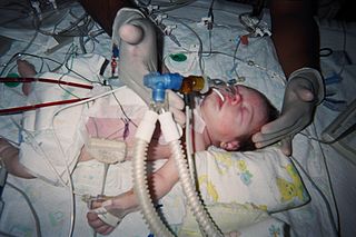
The Jatene procedure, arterial switch operation or arterial switch, is an open heart surgical procedure used to correct dextro-transposition of the great arteries (d-TGA); its development was pioneered by Canadian cardiac surgeon William Mustard and it was named for Brazilian cardiac surgeon Adib Jatene, who was the first to use it successfully. It was the first method of d-TGA repair to be attempted, but the last to be put into regular use because of technological limitations at the time of its conception. Use of the arterial switch is historically preceded by two atrial switch methods: the Senning and Mustard procedures.

Right ventricular hypertrophy (RVH) is a condition defined by an abnormal enlargement of the cardiac muscle surrounding the right ventricle. The right ventricle is one of the four chambers of the heart. It is located towards the lower-end of the heart and it receives blood from the right atrium and pumps blood into the lungs.
Tricuspid insufficiency (TI), more commonly called tricuspid regurgitation (TR), is a type of valvular heart disease in which the tricuspid valve of the heart, located between the right atrium and right ventricle, does not close completely when the right ventricle contracts (systole). TR allows the blood to flow backwards from the right ventricle to the right atrium, which increases the volume and pressure of the blood both in the right atrium and the right ventricle, which may increase central venous volume and pressure if the backward flow is sufficiently severe.
The following outline is provided as an overview of and topical guide to cardiology:
The LeCompte maneuver is a technique used in open heart surgery, primarily on infants and children. The maneuver entails cutting the main pulmonary artery and moving it anterior to the aorta before reattaching the pulmonary artery during the following reconstruction of the great vessels. It allows the surgeon to reconstruct the right ventricular outflow tract without needing to connect the proximal and distal sections with a graft. It also enables the surgeon to avoid compressing the coronary arteries and relieves compression of the bronchi in cases where the pulmonary artery is severely dilated or aneurysmal. If both pulmonary arteries are not mobilized adequately, they can become stretched, leading to pulmonic stenosis.
