Cytochrome f

Last updated
Apocytochr_F_C
PDB 1ci3 EBI.jpg
cytochrome f from the b6f complex of Phormidium laminosum
Identifiers
Symbol Apocytochr_F_C
Pfam PF01333
Pfam clan CL0105
InterPro IPR002325
PROSITE PDOC00169
SCOP 1ctm
SUPERFAMILY 1ctm
TCDB 3.D.3
OPM superfamily 92
OPM protein 3h1j

Cytochrome f is the largest subunit of cytochrome b6f complex (plastoquinolplastocyanin reductase; EC 1.10.99.1). In its structure and functions, the cytochrome b6f complex bears extensive analogy to the cytochrome bc1 complex of mitochondria and photosynthetic purple bacteria. Cytochrome f (cyt f) plays a role analogous to that of cytochrome c1, in spite of their different structures. [1]

Cytochrome b<sub>6</sub>f complex enzyme

The cytochrome b6f complex is an enzyme found in the thylakoid membrane in chloroplasts of plants, cyanobacteria, and green algae, that catalyzes the transfer of electrons from plastoquinol to plastocyanin. The reaction is analogous to the reaction catalyzed by cytochrome bc1 of the mitochondrial electron transport chain. During photosynthesis, the cytochrome b6f complex is one step along the chain that transfers electrons from Photosystem II to Photosystem I, and at the same time pumps protons into the thylakoid space that contribute to create an electrochemical (energy) gradient which is later used to synthesize ATP from ADP.

The Enzyme Commission number is a numerical classification scheme for enzymes, based on the chemical reactions they catalyze. As a system of enzyme nomenclature, every EC number is associated with a recommended name for the respective enzyme.

Contents

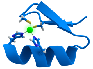
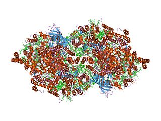
The 3D structure of Brassica rapa (Turnip) cyt f has been determined. [2] The lumen-side segment of cyt f includes two structural domains: a small one above a larger one that, in turn, is on top of the attachment to the membrane domain. The large domain consists of an anti-parallel beta-sandwich and a short haem-binding peptide, which form a three-layer structure. The small domain is inserted between beta-strands F and G of the large domain and is an all-beta domain. The haem nestles between two short helices at the N terminus of cyt f. Within the second helix is the sequence motif for the c-type cytochromes, CxxCH (residues 21-25), which is covalently attached to the haem through thioether bonds to Cys-21 and Cys-24. His-25 is the fifth haem iron ligand. The sixth haem iron ligand is the alpha-amino group of Tyr-1 in the first helix. [2] Cyt f has an internal network of water molecules that may function as a proton wire. [2] The water chain appears to be a conserved feature of cyt f.

Turnip root vegetable

The turnip or white turnip is a root vegetable commonly grown in temperate climates worldwide for its white, fleshy taproot. The word turnip is a compound of tur- as in turned/rounded on a lathe and neep, derived from Latin napus, the word for the plant. Small, tender varieties are grown for human consumption, while larger varieties are grown as feed for livestock. In the north of England, Scotland, Ireland, Cornwall and eastern Canada (Newfoundland), turnip often refers to rutabaga, a larger, yellow root vegetable in the same genus (Brassica) also known as swede.

Protein structure three-dimensional arrangement of atoms in an amino acid-chain molecule

Protein structure is the three-dimensional arrangement of atoms in an amino acid-chain molecule. Proteins are polymers – specifically polypeptides – formed from sequences of amino acids, the monomers of the polymer. A single amino acid monomer may also be called a residue indicating a repeating unit of a polymer. Proteins form by amino acids undergoing condensation reactions, in which the amino acids lose one water molecule per reaction in order to attach to one another with a peptide bond. By convention, a chain under 30 amino acids is often identified as a peptide, rather than a protein. To be able to perform their biological function, proteins fold into one or more specific spatial conformations driven by a number of non-covalent interactions such as hydrogen bonding, ionic interactions, Van der Waals forces, and hydrophobic packing. To understand the functions of proteins at a molecular level, it is often necessary to determine their three-dimensional structure. This is the topic of the scientific field of structural biology, which employs techniques such as X-ray crystallography, NMR spectroscopy, and dual polarisation interferometry to determine the structure of proteins.

Alpha helix type of secondary structure

The alpha helix (α-helix) is a common motif in the secondary structure of proteins and is a right hand-helix conformation in which every backbone N−H group donates a hydrogen bond to the backbone C=O group of the amino acid located three or four residues earlier along the protein sequence.

Related Research Articles

Transmembrane protein protein spanning across a biological membrane

A transmembrane protein (TP) is a type of integral membrane protein that spans the entirety of the cell membrane to which it is permanently attached. Many transmembrane proteins function as gateways to permit the transport of specific substances across the membrane. They frequently undergo significant conformational changes to move a substance through the membrane.

Zinc finger

A zinc finger is a small protein structural motif that is characterized by the coordination of one or more zinc ions (Zn2+) in order to stabilize the fold. Originally coined to describe the finger-like appearance of a hypothesized structure from Xenopus laevis transcription factor IIIA, the zinc finger name has now come to encompass a wide variety of differing protein structures. Xenopus laevis TFIIIA was originally demonstrated to contain zinc and require the metal for function in 1983, the first such reported zinc requirement for a gene regulatory protein. It often apears as a metal-binding domain in multi-domain proteins.

DNA-binding protein proteins that have DNA-binding domains and thus have a specific or general affinity for single- or double-stranded DNA

DNA-binding proteins are proteins that have DNA-binding domains and thus have a specific or general affinity for single- or double-stranded DNA. Sequence-specific DNA-binding proteins generally interact with the major groove of B-DNA, because it exposes more functional groups that identify a base pair. However, there are some known minor groove DNA-binding ligands such as netropsin, distamycin, Hoechst 33258, pentamidine, DAPI and others.

Selective estrogen receptor modulator A structurally diverse group of compounds distinguished from estrogens by their ability to bind and activate estrogen receptors but act as either an agonist or antagonist depending on the tissue type and hormonal milieu. They are classified as eithe

Selective estrogen receptor modulators (SERMs) are a class of drugs that act on the estrogen receptor (ER). A characteristic that distinguishes these substances from pure ER agonists and antagonists is that their action is different in various tissues, thereby granting the possibility to selectively inhibit or stimulate estrogen-like action in various tissues.

Ferredoxins are iron–sulfur proteins that mediate electron transfer in a range of metabolic reactions. The term "ferredoxin" was coined by D.C. Wharton of the DuPont Co. and applied to the "iron protein" first purified in 1962 by Mortenson, Valentine, and Carnahan from the anaerobic bacterium Clostridium pasteurianum.

Nitrophorins are hemoproteins found in the saliva of blood-feeding insects. Saliva of the blood-sucking bug Rhodnius prolixus contains at least seven homologous nitrophorins, designated NP1 to NP7 in order of their relative abundance in the glands. As isolated, nitrophorins contain nitric oxide (NO) ligated to the ferric heme iron (Fe3+). Histamine, which is released by the host in response to tissue damage, is another nitrophorin ligand. Nitrophorins transport NO to the feeding site. Dilution, binding of histamine and increase in pH (from pH ~5 in salivary gland to pH ~7.4 in the host tissue) facilitate the release of NO into the tissue where it induces vasodilatation.

Rieske protein

Rieske proteins are iron-sulfur protein (ISP) components of cytochrome bc1 complexes and cytochrome b6f complexes and responsible for electron transfer in some biological systems. John S. Rieske and co-workers first discovered and isolated the proteins in 1964. It is a unique [2Fe-2S] cluster in that one of the two Fe atoms is coordinated by two histidine residues rather than two cysteine residues. They have since been found in plants, animals, and bacteria with widely ranging electron reduction potentials from -150 to +400 mV.

Basic helix-loop-helix

A basic helix-loop-helix (bHLH) is a protein structural motif that characterizes one of the largest families of dimerizing transcription factors.

Amatoxin is the collective name of a subgroup of at least eight related toxic compounds found in several genera of poisonous mushrooms, most notably the death cap and several other members of the genus Amanita, as well as some Conocybe, Galerina and Lepiota mushroom species. Amatoxins are lethal in even small doses. Unlike many ingested poisons they are not destroyed by heat so cooking the poisonous mushrooms does not diminish their lethality which can be as little as eating half a mushroom.

Protein contact map

A protein contact map represents the distance between all possible amino acid residue pairs of a three-dimensional protein structure using a binary two-dimensional matrix. For two residues and , the element of the matrix is 1 if the two residues are closer than a predetermined threshold, and 0 otherwise. Various contact definitions have been proposed: The distance between the Cα-Cα atom with threshold 6-12 Å; distance between Cβ-Cβ atoms with threshold 6-12 Å ; and distance between the side-chain centers of mass.

The Cys-loop ligand-gated ion channel superfamily is composed of nicotinic acetylcholine, GABAA, GABAA, glycine, 5-HT3, and zinc-activated (ZAC) receptors that are composed of five protein subunits that form a pentameric arrangement around a central pore. There are usually 2 alpha subunits and 3 other beta, gamma, or delta subunits (some consist of 5 alpha subunits). The name "Cys-loop" is because this type of receptors possess a characteristic loop formed by 13 highly conserved amino acids between two cysteine (Cys) residues which form a disulfide bond, near the N-terminal extracellular domain.

LSm

In molecular biology, LSm proteins are a family of RNA-binding proteins found in virtually every cellular organism. LSm is a contraction of 'like Sm', because the first identified members of the LSm protein family were the Sm proteins. LSm proteins are defined by a characteristic three-dimensional structure and their assembly into rings of six or seven individual LSm protein molecules, and play a large number of various roles in mRNA processing and regulation.

Heme A chemical compound

Heme A is a heme, a coordination complex consisting of a macrocyclic ligand called a porphyrin, chelating an iron atom. Heme A is a biomolecule and is produced naturally by many organisms. Heme A, often appears a dichroic green/red when in solution, is a structural relative of heme B, a component of hemoglobin, the red pigment in blood.

In enzymology, nitrile hydratases are mononuclear iron or non-corrinoid cobalt enzymes that catalyse the hydration of diverse nitriles to their corresponding amides

MMP1 protein-coding gene in the species Homo sapiens

Matrix metalloproteinase-1 (MMP-1) also known as interstitial collagenase and fibroblast collagenase is an enzyme that in humans is encoded by the MMP1 gene. The gene is part of a cluster of MMP genes which localize to chromosome 11q22.3. MMP-1 was the first vertebrate collagenase both purified to homogeneity as a protein, and cloned as a cDNA.

Cytochrome b559

Cytochrome b559 is an important component of Photosystem II.

Cytochrome c family

Cytochromes c are proteins containing one or more heme groups that are covalently attached to the peptide backbone via one or two thioether bonds. These bonds are in most cases part of a specific Cys-X-X-Cys-His (CXXCH) binding motif, where X denotes a miscellaneous amino acid. Two thioether bonds of cysteine residues bind to the vinyl sidechains of heme, and the histidine residue coordinates one axial binding site of the heme iron. Less common binding motifs can include a single thioether linkage, a lysine or a methionine instead of the axial histidine or a CXnCH binding motif with n>2. The second axial site of the iron can be coordinated by amino acids of the protein, substrate molecules or water. Cytochromes c possess a wide range of properties and function as electron transfer proteins or catalyse chemical reactions involving redox processes. A prominent member of this family is mitochondrial cytochrome c.

Azurin

Azurin is a small, periplasmic, bacterial blue copper protein found in Pseudomonas, Bordetella, or Alcaligenes bacteria. Azurin moderates single-electron transfer between enzymes associated with the cytochrome chain by undergoing oxidation-reduction between Cu(I) and Cu(II). Each monomer of an azurin tetramer has a molecular weight of approximately 14kDa, contains a single copper atom, is intensively blue, and has a fluorescence emission band centered at 308 nm.

References

  1. Prince RC, George GN (June 1995). "Cytochrome f revealed". Trends Biochem. Sci. 20 (6): 217–8. doi:10.1016/S0968-0004(00)89018-0. PMID   7631417.
  2. 1 2 3 Martinez SE, Huang D, Ponomarev M, Cramer WA, Smith JL (June 1996). "The heme redox center of chloroplast cytochrome f is linked to a buried five-water chain". Protein Sci. 5 (6): 1081–92. doi:10.1002/pro.5560050610. PMC   2143431 Lock-green.svg. PMID   8762139.

Further reading

Digital object identifier Character string used as a permanent identifier for a digital object, in a format controlled by the International DOI Foundation

In computing, a Digital Object Identifier or DOI is a persistent identifier or handle used to uniquely identify objects, standardized by the International Organization for Standardization (ISO). An implementation of the Handle System, DOIs are in wide use mainly to identify academic, professional, and government information, such as journal articles, research reports and data sets, and official publications though they also have been used to identify other types of information resources, such as commercial videos.

Medical Subject Headings (MeSH) is a comprehensive controlled vocabulary for the purpose of indexing journal articles and books in the life sciences; it serves as a thesaurus that facilitates searching. Created and updated by the United States National Library of Medicine (NLM), it is used by the MEDLINE/PubMed article database and by NLM's catalog of book holdings. MeSH is also used by ClinicalTrials.gov registry to classify which diseases are studied by trials registered in ClinicalTrials.

This article incorporates text from the public domain Pfam and InterPro IPR002325

The public domain consists of all the creative works to which no exclusive intellectual property rights apply. Those rights may have expired, been forfeited, expressly waived, or may be inapplicable.

Pfam Database of protein families

Pfam is a database of protein families that includes their annotations and multiple sequence alignments generated using hidden Markov models. The most recent version, Pfam 32.0, was released in September 2018 and contains 17,929 families.

InterPro is a database of protein families, domains and functional sites in which identifiable features found in known proteins can be applied to new protein sequences in order to functionally characterise them.