A ligament is the fibrous connective tissue that connects bones to other bones. It is also known as articular ligament, articular larua, fibrous ligament, or true ligament. Other ligaments in the body include the:

A tendon or sinew is a tough band of dense fibrous connective tissue that connects muscle to bone. It sends the mechanical forces of muscle contraction to the skeletal system, while withstanding tension.

In humans and other primates, the knee joins the thigh with the leg and consists of two joints: one between the femur and tibia, and one between the femur and patella. It is the largest joint in the human body. The knee is a modified hinge joint, which permits flexion and extension as well as slight internal and external rotation. The knee is vulnerable to injury and to the development of osteoarthritis.
Soft tissue is all the tissue in the body that is not hardened by the processes of ossification or calcification such as bones and teeth. Soft tissue connects, surrounds or supports internal organs and bones, and includes muscle, tendons, ligaments, fat, fibrous tissue, lymph and blood vessels, fasciae, and synovial membranes.

The ankle, or the talocrural region, or the jumping bone (informal) is the area where the foot and the leg meet. The ankle includes three joints: the ankle joint proper or talocrural joint, the subtalar joint, and the inferior tibiofibular joint. The movements produced at this joint are dorsiflexion and plantarflexion of the foot. In common usage, the term ankle refers exclusively to the ankle region. In medical terminology, "ankle" can refer broadly to the region or specifically to the talocrural joint.

The anterior cruciate ligament (ACL) is one of a pair of cruciate ligaments in the human knee. The two ligaments are also called "cruciform" ligaments, as they are arranged in a crossed formation. In the quadruped stifle joint, based on its anatomical position, it is also referred to as the cranial cruciate ligament. The term cruciate translates to cross. This name is fitting because the ACL crosses the posterior cruciate ligament to form an “X”. It is composed of strong, fibrous material and assists in controlling excessive motion. This is done by limiting mobility of the joint. The anterior cruciate ligament is one of the four main ligaments of the knee, providing 85% of the restraining force to anterior tibial displacement at 30 and 90° of knee flexion. The ACL is the most injured ligament of the four located in the knee.

A trabecula is a small, often microscopic, tissue element in the form of a small beam, strut or rod that supports or anchors a framework of parts within a body or organ. A trabecula generally has a mechanical function, and is usually composed of dense collagenous tissue. It can be composed of other material such as muscle and bone. In the heart, muscles form trabeculae carneae and septomarginal trabeculae. Cancellous bone is formed from groupings of trabeculated bone tissue.

An implant is a medical device manufactured to replace a missing biological structure, support a damaged biological structure, or enhance an existing biological structure. For example, an implant may be a rod, used to strengthen weak bones. Medical implants are human-made devices, in contrast to a transplant, which is a transplanted biomedical tissue. The surface of implants that contact the body might be made of a biomedical material such as titanium, silicone, or apatite depending on what is the most functional. In some cases implants contain electronics, e.g. artificial pacemaker and cochlear implants. Some implants are bioactive, such as subcutaneous drug delivery devices in the form of implantable pills or drug-eluting stents.

Anterior cruciate ligament reconstruction is a surgical tissue graft replacement of the anterior cruciate ligament, located in the knee, to restore its function after an injury. The torn ligament can either be removed from the knee, or preserved before reconstruction through an arthroscopic procedure. ACL repair is also a surgical option. This involves repairing the ACL by re-attaching it, instead of performing a reconstruction. Theoretical advantages of repair include faster recovery and a lack of donor site morbidity, but randomised controlled trials and long-term data regarding re-rupture rates using contemporary surgical techniques are lacking.

Distraction osteogenesis (DO), also called callus distraction, callotasis and osteodistraction, is a process used in orthopedic surgery, podiatric surgery, and oral and maxillofacial surgery to repair skeletal deformities and in reconstructive surgery. The procedure involves cutting and slowly separating bone, allowing the bone healing process to fill in the gap.
Hand surgery deals with both surgical and non-surgical treatment of conditions and problems that may take place in the hand or upper extremity including injury and infection. Hand surgery may be practiced by post graduates of orthopedic surgery and plastic surgery.

The flexor retinaculum is a fibrous band on the palmar side of the hand near the wrist. It arches over the carpal bones of the hands, covering them and forming the carpal tunnel.
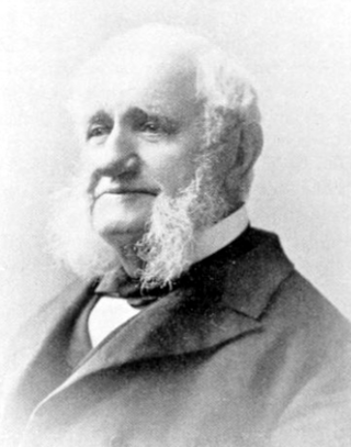
Henry Gassett Davis was an American orthopedic surgeon.
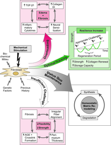
The Mechanostat is a term describing the way in which mechanical loading influences bone structure by changing the mass and architecture to provide a structure that resists habitual loads with an economical amount of material. As changes in the skeleton are accomplished by the processes of formation and resorption, the mechanostat models the effect of influences on the skeleton by those processes, through their effector cells, osteocytes, osteoblasts, and osteoclasts. The term was invented by Harold Frost: an orthopaedic surgeon and researcher described extensively in articles referring to Frost and Webster Jee's Utah Paradigm of Skeletal Physiology in the 1960s. The Mechanostat is often defined as a practical description of Wolff's law described by Julius Wolff (1836–1902), but this is not completely accurate. Wolff wrote his treatises on bone after images of bone sections were described by Culmann and von Meyer, who suggested that the arrangement of the struts (trabeculae) at the ends of the bones were aligned with the stresses experienced by the bone. It has since been established that the static methods used for those calculations of lines of stress were inappropriate for work on what were, in effect, curved beams, a finding described by Lance Lanyon, a leading researcher in the area as "a triumph of a good idea over mathematics." While Wolff pulled together the work of Culmann and von Meyer, it was the French scientist Roux, who first used the term "functional adaptation" to describe the way that the skeleton optimized itself for its function, though Wolff is credited by many for that.
Histology is the study of the minute structure, composition, and function of tissues. Mature human vocal cords are composed of layered structures which are quite different at the histological level.
Harold M. Frost was an American orthopedist and surgeon considered to be one of the most important researchers and theorists in the field of bone biology and bone medicine of his time. He published nearly 500 peer-reviewed scientific and clinical articles and 16 books. According to the Science Citation Index, he is one of the most cited investigators in skeletal research.

Injuries to the arm, forearm or wrist area can lead to various nerve disorders. One such disorder is median nerve palsy. The median nerve controls the majority of the muscles in the forearm. It controls abduction of the thumb, flexion of hand at wrist, flexion of digital phalanx of the fingers, is the sensory nerve for the first three fingers, etc. Because of this major role of the median nerve, it is also called the eye of the hand. If the median nerve is damaged, the ability to abduct and oppose the thumb may be lost due to paralysis of the thenar muscles. Various other symptoms can occur which may be repaired through surgery and tendon transfers. Tendon transfers have been very successful in restoring motor function and improving functional outcomes in patients with median nerve palsy.
Eccentric training is a type of strength training that involves using the target muscles to control weight as it moves in a downward motion. This type of training can help build muscle, improve athletic performance, and reduce the risk of injury. An eccentric contraction is the motion of an active muscle while it is lengthening under load. Eccentric training is repetitively doing eccentric muscle contractions. For example, in a biceps curl the action of lowering the dumbbell back down from the lift is the eccentric phase of that exercise – as long as the dumbbell is lowered slowly rather than letting it drop.
Materials that are used for biomedical or clinical applications are known as biomaterials. The following article deals with fifth generation biomaterials that are used for bone structure replacement. For any material to be classified for biomedical applications, three requirements must be met. The first requirement is that the material must be biocompatible; it means that the organism should not treat it as a foreign object. Secondly, the material should be biodegradable ; the material should harmlessly degrade or dissolve in the body of the organism to allow it to resume natural functioning. Thirdly, the material should be mechanically sound; for the replacement of load-bearing structures, the material should possess equivalent or greater mechanical stability to ensure high reliability of the graft.

Orthopedic surgery is the branch of surgery concerned with conditions involving the musculoskeletal system. Orthopedic surgeons use both surgical and nonsurgical means to treat musculoskeletal injuries, sports injuries, degenerative diseases, infections, bone tumours, and congenital limb deformities. Trauma surgery and traumatology is a sub-specialty dealing with the operative management of fractures, major trauma and the multiply-injured patient.