Extranodal NK/T-cell lymphoma, nasal type

Last updated
Extranodal NK-T-cell lymphoma
Other namesAngiocentric lymphoma, Nasal-type NK lymphoma, NK/T-cell lymphoma, Polymorphic/malignant midline reticulosis
Histopathology of extranodal NK-T cell lymphoma, nasal type.png
Histopathology of extranodal NK-T cell lymphoma, nasal type (H&E stain). [1] These lymphoma cells are typically monotonous, with folded nuclei, indistinct nucleoli and moderate amount of cytoplasm. [2]
Specialty Hematology and Oncology
Causes Epstein–Barr virus

Extranodal NK/T-cell lymphoma, nasal type (ENKTCL-NT) (also termed angiocentric lymphoma, nasal-type NK lymphoma, NK/T-cell lymphoma, polymorphic/malignant midline reticulosis, [3] and lethal midline granuloma [4] ) is a rare type of lymphoma that commonly involves midline areas of the nasal cavity, oral cavity, and/or pharynx [5] At these sites, the disease often takes the form of massive, necrotic, and extremely disfiguring lesions. However, ENKTCL-NT can also involve the eye, larynx, lung, gastrointestinal tract, skin, and various other tissues. [6] ENKTCL-NT mainly affects adults; it is relatively common in Asia and to lesser extents Mexico, Central America, and South America but is rare in Europe and North America. [7] In Korea, ENKTCL-NT often involves the skin and is reported to be the most common form of cutaneous lymphoma after mycosis fungoides. [8]

Contents

ENKTCL-NT is classified as an Epstein-Barr virus-associated lymphoproliferative disease. [9] It is due to the malignant transformation of either one of two types of lymphocytes, NK cells or a T cell variant termed cytotoxic T cells, that are infected with the Epstein–Barr virus (EBV). Typically, the viral infection, which affects >90% of the world population, occurs years before evidence of ENKTCL-NT, is carried in cells in a latent, asymptomatic form, and for unclear reasons becomes active in causing the disease. Following the virus's activation, the infected cells acquire numerous genetic abnormalities which may play an important role in the development and/or progression of ENKTCL-NT. [10]

Epstein-Barr virus-positive nodal NK/T cell lymphoma (EBV+ nodal NKTCL) was considered to be one form of ENKTCL-NT since it is a malignancy of EBV-infected NK or T cells. However, EBV+ nodal NKTCL is manifested primarily by its involvement in lymph nodes; it also has clinical, pathological, pathophysiological, and genetic features that differ significantly from those of ENKTCL-NT. The World Health Organization, 2016, therefore reclassified this lymphoma as a variant of a disease to which its features more closely resemble, peripheral T-cell lymphoma not otherwise specified. [9]

While a rare disease, particularly in North America, ENKTCL-NT has recently gained much interest. Clinical studies have found that newer chemotherapeutic regimens greatly improved survival in cases of early disease. While, survival in advanced cases is still extremely poor, generally being only a few months, [11] recent studies suggest that new regimens directed at gene mutation and expression abnormalities may improve survival. [11] [12] Further study of these new regimens has important implications not only for ENKTCL-NT but also for other NK/T cell malignancies.

Presentation

Extranodal NK/T-cell lymphoma, nasal type occurs primarily in Asians and South Americans; it is comparatively uncommon in other areas. Affected patients (median age 50–60 years old; males predominate) most often (~80% of cases) present with nasal bleeding, upper airway obstruction, perforation of the hard palate, and/or disfiguring, necrotic lesions of the nasal cavity, nasopharynx (including Waldeyer's tonsillar ring), paranasal sinuses, palate, [13] and/or eye socket. [14] Less often, patients present with these findings plus signs and symptoms involving extranasal sites such as the skin, upper respiratory tract, gastrointestinal tract, uterus, testes, and/or elsewhere. [15] Rarely, individuals present with evidence of involvement in the later sites without those involving the head/neck area. On further study these individuals may be found to have occult involvement in the head and neck or to develop such involvement. However, ~10 present of patients present with only skin lesions such as a solitary or multiple subcutaneous masses (which may be ulcerated) in the arms or legs [7] while another ~10% present with masses in the lower gastrointestinal tract (which may be accompanied by bleeding or obstruction), salivary glands, testes, muscles, or other organs without evidence of lesions in the head/neck areas. In these cases, there is relatively little involvement of lymph nodes except as a result of direct invasion from non-nodal sites. [14] Thirty-five to forty-five percent of patients present with a history of malaise, fever, night sweats, and/or weight loss. Most (70–75%) patients are diagnosed with early stage I or II disease while the rest have far more serious stage III or IV disease. Rarely, patients with stage III or IV disease have evidence of a life-threatening complication, hemophagocytic lymphohistiocytosis. [16] Also in rare cases, patients evidence a widespread disease that includes malignant cell infiltrations in the liver, spleen, lymph nodes, bone marrow, and/or blood. These case are, or may soon progress to, a related but potentially fatal disease, aggressive NK-cell leukemia. [14]

About 45% of patients present with elevated levels of serum lactate dehydrogenase; elevation in this serum enzyme is a poor prognostic indicator. [16] Patients with ENKTCL-NT also have elevated levels of plasma EBV DNA. Quantification of these levels at diagnosis correlates with the extent of their tumor load while serially assaying these levels during treatment gives evidence of the tumors response to treatment and residual disease. [14] Rarely, patients show laboratory evidence of hemophagocytic lymphohistiocytosis such as: decreased circulating red blood cells, leukocytes, and/or platelets; increased serum levels of liver-derived enzymes, ferritin, and/or triglycerides; decreased serum levels of fibrinogen; and/or hemophagocytosis, i.e. engulfment of blood cells by tissue histiocytes in the liver, spleen, bone morrow, and/or other tissues. [17] or aggressive NK-cell leukemia (e.g. decreased circulating red blood cells, leukocytes, and/or platelets, increased circulating large, granule-containing malignant NK cells, and infiltrations of the latter cells in bone marrow and other tissues). [14]

Pathogenesis

Disease location

ENKTCL-NT is a disease of malignant NK or, very much less often, cytotoxic T cells. Unlike most other lymphomas, which typically develop in and involve lymphatic tissues (particularly lymph nodes and spleen), ENKTCL-NT commonly develops in non-lymphatic tissues. This difference in distribution probably reflects the occupancy of the T cell and B cell precursors to most lymphomas in lymphatic tissues versus the frequent occupancy of the NK and cytotoxic T cells precursors to ENTCL-NT in non-lymphatic tissues. [14]

Genes

ENKTCL-NT is thought to arise from the expression of EBV genes in the infected NK or cytotoxic T cells and the ability of these genes to cause the cells they infect to overexpress and acquire mutations in key genes that regulate cell growth, immortalization, invasiveness, and ability to evade normal control mechanisms, particularly immune surveillance. Since these gene-related abnormalities are multiple and vary between patients, it is not clear which contribute to the development and/or progression of ENKTCL-NT. Clinical studies are therefore examining targeted therapy tactics to determine which gene abnormalities contribute to, and which drugs targeting these abnormalities are useful in treating, ENKTCL-NT. [18]

EBV genes

Infected cells carry ~10 cytosolic EBV episomes, i.e. gene-bearing viral DNA particles. In the premalignant precursor NK and cytotoxic T cells of ENKTCL-NT, these episomes express only some of their many latency genes, i.e. genes which promote the virus's latency rather than lytic phase of infectivity. EBV has three different latency phases, I, II, and III, in each of which different sets of latency genes are expressed to establish different controls on the cells which they infect. In the premalignant cells of ENKTCL-NT, EBV express latency II genes such as EBNA-1, LMP-1, LMP-2A, and LMP-2B protein-producing genes; EBER-1 and EBER-2 non-coding RNA-producing genes (see EBV non-coding RNAs); and certain BART microRNA-producing genes (see EBV microRNAs). LMP1 protein induces infected cells to overexpress genes that produce cMyc, [12] NF-κB, and BCL2 proteins which when overexpressed block these cells' apoptosis (i.e. cell death) response to injury or the host's immune system and promote their survival and proliferation; [9] LMP2A and LMP2B proteins induce infected cells to overexpress the genes that make AKT and B cell receptor proteins and to activate the NF-κ pathway [11] which when over-activated blocks these cells' apoptosis response and promotes their survival and proliferation; EBER 1 and 2 non-coding RNAs induce infected cells to overexpress the gene that makes the interleukin 10 protein which when overexpressed may promote its parent cells to proliferate and avoid the host's immune system; [9] and certain BART microRNAs may help infected cells avoid attack by the hosts immune system [10] and modify their notch signaling pathway thereby promoting their proliferation. [19] In consequence, the EBV latency II genes force infected cells to become immortal, proliferate excessively, invade tissues, and avoid attack by the hosts' immune system. Due at least in part to these imposed factors, the infected cells may acquire other genetic abnormalities that further promote their malignant behavior. [14] [18]

Infected cell genes

The rapidly proliferating and immortalized EBV-infected NK/T cells accumulate numerous changes in the expression or activity of their genes by acquisition of chromosome deletions, gene mutations, and changes in gene expression.[ citation needed ]

Chromosomes

Deletions in the long (i.e. "q") arm at position 21–25 (notated as 6q21-25) from one of the two chromosome 6's was an early finding in occasional cases of ENKTCL-NT. This deletion removes one of the two copies of several tumor suppressor genes (i.e. genes that protect cells from becoming malignant) such as HACE1, PRDM1, FOXO3, and PTPRK. Subsequent studies showed that the disease is also occasionally associated with losses in the short arm of chromosome 8 at position 11.23 (8p11.23) which for unclear reasons are associated with a poor prognosis, and occasional losses at position 11l.2 in the q arm of chromosome 14 (14q11.2) which correlates with the ENKTCL-NT malignancy being of cytotoxic T cell origin. [12] EBV-infected NK and T cells may also occasionally develop chromosome segregation errors during mitosis and consequently divide into daughter cells which possess too few or too many chromosomes and thereby exhibit chaotic losses or increases in the expression of the genes located on these chromosomes. [12]

Mutated genes

Second generation sequencing methods have uncovered numerous genes which are mutated in the malignant cells of ENKTCL-NT. These mutated genes and their product proteins have the following a) mutation rates in ENKTCL-NT; b) normal functions; c) gains or losses of activity; d) pro-malignant effects on EN/T cells and e) clinical impacts on the course of ENKTCL-NT:

GeneProductMutation rateFunctionMutation typeInfluence on cell functionClinical impact on ENKTCL-NT
TP53 p53 13–62% tumor suppressor gain promotes cell proliferation, survival, migration, invasiveness, and metastasiscorrelates with advanced stage and poor prognosis [18]
DDX3X DDX3X12–20%tumor suppressor loss lost ability to inhibit proliferationcorrelates with advanced stage and poor prognosis [18]
STAT3 STAT38–26% JAK-STAT signaling pathway componentgainpromotes cell proliferation and survivalunknown [18]
STAT5B STAT5B~2–6%JAK-STAT signaling pathway componentgainpromotes cell proliferation and survivalunknown [18]
JAK3 JAK30–35%JAK-STAT signaling pathway componentgainpromotes cell proliferation and survivalunknown [18]
MGA MAX dimerization protein~8%tumor suppressorlossunknownunknown [18]
MLL2 MLL27–80% histone methyltransferase, tumor suppressorlossreduces cellular differentiation, possibly promoting cell proliferation and survivalunknown [18]
BCOR BCL-6 corepressor21–32%inhibits BCL-5, may regulate apoptosis lossmay increase cell survivalunknown [18]
ECSIT ECSIT19%element in TGF-β/BMP/signaling pathwaysgainactivates NF-κB to promote cell survival and prolifarationcorrelates with advanced stage and poor prognosis [12]
ARID1A ARID1A4–8%a SWI/SNF protein that regulates expression of other proteinslossunknownunknown [18]
MCL1 MCL1most casesa SWI/SNF protein that regulates expression of other proteinslossunknownunknown [18]

In the above table, ARID1A protein stands for AT-rich interactive domain-containing protein 1A and ECSIT protein stands for evolutionarily conserved signaling intermediate in Toll pathway; mitochondrial. A gain of function mutation in the ECSIT gene that changes the amino acid at the 140 position in its product protein from valine to alanine (i.e. V140A) is associated with a high incidence of ENKTCL-NT being complicated by the development of life-threatening Hemophagocytic lymphohistiocytosis and thereby a relatively high mortality rate. [5] Numerous other genes are rarely (i.e. ≤2% of cases) mutated in ENKTCL-NT. These include JAK1, MLL3, ARID1A, EP300, ASXL3, MSN, FAT4, NARS, IL6R, MGAM, CHPF2, (see [20] ) and MIR17HG ((see [21] ). [18]

Overexpressed genes

ENKTCL-NT malignant cells overexpress NF-κB, a cellular signaling transcription factor that when up-regulated promotes these cells' proliferation and survival. They also overexpress: 1) aurora kinase A, a serine/threonine-specific protein kinase that when up-regulated in the cancer setting promotes these cells' invasiveness and to develop chromosome segregation errors during mitosis that result in daughter cells having too few or too many chromosome; 2) members of the inhibitor of apoptosis family of proteins including survivin, [12] Bcl-xL, and MCL1 [22] which when up-regulated suppress programmed cell death to promote these cell's survival and resistance to attack by the host immune system; [23] [24] 3) multidrug resistance protein 1, a surface membrane protein that when up-regulated causes these cells to greatly increases the export of anthracyclines such as Adriamycin and Daunomycin thereby rendering them resistant to this class of chemotherapy drugs; 4) EZH2, a histone methyltransferase that when up-regulated indirectly promotes these cells' growth; 5) runt-related transcription factor 3 that when up-regulated indirectly promotes the survival and proliferation of these cells; [12] and 6) programmed death-ligand 1 (PD-L1), that when up-regulated increases the ability of these cells to avoid attack by the host's immune system. [25]

Signaling pathways

In consequence of, or addition to the cited genetic abnormalities, ENKTCL-NT malignant cells have overly active the; JAK-STAT signaling pathway that in the cancer setting promotes cell proliferation, survival, and other pro-malignant behaviors; [14] platelet-derived growth factor signaling pathway that in the cancer setting promotes cell survival and proliferation; Notch signaling pathway that in the cancer setting promotes cellular differentiation and proliferation; and NF-κB signaling that in the cancer setting promotes cell survival and proliferation. Studies suggest that that overactive VEGF receptor and Protein kinase B signaling pathways may also play a role in the pathogenesis of ENKTCL-NT. [12] )

Epigenetic abnormalities

Studies on cultured malignant NK cells and/or patient tissue specimens find that numerous genes are hypermethylated at their promoter sites and therefore are silenced, i.e. make less or none of their protein products. This silencing has been detected in numerous proteins expressed by cultured NK cells (e.g. BCL2L11, DAPK1, PTPN6, TET2, SOCS6, PRDM1, AIM1, HACE, p15, p16, p73, MLH1, RARB, and ASNS ) and the MIR146A gene for its miR-146a microRNA product. Studies conducted on the expression of microRNAs in cultured malignant NK cells have also revealed that many are either over- or under-expressed compared to non-malignant cultured NK cells. This dysregulation of these microRNA genes may reflect the action of products expressed by certain EBV genes and/or the overexpression of the infected cells' MYC gene. In all cases, the epigenetic dysregulation of these genes requires further study to determine its significance for the development and progression of ENKTCL-NT. [12]

Histology

On microscopic examination, involved tissues show commonly show areas of necrosis and cellular infiltrates that are centered around and often injure or destroy small blood vessels. The infiltrates contain large granule-containing lymphocytes that express cell surface CD2, cytoplasmic CD3ε, and cell surface CD56 as well the cytoplasmic intracellular proteins, perforin, granzyme B, and T cell intracellular antigen-1 (TIA-1). These cells exhibit evidence of EBV infection as determined by in situ hybridization assays to detect one of the virus's latent products, typically EBER-1/2 micoRNAs. [14] Identification of the genetic abnormalities cited above in the cells may be of help in establishing the diagnoses and be of use for selecting novel therapeutic approaches to individual patients. [12] Non-malignant inflammatory white blood cells, including eosinophils, are also commonly found in these infiltrates. [14]

Diagnosis

The diagnosis of ENKTCL-NT depends on histological findings that biopsied tissue infiltrates contain lymphocytes that express CD3ε, cytotoxic molecules (granzyme B, perforin, TIA1), and EBV. [12] Bone marrow examination is recommended to determine its involvement in this disorder. Whole body PET-CT scans are recommended to determine the extent of disease at presentation as well as to follow the effects of therapeutic interventions. The tumor load of each individual's disease as well as response to therapies has also been estimated by assaying plasma levels of EBV DNA. [14] ENKTCL-NT can be mimicked by two benign diseases which involve the excessive proliferation of non-malignant NK cells in the GI tract viz., Natural killer cell enteropathy, a disease wherein NK cell infiltrative lesions occur in the intestine, colon, stomach, and/or esophagus, and lymphomatoid gastropathy, a disease wherein these cells infiltrative lesions are limited to the stomach. [26] Another lymphoproliferative disorder of the GI tract, indolent T cell lymphoproliferative disorder of the gastrointestinal tract may also mimic ENKTCL-NT. This chronic disorder involves the proliferation of CD+4, CD8+, CD4-/CD8-, or CD4+/CD8+ T cells in the mucosal layers of the GI tract to give a variety of GI tract symptoms. While generally a persistent and benign disorder, a small but significant percentage of cases have progressed to aggressive lymphomas. [27] [28]

Course of ENKTCL-NT

The course of the untreated disease is heavily dependent on its clinical stage at diagnosis. Patients presenting with highly localized stage I nasal disease usually have nasal but no other symptoms; these individuals commonly show no progression of their disease over long periods of time. Other patients with limited (i.e. stage I or II) disease involving other sites in the head area are more likely to have a relatively slow progression of their disease while patients with stage III or IV disease have a more rapidly progressive disease with a poor prognosis. Patients presenting with ENKTCL-NT that does not involve the head area typically have a disseminated and aggressively progressive disease with a very poor prognosis. [13] Patients with stage I or II localized disease that have been treated with the recently defined chemotherapeutic protocols have 5 year survivals of ~70–89% [11] while those with advanced stage III or IV disseminated disease treated with these protocols have 5 year survivals of 50%. [25] Patients who relapse or are resistant to these protocols have had overall survivals of just a few months. [11]

Three prognostic models, NK-PI, PINK (i.e. prognostic index of natural killer lymphomas), and PINK-E) for ENKTCL-NT have evolved over the past 12 years. The latest model, PINK-E, which applies to patients treated with recently defined regimens, lists 5 risk factors (age >60, state III or IV disease, no nasal involvement, distant lymph node involvement, and detectable blood levels of EBV DNA) to define patients as low, intermediate, and high risk based on their having 0–1, 2, or 3–5 risk factors, respectively. Overall 3 year survival in these 3 respective groups were 81, 55, and 28%. [25] Patients, particularly those in the advanced poor risk groups may develop hemophagocytic lymphohistiocytosis or have their disease progress to aggressive NK-cell leukemia. Both conditions are life-threatening and far less responsive to treatment. [14]

Treatment

The treatment of ENKTCL- NT employs chemotherapy plus, where indicated, radiotherapy. Early chemotherapies relied on CHOP (i.e. cyclophosphamide, an anthracycline (primarily adriamycin), vincristine, and prednisolone) or chop-like regimens. These were only marginally successful because, as it was later discovered, the malignant NK cells in ENKTCL-NT over-express multidrug resistance protein 1. This protein exports various molecules, including anthracyclines and vincristine, from its parent cells and thereby renders these cells resistant to adriamycin [14] and vincristine [29] and therefore to CHOP and CHOP-like regimens. [14] Subsequent studies discovered that L-asparaginase [14] (NK cells do not express L-asaraginase [11] ) and, to a lesser extent, platinum-based antineoplastic drugs (e.g.carboplatin) [16] were active on theses cells. Accordingly, several chemotherapeutic regimens were tested and found to give much better results than previous regimens. However, these regimens have bot undergone phase 3 clinical trials that examine their effectiveness relative to other regimens. The following regimens are recommended by many studies and the European Society for Medical Oncology Clinical Practice guidelines [16] or National Comprehensive Cancer Network: [30]

Experimental drugs

There are numerous regimens that use non-chemotherapeutic agents to target specific elements known or thought to be involved in the survival of the malignant cells in a significant percentage of ENKTCL-NT cases. The targets should be determined as overexpressed or present in the malignant tissues of each case before treatment. [16] The targets, therapeutic agents, and some phase 1 clinical trials (testing for appropriate dosages, safety, and side effects) and/or phase 2 clinical trials (testing for efficacy and safety) include:

Small molecule inhibitors of JAK3 (e.g. tofacitinib), JAK1/JAK2 (e.g. AZD1480), STAT3 (e.g. WP1066), and DDX3X (e.g. RK-33) are being study in pre-clinical in vitro experiments as potential inhibitors of malignant NK/T cell proliferation and survival. They are in further studies to test them as potential therapeutic agents in ENKTCL-NT patients that have activating mutations or overexpression of the cited targets. [18]

See also

Related Research Articles

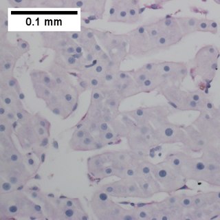
Post-transplant lymphoproliferative disorder (PTLD) is the name given to a B cell proliferation due to therapeutic immunosuppression after organ transplantation. These patients may develop infectious mononucleosis-like lesions or polyclonal polymorphic B-cell hyperplasia. Some of these B cells may undergo mutations which will render them malignant, giving rise to a lymphoma.

Lymphoid leukemias are a group of leukemias affecting circulating lymphocytes, a type of white blood cell. The lymphocytic leukemias are closely related to lymphomas of the lymphocytes, to the point that some of them are unitary disease entities that can be called by either name. Such diseases are all lymphoproliferative disorders. Most lymphoid leukemias involve a particular subtype of lymphocytes, the B cells.

<span class="mw-page-title-main">Primary effusion lymphoma</span> Medical condition

Primary effusion lymphoma (PEL) is classified as a diffuse large B cell lymphoma. It is a rare malignancy of plasmablastic cells that occurs in individuals that are infected with the Kaposi's sarcoma-associated herpesvirus. Plasmablasts are immature plasma cells, i.e. lymphocytes of the B-cell type that have differentiated into plasmablasts but because of their malignant nature do not differentiate into mature plasma cells but rather proliferate excessively and thereby cause life-threatening disease. In PEL, the proliferating plasmablastoid cells commonly accumulate within body cavities to produce effusions, primarily in the pleural, pericardial, or peritoneal cavities, without forming a contiguous tumor mass. In rare cases of these cavitary forms of PEL, the effusions develop in joints, the epidural space surrounding the brain and spinal cord, and underneath the capsule which forms around breast implants. Less frequently, individuals present with extracavitary primary effusion lymphomas, i.e., solid tumor masses not accompanied by effusions. The extracavitary tumors may develop in lymph nodes, bone, bone marrow, the gastrointestinal tract, skin, spleen, liver, lungs, central nervous system, testes, paranasal sinuses, muscle, and, rarely, inside the vasculature and sinuses of lymph nodes. As their disease progresses, however, individuals with the classical effusion-form of PEL may develop extracavitary tumors and individuals with extracavitary PEL may develop cavitary effusions.

<span class="mw-page-title-main">T-cell lymphoma</span> Medical condition

T-cell lymphoma is a rare form of cancerous lymphoma affecting T-cells. Lymphoma arises mainly from the uncontrolled proliferation of T-cells and can become cancerous.

<span class="mw-page-title-main">Intravascular lymphomas</span> Medical condition

Intravascular lymphomas (IVL) are rare cancers in which malignant lymphocytes proliferate and accumulate within blood vessels. Almost all other types of lymphoma involve the proliferation and accumulation of malignant lymphocytes in lymph nodes, other parts of the lymphatic system, and various non-lymphatic organs but not in blood vessels.

<span class="mw-page-title-main">Diffuse large B-cell lymphoma</span> Type of blood cancer

Diffuse large B-cell lymphoma (DLBCL) is a cancer of B cells, a type of lymphocyte that is responsible for producing antibodies. It is the most common form of non-Hodgkin lymphoma among adults, with an annual incidence of 7–8 cases per 100,000 people per year in the US and UK. This cancer occurs primarily in older individuals, with a median age of diagnosis at ~70 years, although it can occur in young adults and, in rare cases, children. DLBCL can arise in virtually any part of the body and, depending on various factors, is often a very aggressive malignancy. The first sign of this illness is typically the observation of a rapidly growing mass or tissue infiltration that is sometimes associated with systemic B symptoms, e.g. fever, weight loss, and night sweats.

<span class="mw-page-title-main">Aggressive NK-cell leukemia</span> Medical condition

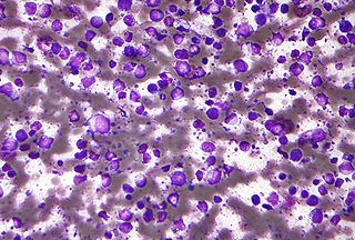
Aggressive NK-cell leukemia is a disease with an aggressive, systemic proliferation of natural killer cells and a rapidly declining clinical course.

X-linked lymphoproliferative disease is a lymphoproliferative disorder, usually caused by SH2DIA gene mutations in males. XLP-positive individuals experience immune system deficiencies that render them unable to effectively respond to the Epstein-Barr virus (EBV), a common virus in humans that typically induces mild symptoms or infectious mononucleosis (IM) in patients. There are two currently known variations of the disorder, known as XLP1 and XLP2. XLP1 is estimated to occur in approximately one in every million males, while XLP2 is rarer, estimated to occur in one of every five million males. Due to therapies such as chemotherapy and stem cell transplants, the survival rate of XLP1 has increased dramatically since its discovery in the 1970s.

<span class="mw-page-title-main">Enteropathy-associated T-cell lymphoma</span> Complication of coeliac disease

Enteropathy-associated T-cell lymphoma (EATL), previously termed enteropathy-associated T-cell lymphoma, type I and at one time termed enteropathy-type T-cell lymphoma (ETTL), is a complication of coeliac disease in which a malignant T-cell lymphoma develops in areas of the small intestine affected by the disease's intense inflammation. While a relatively rare disease, it is the most common type of primary gastrointestinal T-cell lymphoma.

<span class="mw-page-title-main">Marginal zone B-cell lymphoma</span> Group of lymphomas

Marginal zone B-cell lymphomas, also known as marginal zone lymphomas (MZLs), are a heterogeneous group of lymphomas that derive from the malignant transformation of marginal zone B-cells. Marginal zone B cells are innate lymphoid cells that normally function by rapidly mounting IgM antibody immune responses to antigens such as those presented by infectious agents and damaged tissues. They are lymphocytes of the B-cell line that originate and mature in secondary lymphoid follicles and then move to the marginal zones of mucosa-associated lymphoid tissue, the spleen, or lymph nodes. Mucosa-associated lymphoid tissue is a diffuse system of small concentrations of lymphoid tissue found in various submucosal membrane sites of the body such as the gastrointestinal tract, mouth, nasal cavity, pharynx, thyroid gland, breast, lung, salivary glands, eye, skin and the human spleen.

Lethal midline granuloma (LMG) is an historical term for a condition in which necrotic and highly destructive lesions develop progressively in the middle of the face, principally the nose and palate. Many cases presented with ulcerations in or perforations of the palate.

<span class="mw-page-title-main">Plasmablastic lymphoma</span> Type of large B-cell lymphoma

Plasmablastic lymphoma (PBL) is a type of large B-cell lymphoma recognized by the World Health Organization (WHO) in 2017 as belonging to a subgroup of lymphomas termed lymphoid neoplasms with plasmablastic differentiation. The other lymphoid neoplasms within this subgroup are: plasmablastic plasma cell lymphoma ; primary effusion lymphoma that is Kaposi's sarcoma-associated herpesvirus positive or Kaposi's sarcoma-associated Herpesvirus negative; anaplastic lymphoma kinase-positive large B-cell lymphoma; and human herpesvirus 8-positive diffuse large B-cell lymphoma, not otherwise specified. All of these lymphomas are malignancies of plasmablasts, i.e. B-cells that have differentiated into plasmablasts but because of their malignant nature: fail to differentiate further into mature plasma cells; proliferate excessively; and accumulate in and injure various tissues and organs.

Epstein–Barr virus–associated lymphoproliferative diseases are a group of disorders in which one or more types of lymphoid cells, i.e. B cells, T cells, NK cells, and histiocytic-dendritic cells, are infected with the Epstein–Barr virus (EBV). This causes the infected cells to divide excessively, and is associated with the development of various non-cancerous, pre-cancerous, and cancerous lymphoproliferative disorders (LPDs). These LPDs include the well-known disorder occurring during the initial infection with the EBV, infectious mononucleosis, and the large number of subsequent disorders that may occur thereafter. The virus is usually involved in the development and/or progression of these LPDs although in some cases it may be an "innocent" bystander, i.e. present in, but not contributing to, the disease.

<span class="mw-page-title-main">Mosquito bite allergy</span> Excessive reactions to mosquito bites

Mosquito bite allergies, also termed hypersensitivity to mosquito bites, are excessive reactions of varying severity to mosquito bites. They are allergic hypersensitivity reactions caused by the non-toxic allergenic proteins contained in the saliva injected by a female mosquito at the time it takes its blood meal, and are not caused by any toxin or pathogen. By general agreement, mosquito bite allergies do not include the ordinary wheal and flare responses to these bites although these reactions are also allergic in nature. Ordinary mosquito bite allergies are nonetheless detailed here because they are the best understood reactions to mosquito bites and provide a basis for describing what is understood about them.

Natural killer cell enteropathy, also termed NK cell enteropathy (NKCE), and a closely related disorder, lymphomatoid gastropathy (LG), are non-malignant diseases in which one type of lymphocyte, the natural killer cell, proliferates excessively in the gastrointestinal tract. This proliferation causes red, sore-like spots, raised lesions, erosions, and ulcers in the mucosal layer surrounding the GI tract lumen. Both disorders cause either no or only vague symptoms of GI tract disturbances such as nausea, vomiting, and bleeding.

Indolent T cell lymphoproliferative disorder of the gastrointestinal tract or Indolent T cell lymphoproliferative disorder of the GI tract (ITCLD-GT) is a rare and recently recognized disorder in which mature T cell lymphocytes accumulation abnormally in the gastrointestinal tract. This accumulation causes various lesions in the mucosal layer lining the GI tract. Individuals with ITCLD-GT commonly complain of chronic GI tract symptoms such as nausea, vomiting, diarrhea, abdominal pain, and rectal bleeding.

Monomorphic epitheliotropic intestinal T cell lymphoma (MEITL) is an extremely rare peripheral T-cell lymphoma that involves the malignant proliferation of a type of lymphocyte, the T cell, in the gastrointestinal tract. Over time, these T cells commonly spread throughout the mucosal lining of a portion of the GI tract, lead to GI tract nodules and ulcerations, and cause symptoms such as abdominal pain, weight loss, diarrhea, obstruction, bleeding, and/or perforation.

Primary testicular diffuse large B-cell lymphoma (PT-DLBCL), also termed testicular diffuse large B-cell lymphoma and diffuse large B-cell lymphoma of the testes, is a variant of the diffuse large B-cell lymphomas (DLBCL). DLBCL are a large and diverse group of B-cell malignancies with the great majority (-85%) being typed as diffuse large B-cell lymphoma, not otherwise specified. PT-DLBCL is a variant of DLBCL, NOS that involves one or, in uncommon cases, both testicles. Other variants and subtypes of DLBCL may involve the testes by spreading to them from their primary sites of origin in other tissues. PT-DLBCL differs from these other DLBCL in that it begins in the testes and then may spread to other sites.

Diffuse large B-cell lymphoma associated with chronic inflammation (DLBCL-CI) is a subtype of the Diffuse large B-cell lymphomas and a rare form of the Epstein–Barr virus-associated lymphoproliferative diseases, i.e. conditions in which lymphocytes infected with the Epstein-Barr virus (EBV) proliferate excessively in one or more tissues. EBV infects ~95% of the world's population to cause no symptoms, minor non-specific symptoms, or infectious mononucleosis. The virus then enters a latency phase in which the infected individual becomes a lifetime asymptomatic carrier of the virus. Some weeks, months, years, or decades thereafter, a very small fraction of these carriers, particularly those with an immunodeficiency, develop any one of various EBV-associated benign or malignant diseases.

Fibrin-associated diffuse large B-cell lymphoma (FA-DLBCL) is an extremely rare form of the diffuse large B-cell lymphomas (DLBCL). DLBCL are lymphomas in which a particular type of lymphocyte, the B-cell, proliferates excessively, invades multiple tissues, and often causes life-threatening tissue damage. DLBCL have various forms as exemplified by one of its subtypes, diffuse large B-cell lymphoma associated with chronic inflammation (DLBCL-CI). DLBCL-CI is an aggressive malignancy that develops in sites of chronic inflammation that are walled off from the immune system. In this protected environment, the B-cells proliferate excessively, acquire malignant gene changes, form tumor masses, and often spread outside of the protected environment. In 2016, the World Health Organization provisionally classified FA-DLBCL as a DLBCL-CI. Similar to DLBCL-CI, FA-DLBCL involves the proliferation of EBV-infected large B-cells in restricted anatomical spaces that afford protection from an individual's immune system. However, FA-DLBCL differs from DLBCL-CI in many other ways, including, most importantly, its comparatively benign nature. Some researchers have suggested that this disease should be regarded as a non-malignant or pre-malignant lymphoproliferative disorder rather than a malignant DLBCL-CI.

References

  1. Takahara M, Kumai T, Kishibe K, Nagato T, Harabuchi Y (2021). "Extranodal NK/T-Cell Lymphoma, Nasal Type: Genetic, Biologic, and Clinical Aspects with a Central Focus on Epstein-Barr Virus Relation". Microorganisms. 9 (7): 1381. doi: 10.3390/microorganisms9071381 . PMC   8304202 . PMID   34202088.{{cite journal}}: CS1 maint: multiple names: authors list (link)
    - "This article is an open access article distributed under the terms and conditions of the Creative Commons Attribution (CC BY) license (https://creativecommons.org/licenses/by/4.0/)."
  2. Mario L. Marques-Piubelli, M.D., Carlos A. Torres-Cabala, M.D., Roberto N. Miranda, M.D. "Extranodal NK / T cell lymphoma, nasal type". Pathology Outlines.{{cite web}}: CS1 maint: multiple names: authors list (link) Last author update: 5 January 2021. Last staff update: 14 October 2021
  3. Rapini, Ronald P.; Bolognia, Jean L.; Jorizzo, Joseph L. (2007). Dermatology: 2-Volume Set. St. Louis: Mosby. ISBN   978-1-4160-2999-1.
  4. Li DM, Lun LD (December 2012). "Mucor irregularis infection and lethal midline granuloma: a case report and review of published literature". Mycopathologia. 174 (5–6): 429–39. doi:10.1007/s11046-012-9559-2. PMID   22744721. S2CID   14415645.
  5. 1 2 Yamaguchi M, Oguchi M, Suzuki R (September 2018). "Extranodal NK/T-cell lymphoma: Updates in biology and management strategies". Best Practice & Research. Clinical Haematology. 31 (3): 315–321. doi:10.1016/j.beha.2018.07.002. PMID   30213402. S2CID   52272644.
  6. Park S, Ko YH (January 2014). "Epstein-Barr virus-associated T/natural killer-cell lymphoproliferative disorders". The Journal of Dermatology. 41 (1): 29–39. doi:10.1111/1346-8138.12322. PMID   24438142. S2CID   42534926.
  7. 1 2 Goodlad JR (June 2017). "Epstein-Barr Virus-associated Lymphoproliferative Disorders in the Skin". Surgical Pathology Clinics. 10 (2): 429–453. doi:10.1016/j.path.2017.01.001. PMID   28477890.
  8. James, William D.; Berger, Timothy G.; et al. (2006). Andrews' Diseases of the Skin: clinical Dermatology. Saunders Elsevier. ISBN   978-0-7216-2921-6.
  9. 1 2 3 4 Rezk SA, Zhao X, Weiss LM (June 2018). "Epstein – Barr virus – associated lymphoid proliferations, a 2018 update". Human Pathology. 79: 18–41. doi:10.1016/j.humpath.2018.05.020. PMID   29885408. S2CID   47010934.
  10. 1 2 Farrell PJ (August 2018). "Epstein-Barr Virus and Cancer". Annual Review of Pathology. 14: 29–53. doi:10.1146/annurev-pathmechdis-012418-013023. PMID   30125149. S2CID   52051261.
  11. 1 2 3 4 5 6 7 Hu B, Oki Y (2018). "Novel Immunotherapy Options for Extranodal NK/T-Cell Lymphoma". Frontiers in Oncology. 8: 139. doi: 10.3389/fonc.2018.00139 . PMC   5937056 . PMID   29761078.
  12. 1 2 3 4 5 6 7 8 9 10 11 de Mel S, Soon GS, Mok Y, Chung TH, Jeyasekharan AD, Chng WJ, Ng SB (June 2018). "The Genomics and Molecular Biology of Natural Killer/T-Cell Lymphoma: Opportunities for Translation". International Journal of Molecular Sciences. 19 (7): 1931. doi: 10.3390/ijms19071931 . PMC   6073933 . PMID   29966370.
  13. 1 2 Kwong YL (2011). "The diagnosis and management of extranodal NK/T-cell lymphoma, nasal-type and aggressive NK-cell leukemia". Journal of Clinical and Experimental Hematopathology. 51 (1): 21–8. doi: 10.3960/jslrt.51.21 . PMID   21628857.
  14. 1 2 3 4 5 6 7 8 9 10 11 12 13 14 15 Tse E, Kwong YL (April 2017). "The diagnosis and management of NK/T-cell lymphomas". Journal of Hematology & Oncology. 10 (1): 85. doi: 10.1186/s13045-017-0452-9 . PMC   5391564 . PMID   28410601.
  15. Shannon-Lowe C, Rickinson AB, Bell AI (October 2017). "Epstein-Barr virus-associated lymphomas". Philosophical Transactions of the Royal Society of London. Series B, Biological Sciences. 372 (1732): 20160271. doi:10.1098/rstb.2016.0271. PMC   5597738 . PMID   28893938.
  16. 1 2 3 4 5 6 7 8 9 Yamaguchi M, Miyazaki K (December 2017). "Current treatment approaches for NK/T-cell lymphoma". Journal of Clinical and Experimental Hematopathology. 57 (3): 98–108. doi:10.3960/jslrt.17018. PMC   6144191 . PMID   28679966.
  17. Jin Z, Wang Y, Wang J, Wu L, Pei R, Lai W, Wang Z (May 2018). "Multivariate analysis of prognosis for patients with natural killer/T cell lymphoma-associated hemophagocytic lymphohistiocytosis". Hematology (Amsterdam, Netherlands). 23 (4): 228–234. doi: 10.1080/10245332.2017.1385191 . PMID   28982299.
  18. 1 2 3 4 5 6 7 8 9 10 11 12 13 14 Zhang Y, Li C, Xue W, Zhang M, Li Z (2018). "Frequent Mutations in Natural Killer/T Cell Lymphoma". Cellular Physiology and Biochemistry. 49 (1): 1–16. doi: 10.1159/000492835 . PMID   30134235.
  19. Dojcinov SD, Fend F, Quintanilla-Martinez L (March 2018). "EBV-Positive Lymphoproliferations of B- T- and NK-Cell Derivation in Non-Immunocompromised Hosts". Pathogens (Basel, Switzerland). 7 (1): 28. doi: 10.3390/pathogens7010028 . PMC   5874754 . PMID   29518976.
  20. "CHPF2 chondroitin polymerizing factor 2 [Homo sapiens (human)] – Gene – NCBI".
  21. "MIR17HG miR-17-92a-1 cluster host gene [Homo sapiens (human)] – Gene – NCBI".
  22. Yasuda H, Sugimoto K, Imai H, Isobe Y, Sasaki M, Kojima Y, Nakamura S, Oshimi K (January 2009). "Expression levels of apoptosis-related proteins and Ki-67 in nasal NK / T-cell lymphoma". European Journal of Haematology. 82 (1): 39–45. doi: 10.1111/j.1600-0609.2008.01152.x . PMID   18778369.
  23. Peery RC, Liu JY, Zhang JT (October 2017). "Targeting survivin for therapeutic discovery: past, present, and future promises". Drug Discovery Today. 22 (10): 1466–1477. doi:10.1016/j.drudis.2017.05.009. hdl: 1805/15547 . PMID   28577912.
  24. 1 2 Kale J, Osterlund EJ, Andrews DW (January 2018). "BCL-2 family proteins: changing partners in the dance towards death". Cell Death and Differentiation. 25 (1): 65–80. doi:10.1038/cdd.2017.186. PMC   5729540 . PMID   29149100.
  25. 1 2 3 4 5 6 Suzuki R (February 2018). "NK/T Cell Lymphoma: Updates in Therapy". Current Hematologic Malignancy Reports. 13 (1): 7–12. doi:10.1007/s11899-018-0430-5. PMID   29368155. S2CID   3805195.
  26. Xia D, Morgan EA, Berger D, Pinkus GS, Ferry JA, Zukerberg LR (January 2019). "NK-Cell Enteropathy and Similar Indolent Lymphoproliferative Disorders: A Case Series With Literature Review". American Journal of Clinical Pathology. 151 (1): 75–85. doi: 10.1093/ajcp/aqy108 . PMID   30212873.
  27. Matnani R, Ganapathi KA, Lewis SK, Green PH, Alobeid B, Bhagat G (March 2017). "Indolent T- and NK-cell lymphoproliferative disorders of the gastrointestinal tract: a review and update". Hematological Oncology. 35 (1): 3–16. doi: 10.1002/hon.2317 . PMID   27353398. S2CID   21364706.
  28. Sharma A, Oishi N, Boddicker RL, Hu G, Benson HK, Ketterling RP, Greipp PT, Knutson DL, Kloft-Nelson SM, He R, Eckloff BW, Jen J, Nair AA, Davila JI, Dasari S, Lazaridis KN, Bennani NN, Wu TT, Nowakowski GS, Murray JA, Feldman AL (May 2018). "Recurrent STAT3-JAK2 fusions in indolent T-cell lymphoproliferative disorder of the gastrointestinal tract". Blood. 131 (20): 2262–2266. doi:10.1182/blood-2018-01-830968. PMC   5958657 . PMID   29592893.
  29. He SM, Li R, Kanwar JR, Zhou SF (2011). "Structural and functional properties of human multidrug resistance protein 1 (MRP1/ABCC1)". Current Medicinal Chemistry. 18 (3): 439–81. doi:10.2174/092986711794839197. PMID   21143116.
  30. add NCCN ref
  31. "Pilot Study of Pembrolizumab in Untreated Extranodal, NK/T Cell Lymphoma, Nasal Type". 12 May 2021.
  32. "Phase I/II Study of Pembrolizumab in Patients with Relapsed or Refractory Extranodal NK/T- Cell Lymphoma (ENKTL), Nasal Type and EBV-associated Diffuse Large B Cell Lymphomas (EBV-DLBCL)". 5 May 2021.
  33. "PD-1 Blockade with Pembrolizumab in Relapsed/Refractory Mature T-cell and NK-cell Lymphomas". 15 April 2019.
  34. "A Phase II Study of Brentuximab Vedotin in Patients with Relapsed or Refractory EBV-and CD30-positive Lymphomas". 31 October 2019.
  35. "An Open Label, Phase 2 Study to Assess the Clinical Efficacy and Safety of Daratumumab in Patients with Relapsed or Refractory Natural Killer/T-Cell Lymphoma, Nasal Type". 18 December 2020.
  36. Rouce, Rayne (8 January 2021). "Administration of Most Closely Matched Third Party Rapidly Generated LMP, BARF1 and EBNA1 Specific CytotoxicT-Lymphocytes to Patients With EBV-Positive Lymphoma and Other EBV-Positive Malignancies".
  37. "A Phase 2 Study to Evaluate the Efficacy and Safety of Postremission Therapy Using VT-EBV-N in EBV Positive Extranodal NK/T Cell Lymphoma Patients". 5 November 2019.
  38. "A Phase 2 Study of Venetoclax and Romidepsin with Safety Lead-In for Treatment of Relapsed/Refractory Mature T-Cell Lymphomas". 7 April 2021.