
A xanthoma is a deposition of yellowish cholesterol-rich material that can appear anywhere in the body in various disease states. They are cutaneous manifestations of lipidosis in which lipids accumulate in large foam cells within the skin. They are associated with hyperlipidemias, both primary and secondary types.

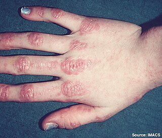
Lichen planus (LP) is a chronic inflammatory and autoimmune disease that affects the skin, nails, hair, and mucous membranes. It is not an actual lichen, but is named for its appearance. It is characterized by polygonal, flat-topped, violaceous papules and plaques with overlying, reticulated, fine white scale, commonly affecting dorsal hands, flexural wrists and forearms, trunk, anterior lower legs and oral mucosa. The hue may be gray-brown in people with darker skin. Although there is a broad clinical range of LP manifestations, the skin and oral cavity remain as the major sites of involvement. The cause is unknown, but it is thought to be the result of an autoimmune process with an unknown initial trigger. There is no cure, but many different medications and procedures have been used in efforts to control the symptoms.
Dermatomyositis (DM) is a long-term inflammatory disorder which affects the skin and the muscles. Its symptoms are generally a skin rash and worsening muscle weakness over time. These may occur suddenly or develop over months. Other symptoms may include weight loss, fever, lung inflammation, or light sensitivity. Complications may include calcium deposits in muscles or skin.

Calcinosis cutis is an uncommon condition marked by calcium buildup in the skin and subcutaneous tissues. Calcinosis cutis can range in intensity from little nodules in one area of the body to huge, crippling lesions affecting a vast portion of the body. Five kinds of the condition are typically distinguished: calciphylaxis, idiopathic calcification, iatrogenic calcification, dystrophic calcification, and metastatic calcification.

Hyperpigmentation is the darkening of an area of skin or nails caused by increased melanin.

Pearly penile papules are benign, small bumps or spots on the human penis. They vary in size from 1–4 mm, are pearly or flesh-colored, smooth and dome-topped or filiform, and appear in one or, several rows around the corona, the ridge of the head of the penis and sometimes on the penile shaft. They are painless, non-cancerous and not harmful. The medical condition of having such papules is called hirsutoid papillomatosis or hirsuties papillaris coronae glandis.
Leukemia cutis is the infiltration of neoplastic leukocytes or their precursors into the skin resulting in clinically identifiable cutaneous lesions. This condition may be contrasted with leukemids, which are skin lesions that occur with leukemia, but which are not related to leukemic cell infiltration. Leukemia cutis can occur in most forms of leukemia, including chronic myeloid leukemia, acute lymphoblastic leukemia, chronic lymphocytic leukemia, acute myeloid leukemia, and prolymphocytic leukemia.

Lichen nitidus is a chronic inflammatory disease of unknown cause characterized by 1–2 mm, discrete and uniform, shiny, flat-topped, pale flesh-colored or reddish-brown papules that may appear as hypopigmented against dark skin. Occasionally, minimal scaling is present or can be induced by rubbing the surface of the papules. The disease usually affects children and young adults and is painless and usually nonpruritic, although protracted itching may occur in some cases. It is sometimes referred to by dermatologists as "mini lichen planus".
Palisaded neutrophilic and granulomatous dermaititis (PNGS) is usually associated with a well-defined connective tissue disease, lupus erythematosus or rheumatoid arthritis most commonly, and often presents with eroded or ulcerated symmetrically distributed umbilicated papules or nodules on the elbows.
Eccrine angiomatous hamartoma (EAH), first described by Lotzbeck in 1859, is a rare benign vascular hamartoma characterized histologically by a proliferation of eccrine and vascular components. EAH exists on a spectrum of cutaneous tumors that include eccrine nevus, mucinous eccrine nevus and EAH. Each diagnostic subtype is characterized by an increase in the number as well as size of mature eccrine glands or ducts, with EAH being distinguished by the added vascular component.

Angioma serpiginosum is characterized by minute, copper-colored to bright red angiomatous puncta that have a tendency to become papular.
Glomeruloid hemangioma is a distinctive vascular tumor first described in 1990 when found to be associated with POEMS syndrome and Castleman disease. Glomeruloid hemangiomas can manifest as wine-red sessile or pedunculated papules, papulonodules, subcutaneous bluish compressible tumors, or small, firm, reddish-violaceous, dome-shaped papules.
Median nail dystrophy, also known as dystrophia unguis mediana canaliformis, median canaliform dystrophy of Heller, and solenonychia consists of longitudinal splitting or canal formation in the midline of the nail, a split which often resembles a fir tree, occurring at the cuticle and proceeding outward as the nail grows.
Erythromelanosis follicularis faciei et colli is an erythematous pigmentary disease involving the follicles, characterized by a reddish-brown, sharply demarcated, symmetrical discoloration involving the preauricular and maxillary regions.

Transient neonatal pustular melanosis (TNPM), also known as pustular melanosis, is a type of neonatal pustular eruption that is a transient rash common in newborns. It is vesiculopustular rash made up of 1–3 mm fluid-filled lesions that rupture, leaving behind a collarette of scale and a brown macule. The lesions are fragile and with no surrounding erythema. This rash occurs only in the newborn stage, usually appearing a few days after birth, but pigmented macules are sometimes already present at birth. The rash usually fades over three to four weeks but may linger for up to three months after birth. It can occur anywhere on the body, including the palms and soles.

Acrokeratosis verruciformis is a rare autosomal dominant disorder appearing at birth or in early childhood, characterized by skin lesions that are small, verrucous, flat papules resembling warts along with palmoplantar punctate keratoses and pits. However sporadic forms, whose less than 10 cases have been reported, presents at a later age, usually after the first decade and generally lack palmoplantar keratoses. Whether acrokeratosis verruciformis and Darier disease are related or distinct entities has been controversial, like Darier's disease, it is associated with defects in the ATP2A2 gene. however the specific mutations found in the ATP2A2 gene in acrokeratosis verruciformis have never been found in Darier's disease.
Actinic granuloma (AG) was first described by O'Brien in 1975 as a rare granulomatous disease. Lesions appear on sun-exposed areas, usually on the face, neck, and scalp, with a slight preference for middle-aged women. They are typically asymptomatic, single or multiple, annular or polycyclic lesions measuring up to 6 cm in diameter, with slow centrifugal expansion, an erythematous elevated edge, and a hypopigmented, atrophic center.
Granuloma multiforme is a cutaneous condition most commonly seen in central Africa, and rarely elsewhere, characterized by skin lesions that are on the upper trunk and arms in sun-exposed areas. It may be confused with tuberculoid leprosy, with which it has clinical similarities. The condition was first noted by Gosset in the 1940s, but it was not until 1964 that Leiker coined the term to describe "a disease resembling leprosy" in his study in Nigeria.
Progressive nodular histiocytosis is a cutaneous condition clinically characterized by the development of two types of skin lesions: superficial papules and deeper larger subcutaneous nodules. Progressive nodular histiocytosis was first reported in 1978 by Taunton et al. It is a subclass of non-Langerhans cell histiocytosis and a subgroup of xanthogranuloma.
Histoid leprosy is a skin condition, a rare form of multibacillary leprosy. It can occur in those with relapsing leprosy after undergoing antibiotic therapy with dapsone, or less frequently in the first infection.
