
Color blindness or color vision deficiency (CVD) is the decreased ability to see color or differences in color. It can impair tasks such as selecting ripe fruit, choosing clothing, and reading traffic lights. Color blindness may make some academic activities more difficult. However, issues are generally minor, and people with colorblindness automatically develop adaptations and coping mechanisms. People with total color blindness (achromatopsia) may also be uncomfortable in bright environments and have decreased visual acuity.

The visible spectrum is the portion of the electromagnetic spectrum that is visible to the human eye. Electromagnetic radiation in this range of wavelengths is called visible light or simply light. A typical human eye will respond to wavelengths from about 380 to about 700 nanometers. In terms of frequency, this corresponds to a band in the vicinity of 400–790 terahertz. These boundaries are not sharply defined and may vary per individual. Under optimal conditions these limits of human perception can extend to 310 nm (ultraviolet) and 1100 nm . The optical spectrum is sometimes considered to be the same as the visible spectrum, but some authors define the term more broadly, to include the ultraviolet and infrared parts of the electromagnetic spectrum as well.

A set of primary colors or primary colours consists of colorants or colored lights that can be mixed in varying amounts to produce a gamut of colors. This is the essential method used to create the perception of a broad range of colors in, e.g., electronic displays, color printing, and paintings. Perceptions associated with a given combination of primary colors can be predicted by an appropriate mixing model that reflects the physics of how light interacts with physical media, and ultimately the retina.
The Ishihara test is a color vision test for detection of red-green color deficiencies. It was named after its designer, Shinobu Ishihara, a professor at the University of Tokyo, who first published his tests in 1917.

Color vision, a feature of visual perception, is an ability to perceive differences between light composed of different frequencies independently of light intensity. Color perception is a part of the larger visual system and is mediated by a complex process between neurons that begins with differential stimulation of different types of photoreceptors by light entering the eye. Those photoreceptors then emit outputs that are propagated through many layers of neurons and then ultimately to the brain. Color vision is found in many animals and is mediated by similar underlying mechanisms with common types of biological molecules and a complex history of evolution in different animal taxa. In primates, color vision may have evolved under selective pressure for a variety of visual tasks including the foraging for nutritious young leaves, ripe fruit, and flowers, as well as detecting predator camouflage and emotional states in other primates.
Dichromacy is the state of having two types of functioning photoreceptors, called cone cells, in the eyes. Organisms with dichromacy are called dichromats. Dichromats require only two primary colors to be able to represent their visible gamut. By comparison, trichromats need three primary colors, and tetrachromats need four. Likewise, every color in a dichromat's gamut can be evoked with monochromatic light. By comparison, every color in a trichromat's gamut can be evoked with a combination of monochromatic light and white light.
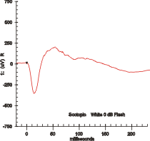
Electroretinography measures the electrical responses of various cell types in the retina, including the photoreceptors, inner retinal cells, and the ganglion cells. Electrodes are placed on the surface of the cornea or on the skin beneath the eye to measure retinal responses. Retinal pigment epithelium (RPE) responses are measured with an EOG test with skin-contact electrodes placed near the canthi. During a recording, the patient's eyes are exposed to standardized stimuli and the resulting signal is displayed showing the time course of the signal's amplitude (voltage). Signals are very small, and typically are measured in microvolts or nanovolts. The ERG is composed of electrical potentials contributed by different cell types within the retina, and the stimulus conditions can elicit stronger response from certain components.

Shinobu Ishihara was a Japanese ophthalmologist who created the Ishihara color test to detect colour blindness. He was an army surgeon.
An anomaloscope is an instrument and color vision test, often used to quantify and characterize color blindness. They are expensive and require specialized knowledge to operate, but are viewed as the gold standard for color vision standards. As a result, they are normally used for academic studies, rather than job pre-screening. They are also used to validate other color vision standards with regards to classification of color vision defects.

A color triangle is an arrangement of colors within a triangle, based on the additive or subtractive combination of three primary colors at its corners.

Alarik Frithiof Holmgren was a Swedish physician, physiologist and professor at Upsala University, most noted for his research of color blindness. He was a vocal opponent of vivisection, and particularly the use of curare to immobilize subjects so they appeared peaceful while enduring great pain.
The Farnsworth Lantern Test, or FALANT, is a color vision test originally developed specifically to screen sailors for tasks requiring color vision, such as identifying signal lights at night. It screens for red-green deficiencies, but not the much rarer blue color deficiency.

The Lagerlunda rail accident occurred in the early hours of November 15, 1875 about 8 km west of Linköping in Östergötland, Sweden. Unclear signalling between a station master and a steam engine driver led to a train leaving the station although another train was approaching on the single line track. 9 people were killed in the head-on collision shortly after. The station master was sentenced to 6 months of prison.
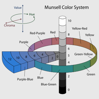
The Farnsworth–Munsell 100 Hue Color Vision test is a color vision test often used to test for color blindness. The system was developed by Dean Farnsworth in the 1940s and it tests the ability to isolate and arrange minute differences in various color targets with constant value and chroma that cover all the visual hues described by the Munsell color system. There are several variations of the test, one featuring 100 color hues and one featuring 15 color hues. Originally taken in an analog environment with physical hue tiles, the test is now taken from computer consoles. An accurate quantification of color vision accuracy is particularly important to designers, photographers and colorists, who all rely on accurate color vision to produce quality content.

EnChroma lenses are specialized glasses designed to address symptoms of red-green color blindness. Studies have shown that these lenses can alter the perception of colors that were already perceived, but they do not fully restore normal color vision. Some initial claims made by the manufacturer have been subject to criticism and described as marketing hype,while research suggests that the lenses may have a limited positive impact on individuals with red-green color blindness.
The City University test is a color vision test used to detect color vision deficiency. The commonly used Ishihara test is used to detect mainly congenital red-green color blindness, but its usefulness is limited in detecting acquired color vision deficiencies.
A color vision test is used for measuring color vision against a standard. These tests are most often used to diagnose color vision deficiencies, though several of the standards are designed to categorize normal color vision into sub-levels. With the large prevalence of color vision deficiencies and the wide range of professions that restrict hiring the colorblind for safety or aesthetic reasons, clinical color vision standards must be designed to be fast and simple to implement. Color vision standards for academic use trade speed and simplicity for accuracy and precision.
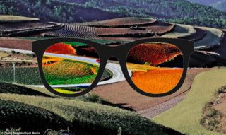
Color blind glasses or color correcting lenses are light filters, usually in the form of glasses or contact lenses, that attempt to alleviate color blindness, by bringing deficient color vision closer to normal color vision or to make certain color tasks easier to accomplish. Despite viral status, the academic literature is generally skeptical of the efficacy of color correcting lenses.

Color tasks are tasks that involve the recognition of colors. Color tasks can be classified according to how the color is interpreted. Cole describes four categories of color tasks:
Congenital red–green color blindness is an inherited condition that is the root cause of the majority of cases of color blindness. It has no significant symptoms aside from its minor to moderate effect on color vision. It is caused by variation in the functionality of the red and/or green opsin proteins, which are the photosensitive pigment in the cone cells of the retina, which mediate color vision. Males are more likely to inherit red–green color blindness than females, because the genes for the relevant opsins are on the X chromosome. Screening for congenital red–green color blindness is typically performed with the Ishihara or similar color vision test. There is no cure for color blindness.
