
The nervous system is a highly complex part of an animal that coordinates its actions and sensory information by transmitting signals to and from different parts of its body. The nervous system detects environmental changes that impact the body, then works in tandem with the endocrine system to respond to such events. Nervous tissue first arose in wormlike organisms about 550 to 600 million years ago. In vertebrates it consists of two main parts, the central nervous system (CNS) and the peripheral nervous system (PNS). The CNS consists of the brain and spinal cord. The PNS consists mainly of nerves, which are enclosed bundles of the long fibers or axons, that connect the CNS to every other part of the body. Nerves that transmit signals from the brain are called motor or efferent nerves, while those nerves that transmit information from the body to the CNS are called sensory or afferent. Spinal nerves serve both functions and are called mixed nerves. The PNS is divided into three separate subsystems, the somatic, autonomic, and enteric nervous systems. Somatic nerves mediate voluntary movement. The autonomic nervous system is further subdivided into the sympathetic and the parasympathetic nervous systems. The sympathetic nervous system is activated in cases of emergencies to mobilize energy, while the parasympathetic nervous system is activated when organisms are in a relaxed state. The enteric nervous system functions to control the gastrointestinal system. Both autonomic and enteric nervous systems function involuntarily. Nerves that exit from the cranium are called cranial nerves while those exiting from the spinal cord are called spinal nerves.

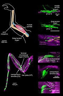
Muscle spindles are stretch receptors within the body of a muscle that primarily detect changes in the length of the muscle. They convey length information to the central nervous system via afferent nerve fibers. This information can be processed by the brain as proprioception. The responses of muscle spindles to changes in length also play an important role in regulating the contraction of muscles, for example, by activating motor neurons via the stretch reflex to resist muscle stretch.

The grey column refers to a somewhat ridge-shaped mass of grey matter in the spinal cord. This presents as three columns: the anterior grey column, the posterior grey column, and the lateral grey column, all of which are visible in cross-section of the spinal cord.

Rodolfo Llinás Riascos is a Colombian neuroscientist. He is currently the Thomas and Suzanne Murphy Professor of Neuroscience and Chairman Emeritus of the department of Physiology & Neuroscience at the NYU School of Medicine. Llinás has published over 800 scientific articles.
Central pattern generators (CPGs) are biological neural circuits that produce rhythmic outputs in the absence of rhythmic input. They are the source of the tightly-coupled patterns of neural activity that drive rhythmic motions like walking, breathing, or chewing. The ability to function without input from higher brain areas still requires modulatory inputs, and their outputs are not fixed. Flexibility in response to sensory input is a fundamental quality of CPG-driven behavior. To be classified as a rhythmic generator, a CPG requires:
- "two or more processes that interact such that each process sequentially increases and decreases, and
- that, as a result of this interaction, the system repeatedly returns to its starting condition."

The scratch reflex is a response to activation of sensory neurons whose peripheral terminals are located on the surface of the body. Some sensory neurons can be activated by stimulation with an external object such as a parasite on the body surface. Alternatively, some sensory neurons can respond to a chemical stimulus that produces an itch sensation. During a scratch reflex, a nearby limb reaches toward and rubs against the site on the body surface that has been stimulated. The scratch reflex has been extensively studied to understand the functioning of neural networks in vertebrates. Despite decades of research, key aspects of the scratch reflex are still unknown, such as the neural mechanisms by which the reflex is terminated.
From the ancient Egyptian mummifications to 18th century scientific research on "globules" and neurons, there is evidence of neuroscience practice throughout the early periods of history. The early civilizations lacked adequate means to obtain knowledge about the human brain. Their assumptions about the inner workings of the mind, therefore, were not accurate. Early views on the function of the brain regarded it to be a form of "cranial stuffing" of sorts. In ancient Egypt, from the late Middle Kingdom onwards, in preparation for mummification, the brain was regularly removed, for it was the heart that was assumed to be the seat of intelligence. According to Herodotus, during the first step of mummification: "The most perfect practice is to extract as much of the brain as possible with an iron hook, and what the hook cannot reach is mixed with drugs." Over the next five thousand years, this view came to be reversed; the brain is now known to be the seat of intelligence, although colloquial variations of the former remain as in "memorizing something by heart".

The mesencephalic nucleus is involved with reflex proprioception of the periodontium and of the muscles of mastication in the jaw that functions to prevent biting down hard enough to lose a tooth. To subserve this reflex protective function, mechanoreceptive nerves in the periodontal ligament sense tooth movement and project to the mesencephalic nucleus. Likewise, afferent fibers from muscle spindles, the sensory organs of skeletal muscle, are stimulated by the stretch of hard contraction of jaw muscles. The temporomandibular joints and the Golgi tendon organs of the jaw muscles do not project to the mesencephalic nucleus. The mesencephalic nucleus is one of four trigeminal nerve nuclei, three sensory and one motor. The other two sensory nuclei are the chief sensory nucleus mediating conscious facial touch and the spinal trigeminal nucleus, mediating pain in the head, and is of importance in headache. The trigeminal motor nucleus innervates the muscles of mastication.
Low-threshold spikes (LTS) refer to membrane depolarizations by the T-type calcium channel. LTS occur at low, negative, membrane depolarizations. They often follow a membrane hyperpolarization, which can be the result of decreased excitability or increased inhibition. LTS result in the neuron reaching the threshold for an action potential. LTS is a large depolarization due to an increase in Ca2+ conductance, so LTS is mediated by calcium (Ca2+) conductance. The spike is typically crowned by a burst of two to seven action potentials, which is known as a low-threshold burst. LTS are voltage dependent and are inactivated if the cell's resting membrane potential is more depolarized than −60mV. LTS are deinactivated, or recover from inactivation, if the cell is hyperpolarized and can be activated by depolarizing inputs, such as excitatory postsynaptic potentials (EPSP). LTS were discovered by Rodolfo Llinás and coworkers in 1980s.
In mammals, the Bötzinger complex (BötC) is a group of neurons located in the rostral ventrolateral medulla, and ventral respiratory column. In the medulla, this group is located caudally to the facial nucleus and ventral to nucleus ambiguus.
Proprioception, also referred to as kinaesthesia, is the sense of self-movement and body position. It is sometimes described as the "sixth sense".
The neuroscience of rhythm refers to the various forms of rhythm generated by the central nervous system (CNS). Nerve cells, also known as neurons in the human brain are capable of firing in specific patterns which cause oscillations. The brain possesses many different types of oscillators with different periods. Oscillators are simultaneously outputting frequencies from .02 Hz to 600 Hz. It is now well known that a computer is capable of running thousands of processes with just one high frequency clock. Humans have many different clocks as a result of evolution. Prior organisms had no need for a fast responding oscillator. This multi-clock system permits quick response to constantly changing sensory input while still maintaining the autonomic processes that sustain life. This method modulates and controls a great deal of bodily functions.
Neural accommodation or neuronal accommodation occurs when a neuron or muscle cell is depolarised by slowly rising current in vitro. The Hodgkin–Huxley model also shows accommodation. Sudden depolarisation of a nerve evokes propagated action potential by activating voltage-gated fast sodium channels incorporated in the cell membrane if the depolarisation is strong enough to reach threshold. The open sodium channels allow more sodium ions to flow into the cell and resulting in further depolarisation, which will subsequently open even more sodium channels. At a certain moment this process becomes regenerative and results in the rapid ascending phase of action potential. In parallel with the depolarisation and sodium channel activation, the inactivation process of the sodium channels is also driven by depolarisation. Since the inactivation is much slower than the activation process, during the regenerative phase of action potential, inactivation is unable to prevent the "chain reaction"-like rapid increase in the membrane voltage.
Krešimir Krnjević is a Canadian-British neurophysiologist.

The classification of peripheral nerves in the peripheral nervous system (PNS) groups the nerves into two main groups, the somatic and the autonomic nervous systems. Together, these two systems provide information regarding the location and status of the limbs, organs, and the remainder of the body to the central nervous system (CNS) via nerves and ganglia present outside of the spinal cord and brain. The somatic nervous system directs all voluntary movements of the skeletal muscles, and can be sub-divided into afferent and efferent neuronal flow. The autonomic nervous system is divided primarily into the sympathetic and parasympathetic nervous systems with a third system, the enteric nervous system, receiving less recognition.
Brain-body interactions are patterns of neural activity in the central nervous system to coordinate the activity between the brain and body. The nervous system consists of central and peripheral nervous systems and coordinates the actions of an animal by transmitting signals to and from different parts of its body. The brain and spinal cord are interwoven with the body and interact with other organ systems through the somatic, autonomic and enteric nervous systems. Neural pathways regulate brain-body interactions and allow to sense and control its body and interact with the environment.


