
An electron microscope is a microscope that uses a beam of electrons as a source of illumination. They use electron optics that are analogous to the glass lenses of an optical light microscope to control the electron beam, for instance focusing them to produce magnified images or electron diffraction patterns. As the wavelength of an electron can be up to 100,000 times smaller than that of visible light, electron microscopes have a much higher resolution of about 0.1 nm, which compares to about 200 nm for light microscopes. Electron microscope may refer to:

A scanning electron microscope (SEM) is a type of electron microscope that produces images of a sample by scanning the surface with a focused beam of electrons. The electrons interact with atoms in the sample, producing various signals that contain information about the surface topography and composition of the sample. The electron beam is scanned in a raster scan pattern, and the position of the beam is combined with the intensity of the detected signal to produce an image. In the most common SEM mode, secondary electrons emitted by atoms excited by the electron beam are detected using a secondary electron detector. The number of secondary electrons that can be detected, and thus the signal intensity, depends, among other things, on specimen topography. Some SEMs can achieve resolutions better than 1 nanometer.

Transmission electron microscopy (TEM) is a microscopy technique in which a beam of electrons is transmitted through a specimen to form an image. The specimen is most often an ultrathin section less than 100 nm thick or a suspension on a grid. An image is formed from the interaction of the electrons with the sample as the beam is transmitted through the specimen. The image is then magnified and focused onto an imaging device, such as a fluorescent screen, a layer of photographic film, or a detector such as a scintillator attached to a charge-coupled device or a direct electron detector.

The Molecular Foundry is a nanoscience user facility located at the Lawrence Berkeley National Laboratory in Berkeley, California, and is one of five Nanoscale Science Research Centers sponsored by the United States Department of Energy.

Thomas Eugene Everhart FREng is an American educator and physicist. His area of expertise is the physics of electron beams. Together with Richard F. M. Thornley he designed the Everhart–Thornley detector. These detectors are still in use in scanning electron microscopes, even though the first such detector was made available as early as 1956.

Transmission Electron Aberration-Corrected Microscope (TEAM) is a collaborative research project between four US laboratories and two companies. The project's main activity is design and application of a transmission electron microscope (TEM) with a spatial resolution below 0.05 nanometers, which is roughly half the size of an atom of hydrogen.
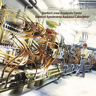
The Stanford Synchrotron Radiation Lightsource, a division of SLAC National Accelerator Laboratory, is operated by Stanford University for the Department of Energy. SSRL is a National User Facility which provides synchrotron radiation, a name given to electromagnetic radiation in the x-ray, ultraviolet, visible and infrared realms produced by electrons circulating in a storage ring at nearly the speed of light. The extremely bright light that is produced can be used to investigate various forms of matter ranging from objects of atomic and molecular size to man-made materials with unusual properties. The obtained information and knowledge is of great value to society, with impact in areas such as the environment, future technologies, health, biology, basic research, and education.

Uranyl acetate is the acetate salt of uranium oxide, a toxic yellow-green powder useful in certain laboratory tests. Structurally, it is a coordination polymer with formula UO2(CH3CO2)2(H2O)·H2O.

The Advanced Light Source (ALS) is a research facility at Lawrence Berkeley National Laboratory in Berkeley, California. One of the world's brightest sources of ultraviolet and soft x-ray light, the ALS is the first "third-generation" synchrotron light source in its energy range, providing multiple extremely bright sources of intense and coherent short-wavelength light for use in scientific experiments by researchers from around the world. It is funded by the US Department of Energy (DOE) and operated by the University of California. In June 2018, Stephen Kevan became the director of the ALS.
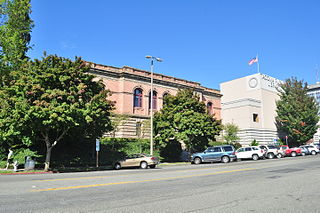
The Tacoma Public Library system serves residents of Tacoma, Washington. It operates eight library branches, which include a central library in downtown Tacoma, two regional locations in north and south Tacoma, and five neighborhood branch locations. Tacoma Public Library has nearly 150,000 registered users, and over 2 million items in circulation. Tacoma Public Library serves a population of 198,100 people.

FEI Company is an American company that designs, manufactures, and supports microscope technology. Headquartered in Hillsboro, Oregon, FEI has over 2,800 employees and sales and service operations in more than 50 countries around the world. Formerly listed on the NASDAQ, it is a subsidiary of Thermo Fisher Scientific.
Stege Marsh, also known as the South Richmond Marshes, is a tidal marshland wetlands area in Richmond, California in western Contra Costa County.

The Miller Institute for Basic Research in Science was established on the University of California, Berkeley, campus in 1955 after Adolph C. Miller and his wife, Mary Sprague Miller, made a donation to the university. It was their wish that the donation be used to establish an institute "dedicated to the encouragement of creative thought and conduct of pure science".

Ondrej L. Krivanek is a Czech/British physicist resident in the United States, and a leading developer of electron-optical instrumentation. He won the Kavli Prize for Nanoscience in 2020 for his substantial innovations in atomic resolution electron microscopy.
Peter Nigel Tripp Unwin FRS is a British scientist at the MRC Laboratory of Molecular Biology, where he was Head of the Neurobiology Division from 1992 until 2008. He is currently also Emeritus Professor of Cell Biology at the Scripps Research Institute.
Tappeh Nurjan is a village in Bagheli-ye Marama Rural District, in the Central District of Gonbad-e Qabus County, Golestan Province, Iran. At the 2006 census, its population was 496, in 112 families.
In situ electron microscopy is an investigatory technique where an electron microscope is used to watch a sample's response to a stimulus in real time. Due to the nature of the high-energy beam of electrons used to image a sample in an electron microscope, microscopists have long observed that specimens are routinely changed or damaged by the electron beam. Starting in the 1960s, and using transmission electron microscopes (TEMs), scientists made deliberate attempts to modify materials while the sample was in the specimen chamber, and to capture images through time of the induced damages.
Joachim Frank ; born September 12, 1940) is a German-American biophysicist at Columbia University and a Nobel laureate. He is regarded as the founder of single-particle cryo-electron microscopy (cryo-EM), for which he shared the Nobel Prize in Chemistry in 2017 with Jacques Dubochet and Richard Henderson. He also made significant contributions to structure and function of the ribosome from bacteria and eukaryotes.
The International Federation of Societies for Microscopy is an international non-governmental organization representing microscopy. It currently has 37 national members and 9 associate members, which are split into three regional committees, the Committee for Asia-Pacific Societies of Microscopy, the European Microscopy Society and the Interamerica Committee for Societies for EM.

Cryogenic electron microscopy (cryoEM) is a cryomicroscopy technique applied on samples cooled to cryogenic temperatures. For biological specimens, the structure is preserved by embedding in an environment of vitreous ice. An aqueous sample solution is applied to a grid-mesh and plunge-frozen in liquid ethane or a mixture of liquid ethane and propane. While development of the technique began in the 1970s, recent advances in detector technology and software algorithms have allowed for the determination of biomolecular structures at near-atomic resolution. This has attracted wide attention to the approach as an alternative to X-ray crystallography or NMR spectroscopy for macromolecular structure determination without the need for crystallization.