
Histology, also microanatomy, is the branch of biology which studies the tissues of animals and plants using microscopy. It is commonly studied using a light microscope or electron microscope, the specimen having been sectioned, stained, and mounted on a microscope slide. Histological studies may be conducted using tissue culture, where live animal cells are isolated and maintained in an artificial environment for various research projects. The ability to visualize or differentially identify microscopic structures is frequently enhanced through the use of staining. Histology is one of the major preclinical subjects in medical school. Medical students are expected to be familiar with the morphological features and function of all cells and tissues of the human body from an early stage of their studies, so histology often stretches over several semesters.

Eosin is the name of several fluorescent acidic compounds which bind to and form salts with basic, or eosinophilic, compounds like proteins containing amino acid residues such as arginine and lysine, and stains them dark red or pink as a result of the actions of bromine on fluorescein. In addition to staining proteins in the cytoplasm, it can be used to stain collagen and muscle fibers for examination under the microscope. Structures that stain readily with eosin are termed eosinophilic.
Romanowsky staining is a prototypical staining technique that was the forerunner of several distinct but similar methods, including Giemsa, Jenner, Wright, Field, May Grunwald stain and Leishman stains, which are used to differentiate cells in pathologic specimens. It was named after the Russian physician Dmitri Leonidovich Romanowsky (1861–1921), who invented it in 1891.
Staining is an auxiliary technique used in microscopy to enhance contrast in the microscopic image. Stains and dyes are frequently used in biology and medicine to highlight structures in biological tissues for viewing, often with the aid of different microscopes. Stains may be used to define and examine bulk tissues, cell populations, or organelles within individual cells.
A lactotropic cell is a cell in the anterior pituitary which produces prolactin in response to hormonal signals including dopamine which is inhibitory and thyrotropin-releasing hormone which is stimulatory. Other regulators include oxytocin, estrogen and progesterone.
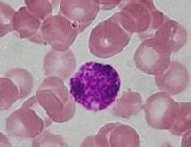
Eosinophilic refers to the staining of certain tissues, cells, or organelles after they have been washed with eosin, a dye.
Basophilic is a technical term used by histologists. It describes the microscopic appearance of cells and tissues, as seen down the microscope, after a histological section has been stained with a basic dye. The most common such dye is haematoxylin.
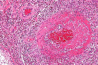
Fibrinoid necrosis is a specific pattern of irreversible, uncontrolled cell death that occurs when antigen-antibody complexes are deposited in the walls of blood vessels along with fibrin. It is common in the immune-mediated vasculitides which are a result of type III hypersensitivity. When stained with hematoxylin and eosin, they appear brightly eosinophilic and smudged.

Eosin methylene blue is a selective stain for gram-negative bacteria. EMB contains dyes that are toxic to gram-positive bacteria. EMB is the selective and differential medium for coliforms. It is a blend of two stains, eosin and methylene blue in the ratio of 6:1. A common application of this stain is in the preparation of EMB agar, a differential microbiological medium, which slightly inhibits the growth of Gram-positive bacteria and provides a color indicator distinguishing between organisms that ferment lactose and those that do not. Organisms that ferment lactose display "nucleated colonies"—colonies with dark centers.
Coagulative necrosis is a type of accidental cell death typically caused by ischemia or infarction. In coagulative necrosis the architecture of dead tissue is preserved for at least a couple of days. It is believed that the injury denatures structural proteins as well as lysosomal enzymes thus blocking the proteolysis of the damaged cells. The lack of lysosomal enzymes allows it to maintain a "coagulated" morphology for some time. Like most types of necrosis if enough viable cells are present around the affected area regeneration will usually occur.

An oncocytoma is a tumor made up of oncocytes, epithelial cells characterized by an excessive amount of mitochondria, resulting in an abundant acidophilic, granular cytoplasm. The cells and the tumor that they compose are often benign but sometimes may be premalignant or malignant.

Karyolysis, and λύσις lysis from λύειν lyein, "to separate") is the complete dissolution of the chromatin of a dying cell due to the enzymatic degradation by endonucleases. The whole cell will eventually stain uniformly with eosin after karyolysis. It is usually associated with karyorrhexis and occurs mainly as a result of necrosis, while in apoptosis after karyorrhexis the nucleus usually dissolves into apoptotic bodies.

Eosin Y is a form of eosin. It is most commonly used as an acidic red stain for highlighting cytoplasm material in samples.

Papanicolaou stain is a multichromatic staining cytological technique developed by George Papanikolaou, the father of cytopathology.

Hematoxylin and eosin stain or haematoxylin and eosin stain is one of the principal stains in histology. It is the most widely used stain in medical diagnosis and is often the gold standard; for example, when a pathologist looks at a biopsy of a suspected cancer, the histological section is likely to be stained with H&E. A combination of hematoxylin and eosin, it produces blues, violets, and reds.

Phosphotungstic acid haematoxylin (PTAH) is a mix of haematoxylin with phosphotungstic acid, used in histology for staining.
Acidophile is a term used by histologists to describe a particular staining pattern of cells and tissues when using haematoxylin and eosin stains. Specifically, the name refers to structures which "love" acid, and take it up readily. More specifically, acidophilia can be described by cationic groups of most often proteins in the cell readily reacting with acidic stains.
Disclosing tablets are chewable tablets used to make dental plaque visible.

Luxol fast blue stain, abbreviated LFB stain or simply LFB, is a commonly used stain to observe myelin under light microscopy, created by Heinrich Klüver and Elizabeth Barrera in 1953. LFB is commonly used to detect demyelination in the central nervous system (CNS), but cannot discern myelination in the peripheral nervous system.
In diagnostic pathology, a hematoxylin body, or LE body, is a dense, homogeneous, basophilic particle, easily stainable with hematoxylin. It consists of degraded nuclear material from an injured cell, along with autoantibodies and a limited amount of cytoplasm.


