
The human leg, in the general word sense, is the entire lower limb of the human body, including the foot, thigh and even the hip or gluteal region. However, the definition in human anatomy refers only to the section of the lower limb extending from the knee to the ankle, also known as the crus or, especially in non-technical use, the shank. Legs are used for standing, and all forms of locomotion including recreational such as dancing, and constitute a significant portion of a person's mass. Female legs generally have greater hip anteversion and tibiofemoral angles, but shorter femur and tibial lengths than those in males.

The sacroiliac joint or SI joint (SIJ) is the joint between the sacrum and the ilium bones of the pelvis, which are connected by strong ligaments. In humans, the sacrum supports the spine and is supported in turn by an ilium on each side. The joint is strong, supporting the entire weight of the upper body. It is a synovial plane joint with irregular elevations and depressions that produce interlocking of the two bones. The human body has two sacroiliac joints, one on the left and one on the right, that often match each other but are highly variable from person to person.
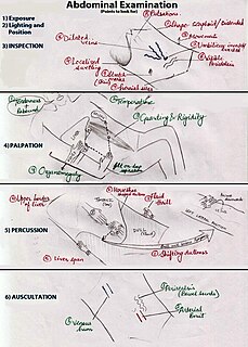
An abdominal examination is a portion of the physical examination which a physician or nurse uses to clinically observe the abdomen of a patient for signs of disease. The physical examination typically occurs after a thorough medical history is taken, that is, after the physician asks the patient the course of their symptoms. The abdominal examination is conventionally split into four different stages: first, inspection of the patient and the visible characteristics of their abdomen. Auscultation (listening) of the abdomen with a stethoscope. Palpation of the patient's abdomen. Finally, percussion (tapping) of the patient's abdomen and abdominal organs. Depending on the need to test for specific diseases such as ascites, special tests may be performed as a part of the physical examination. An abdominal examination may be performed because the physician suspects a disease of the organs inside the abdominal cavity, or simply as a part of a complete physical examination for other conditions. In a complete physical examination, the abdominal exam classically follows the respiratory examination and cardiovascular examination.

The gracilis muscle is the most superficial muscle on the medial side of the thigh. It is thin and flattened, broad above, narrow and tapering below.

A hip dislocation is when the thighbone (femur) separates from the hip bone (pelvis). Specifically it is when the ball–shaped head of the femur separates from its cup–shaped socket in the hip bone, known as the acetabulum. The joint of the femur and pelvis is very stable, secured by both bony and soft-tissue constraints. With that, dislocation would require significant force which typically results from significant trauma such as from a motor vehicle collision or from a fall from elevation. Hip dislocations can also occur following a hip replacement or from a developmental abnormality known as hip dysplasia.

An anoscopy is an examination using a small, rigid, tubular instrument called an anoscope. This is inserted a few inches into the anus in order to evaluate problems of the anal canal. Anoscopy is used to diagnose hemorrhoids, anal fissures, and some cancers.

Motion, the process of movement, is described using specific anatomical terms. Motion includes movement of organs, joints, limbs, and specific sections of the body. The terminology used describes this motion according to its direction relative to the anatomical position of the body parts involved. Anatomists and others use a unified set of terms to describe most of the movements, although other, more specialized terms are necessary for describing unique movements such as those of the hands, feet, and eyes.
The knee examination, in medicine and physiotherapy, is performed as part of a physical examination, or when a patient presents with knee pain or a history that suggests a pathology of the knee joint.
In medicine, physiotherapy, chiropractic, and osteopathy the hip examination, or hip exam, is undertaken when a patient has a complaint of hip pain and/or signs and/or symptoms suggestive of hip joint pathology. It is a physical examination maneuver.

In medicine, Fowler's position is a standard patient position in which the patient is seated in a semi-sitting position and may have knees either bent or straight. Variations in the angle are denoted by high Fowler, indicating an upright position at approximately 90 degrees and semi-Fowler, 30 to 45 degrees; and low Fowler, where the head is slightly elevated." It is an intervention used to promote oxygenation via maximum chest expansion and is implemented during events of respiratory distress. Fowler's position facilitates the relaxing of tension of the abdominal muscles, allowing for improved breathing. In immobile patients and infants, the Fowler's position alleviates compression of the chest that occurs due to gravity. Fowler's position increases comfort during eating and other activities, is used in postpartum women to improve uterine drainage, and in infants when signs of respiratory distress are present. Fowler's position is also used when oral or nasal gastric feeding tubes have been implemented as it minimizes the risk of aspiration. Peristalsis and swallowing are aided by the effect of gravitational pull.

The acetabular labrum is a ring of cartilage that surrounds the acetabulum of the hip. The anterior portion is most vulnerable when the labrum tears.

Neurogenic claudication (NC), also known as pseudoclaudication, is the most common symptom of lumbar spinal stenosis (LSS) and describes intermittent leg pain from impingement of the nerves emanating from the spinal cord. Neurogenic means that the problem originates within the nervous system. Claudication, from the Latin word for to limp, refers to painful cramping or weakness in the legs. NC should therefore be distinguished from vascular claudication, which stems from a circulatory problem rather than a neural one.
A GALS screen is an examination used by doctors and other healthcare professionals to detect locomotor abnormalities and functional disability relating to gait, arms, legs and the spine.
Scissor gait is a form of gait abnormality primarily associated with spastic cerebral palsy. That condition and others like it are associated with an upper motor neuron lesion.
The Trendelenburg Test or Brodie–Trendelenburg test is a test which can be carried out as part of a physical examination to determine the competency of the valves in the superficial and deep veins of the legs in patients with varicose veins.

A patellar dislocation is a knee injury in which the patella (kneecap) slips out of its normal position. Often the knee is partly bent, painful and swollen. The patella is also often felt and seen out of place. Complications may include a patella fracture or arthritis.

The Thomas test is a physical examination test, named after the Welsh orthopaedic surgeon, Hugh Owen Thomas (1834–1891), to rule out hip flexion contracture and psoas syndrome
Ober's test is used in physical examination to identify tightness of the iliotibial band . During the test, the patient lies on his/her side with the unaffected leg on the bottom with their shoulder and pelvis in line. The lower hip and knee can be in a flexed position to take out any lordosis of the lumbar spine.
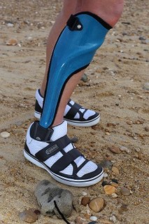
Orthotics is a medical specialty that focuses on the design and application of orthoses. An orthosis is "an externally applied device used to influence the structural and functional characteristics of the neuromuscular and skeletal system".
Surgical positioning is the practice of placing a patient in a particular physical position during surgery. The goal in selecting and adjusting a particular surgical position is to maintain the patient's safety while allowing access to the surgical site. Often a patient must be placed in an unnatural position to gain access to the surgical site.


