Vestibulo–ocular reflex

Last updated
The vestibulo-ocular reflex. A rotation of the head is detected, which triggers an inhibitory signal to the extraocular muscles on one side and an excitatory signal to the muscles on the other side. The result is a compensatory movement of the eyes. Simple vestibulo-ocular reflex.PNG
The vestibulo-ocular reflex. A rotation of the head is detected, which triggers an inhibitory signal to the extraocular muscles on one side and an excitatory signal to the muscles on the other side. The result is a compensatory movement of the eyes.

The vestibulo-ocular reflex (VOR) is a reflex that acts to stabilize gaze during head movement, with eye movement due to activation of the vestibular system. The reflex acts to stabilize images on the retinas of the eye during head movement. Gaze is held steadily on a location by producing eye movements in the direction opposite that of head movement. For example, when the head moves to the right, the eyes move to the left, meaning the image a person sees stays the same even though the head has turned. Since slight head movement is present all the time, VOR is necessary for stabilizing vision: people with an impaired reflex find it difficult to read using print, because the eyes do not stabilise during small head tremors, and also because damage to reflex can cause nystagmus. [1]

Contents

The VOR does not depend on what is seen. It can also be activated by hot or cold stimulation of the inner ear, where the vestibular system sits, and works even in total darkness or when the eyes are closed.[ citation needed ] However, in the presence of light, the fixation reflex is also added to the movement. [2]

In lower animals, the organs that coordinate balance and movement are not independent from eye movement. A fish, for instance, moves its eyes by reflex when its tail is moved. Humans have semicircular canals, neck muscle "stretch" receptors, and the utricle (gravity organ). Though the semicircular canals cause most of the reflexes which are responsive to acceleration, the maintaining of balance is mediated by the stretch of neck muscles and the pull of gravity on the utricle (otolith organ) of the inner ear. [2]

The VOR has both rotational and translational aspects. When the head rotates about any axis (horizontal, vertical, or torsional) distant visual images are stabilized by rotating the eyes about the same axis, but in the opposite direction. [3] When the head translates, for example during walking, the visual fixation point is maintained by rotating gaze direction in the opposite direction, [4] by an amount that depends on distance. [5]

Function

Vestibulo-ocular reflex EN.svg

The vestibulo-ocular reflex is driven by signals arising from the vestibular system of the inner ear. The semicircular canals detect head rotation and provide the rotational component, whereas the otoliths detect head translation and drive the translational component. The signal for the horizontal rotational component travels via the vestibular nerve through the vestibular ganglion and end in the vestibular nuclei in the brainstem. From these nuclei, fibers cross to the abducens nucleus of the opposite side of the brain. Here, fibres synapse with 2 additional pathways. One pathway projects directly to the lateral rectus muscle of the eye via the abducens nerve. Another nerve tract projects from the abducens nucleus by the medial longitudinal fasciculus to the oculomotor nucleus of the opposite side, which contains motor neurons that drive eye muscle activity, specifically activating the medial rectus muscle of the eye through the oculomotor nerve.

Another pathway (not in picture) directly projects from the vestibular nucleus through the ascending tract of Deiter's to the medial rectus muscle motor neuron of the same side. In addition there are inhibitory vestibular pathways to the ipsilateral abducens nucleus. However no direct vestibular neuron to medial rectus motoneuron pathway exists. [6]

Similar pathways exist for the vertical and torsional components of the VOR.

In addition to these direct pathways, which drive the velocity of eye rotation, there is an indirect pathway that builds up the position signal needed to prevent the eye from rolling back to center when the head stops moving. This pathway is particularly important when the head is moving slowly because here position signals dominate over velocity signals. David A. Robinson discovered that the eye muscles require this dual velocity-position drive, and also proposed that it must arise in the brain by mathematically integrating the velocity signal and then sending the resulting position signal to the motoneurons. Robinson was correct: the 'neural integrator' for horizontal eye position was found in the nucleus prepositus hypoglossi [7] in the medulla, and the neural integrator for vertical and torsional eye positions was found in the interstitial nucleus of Cajal [8] in the midbrain. The same neural integrators also generate eye position for other conjugate eye movements such as saccades and smooth pursuit.

Example

For instance, if the head is turned clockwise as seen from above, then excitatory impulses are sent from the semicircular canal on the right side via the vestibular nerve through Scarpa's ganglion and end in the right vestibular nuclei in the brainstem. From this nuclei excitatory fibres cross to the left abducens nucleus. There they project and stimulate the lateral rectus of the left eye via the abducens nerve. In addition, by the medial longitudinal fasciculus and oculomotor nuclei, they activate the medial rectus muscles on the right eye. As a result, both eyes will turn counter-clockwise.

Furthermore, some neurons from the right vestibular nucleus directly stimulate the right medial rectus motor neurons, and inhibits the right abducens nucleus.

Speed

The vestibulo-ocular reflex needs to be fast: for clear vision, head movement must be compensated almost immediately; otherwise, vision corresponds to a photograph taken with a shaky hand. Signals are sent from the semicircular canals using only three neurons, called the three neuron arc. This results in eye movements that lag head movement by less than 10 ms. [9] The vestibulo-ocular reflex is one of the fastest reflexes in the human body.

VOR suppression

When a person tracks the movement of something with both their eyes and head together, the VOR is counterproductive to the goal of keeping the gaze and head angle aligned. Research indicates that there exists mechanisms in the brain to suppress the VOR using the active visual (retinal) feedback obtained by watching the object in motion. [10] In the absence of visual feedback, such as when the object passes behind an opaque barrier, humans can continue to visually track the apparent position of the object using anticipatory (extra-retinal) systems within the brain, and the VOR is also suppressed during this activity. The VOR can even be cognitively suppressed, such as when following an imagined target with the eyes and head together, although the effect tends to be less dramatic than with visual feedback. [11]

Gain

The "gain" of the VOR is defined as the change in the eye angle divided by the change in the head angle during the head turn. Ideally the gain of the rotational VOR is 1.0. The gain of the horizontal and vertical VOR is usually close to 1.0, but the gain of the torsional VOR (rotation around the line of sight) is generally low. [3] The gain of the translational VOR has to be adjusted for distance, because of the geometry of motion parallax. When the head translates, the angular direction of near targets changes faster than the angular direction of far targets. [5]

If the gain of the VOR is wrong (different from 1)—for example, if eye muscles are weak, or if a person puts on a new pair of eyeglasses—then head movement results in image motion on the retina, resulting in blurred vision. Under such conditions, motor learning adjusts the gain of the VOR to produce more accurate eye motion. This is what is referred to as VOR adaptation.

Ethanol consumption can disrupt the VOR, reducing dynamic visual acuity. [12]

Clinical significance

Testing

This reflex can be tested by the rapid head impulse test or Halmagyi–Curthoys test, in which the head is rapidly moved to the side with force, and is controlled if the eyes succeed to remain to look in the same direction. When the function of the right balance system is reduced, by a disease or by an accident, a quick head movement to the right cannot be sensed properly anymore. As a consequence, no compensatory eye movement is generated, and the patient cannot fixate a point in space during this rapid head movement.

The head impulse test can be done at the bed side and used as a screening tool for problems with a person's vestibular system. [13] It can also be diagnostically tested by doing a video-head impulse test (VHIT). In this diagnostic test, a person wears highly sensitive goggles that detect rapid changes in eye movement. This test can provide site-specific information on vestibular system and its function. [14]

Another way of testing the VOR response is a caloric reflex test, which is an attempt to induce nystagmus (compensatory eye movement in the absence of head motion) by pouring cold or warm water into the ear. Also available is bi-thermal air caloric irrigations, in which warm and cool air is administered into the ear.[ citation needed ]

The vestibulo-ocular reflex can be tested by the aforementioned caloric reflex test; this plays an important part in confirming diagnosis of brainstem death. A code of practice must be followed in this process, namely that of the Academy of Medical Royal Colleges. [15]

Cervico-ocular reflex

Summary: Cervico-ocular reflex, also known by its acronym COR, involves the achievement of stabilization of a visual target, [16] and image on the retina, through adjustments of gaze impacted by neck and, or head movements or rotations. The process works in conjunction with the vestibulo-ocular reflex (VOR). [17] It is conspicuous in certain animals that cannot move their eyes much, such as owls. [18]

See also

Related Research Articles

<span class="mw-page-title-main">Sense of balance</span> Physiological sense regarding posture

The sense of balance or equilibrioception is the perception of balance and spatial orientation. It helps prevent humans and nonhuman animals from falling over when standing or moving. Equilibrioception is the result of a number of sensory systems working together; the eyes, the inner ears, and the body's sense of where it is in space (proprioception) ideally need to be intact.

<span class="mw-page-title-main">Abducens nerve</span> Cranial nerve VI, for eye movements

The abducens nerve or abducent nerve, also known as the sixth cranial nerve, cranial nerve VI, or simply CN VI, is a cranial nerve in humans and various other animals that controls the movement of the lateral rectus muscle, one of the extraocular muscles responsible for outward gaze. It is a somatic efferent nerve.

<span class="mw-page-title-main">Oculomotor nerve</span> Cranial nerve III, for eye movements

The oculomotor nerve, also known as the third cranial nerve, cranial nerve III, or simply CN III, is a cranial nerve that enters the orbit through the superior orbital fissure and innervates extraocular muscles that enable most movements of the eye and that raise the eyelid. The nerve also contains fibers that innervate the intrinsic eye muscles that enable pupillary constriction and accommodation. The oculomotor nerve is derived from the basal plate of the embryonic midbrain. Cranial nerves IV and VI also participate in control of eye movement.

<span class="mw-page-title-main">Vestibular system</span> Sensory system that facilitates body balance

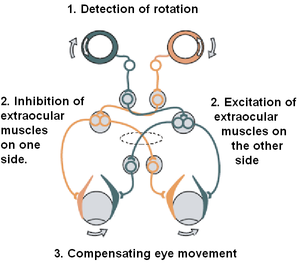
The vestibular system, in vertebrates, is a sensory system that creates the sense of balance and spatial orientation for the purpose of coordinating movement with balance. Together with the cochlea, a part of the auditory system, it constitutes the labyrinth of the inner ear in most mammals.

<span class="mw-page-title-main">Medial longitudinal fasciculus</span> Nerve tracts in the brainstem

The medial longitudinal fasciculus (MLF) is an area of crossed over tracts, on each side of the brainstem. These bundles of axons are situated near the midline of the brainstem. They are made up of both ascending and descending fibers that arise from a number of sources and terminate in different areas, including the superior colliculus, the vestibular nuclei, and the cerebellum. It contains the interstitial nucleus of Cajal, responsible for oculomotor control, head posture, and vertical eye movement.

<span class="mw-page-title-main">Accommodation reflex</span> Reflex action of the human eye

The accommodation reflex is a reflex action of the eye, in response to focusing on a near object, then looking at a distant object, comprising coordinated changes in vergence, lens shape (accommodation) and pupil size. It is dependent on cranial nerve II, superior centers (interneuron) and cranial nerve III. The change in the shape of the lens is controlled by ciliary muscles inside the eye. Changes in contraction of the ciliary muscles alter the focal distance of the eye, causing nearer or farther images to come into focus on the retina; this process is known as accommodation. The reflex, controlled by the parasympathetic nervous system, involves three responses: pupil constriction, lens accommodation, and convergence.

<span class="mw-page-title-main">Eye movement</span> Movement of the eyes

Eye movement includes the voluntary or involuntary movement of the eyes. Eye movements are used by a number of organisms to fixate, inspect and track visual objects of interests. A special type of eye movement, rapid eye movement, occurs during REM sleep.

<span class="mw-page-title-main">Medial rectus muscle</span> Extraocular muscle that rotates the eye medially

The medial rectus muscle is a muscle in the orbit near the eye. It is one of the extraocular muscles. It originates from the common tendinous ring, and inserts into the anteromedial surface of the eye. It is supplied by the inferior division of the oculomotor nerve (III). It rotates the eye medially (adduction).

<span class="mw-page-title-main">Extraocular muscles</span> Seven extrinsic muscles of the human eye

The extraocular muscles, or extrinsic ocular muscles, are the seven extrinsic muscles of the human eye. Six of the extraocular muscles, the four recti muscles, and the superior and inferior oblique muscles, control movement of the eye and the other muscle, the levator palpebrae superioris, controls eyelid elevation. The actions of the six muscles responsible for eye movement depend on the position of the eye at the time of muscle contraction.

Oscillopsia is a visual disturbance in which objects in the visual field appear to oscillate. The severity of the effect may range from a mild blurring to rapid and periodic jumping. Oscillopsia is an incapacitating condition experienced by many patients with neurological disorders. It may be the result of ocular instability occurring after the oculomotor system is affected, no longer holding images steady on the retina. A change in the magnitude of the vestibulo-ocular reflex due to vestibular disease can also lead to oscillopsia during rapid head movements. Oscillopsia may also be caused by involuntary eye movements such as nystagmus, or impaired coordination in the visual cortex and is one of the symptoms of superior canal dehiscence syndrome. Those affected may experience dizziness and nausea. Oscillopsia can also be used as a quantitative test to document aminoglycoside toxicity. Permanent oscillopsia can arise from an impairment of the ocular system that serves to maintain ocular stability. Paroxysmal oscillopsia can be due to an abnormal hyperactivity in the peripheral ocular or vestibular system.

<span class="mw-page-title-main">Abducens nucleus</span>

The abducens nucleus is the originating nucleus from which the abducens nerve (VI) emerges—a cranial nerve nucleus. This nucleus is located beneath the fourth ventricle in the caudal portion of the pons near the midline, medial to the sulcus limitans.

<span class="mw-page-title-main">Flocculus</span>

The flocculus is a small lobe of the cerebellum at the posterior border of the middle cerebellar peduncle anterior to the biventer lobule. Like other parts of the cerebellum, the flocculus is involved in motor control. It is an essential part of the vestibulo-ocular reflex, and aids in the learning of basic motor skills in the brain.

<span class="mw-page-title-main">Caloric reflex test</span> Test of the vestibulo-ocular reflex

In medicine, the caloric reflex test is a test of the vestibulo-ocular reflex that involves irrigating cold or warm water or air into the external auditory canal. This method was developed by Robert Bárány, who won a Nobel prize in 1914 for this discovery.

<span class="mw-page-title-main">Vestibulospinal tract</span> Neural tract in the central nervous system

The vestibulospinal tract is a neural tract in the central nervous system. Specifically, it is a component of the extrapyramidal system and is classified as a component of the medial pathway. Like other descending motor pathways, the vestibulospinal fibers of the tract relay information from nuclei to motor neurons. The vestibular nuclei receive information through the vestibulocochlear nerve about changes in the orientation of the head. The nuclei relay motor commands through the vestibulospinal tract. The function of these motor commands is to alter muscle tone, extend, and change the position of the limbs and head with the goal of supporting posture and maintaining balance of the body and head.

<span class="mw-page-title-main">Cranial nerve examination</span> Type of neurological examination

The cranial nerve exam is a type of neurological examination. It is used to identify problems with the cranial nerves by physical examination. It has nine components. Each test is designed to assess the status of one or more of the twelve cranial nerves (I-XII). These components correspond to testing the sense of smell (I), visual fields and acuity (II), eye movements and pupils, sensory function of face (V), strength of facial (VII) and shoulder girdle muscles (XI), hearing and balance, taste, pharyngeal movement and reflex, tongue movements (XII).

Conjugate gaze palsies are neurological disorders affecting the ability to move both eyes in the same direction. These palsies can affect gaze in a horizontal, upward, or downward direction. These entities overlap with ophthalmoparesis and ophthalmoplegia.

Listing's law, named after German mathematician Johann Benedict Listing (1808–1882), describes the three-dimensional orientation of the eye and its axes of rotation. Listing's law has been shown to hold when the head is stationary and upright and gaze is directed toward far targets, i.e., when the eyes are either fixating, making saccades, or pursuing moving visual targets.

The term gaze is frequently used in physiology to describe coordinated motion of the eyes and neck. The lateral gaze is controlled by the paramedian pontine reticular formation (PPRF). The vertical gaze is controlled by the rostral interstitial nucleus of medial longitudinal fasciculus and the interstitial nucleus of Cajal.

The righting reflex, also known as the labyrinthine righting reflex, is a reflex that corrects the orientation of the body when it is taken out of its normal upright position. It is initiated by the vestibular system, which detects that the body is not erect and causes the head to move back into position as the rest of the body follows. The perception of head movement involves the body sensing linear acceleration or the force of gravity through the otoliths, and angular acceleration through the semicircular canals. The reflex uses a combination of visual system inputs, vestibular inputs, and somatosensory inputs to make postural adjustments when the body becomes displaced from its normal vertical position. These inputs are used to create what is called an efference copy. This means that the brain makes comparisons in the cerebellum between expected posture and perceived posture, and corrects for the difference. The reflex takes 6 or 7 weeks to perfect, but can be affected by various types of balance disorders.

<span class="mw-page-title-main">Corticomesencephalic tract</span>

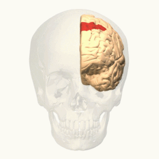
In neuroanatomy, corticomesencephalic tract is a descending nerve tract that originates in the frontal eye field and terminate in the midbrain. Its fibers mediate conjugate eye movement.

References

  1. "Vestibular nystagmus". www.dizziness-and-balance.com.
  2. 1 2 "Sensory Reception: Human Vision: Structure and function of the Human Eye" vol. 27, p. 179 Encyclopædia Britannica, 1987
  3. 1 2 Crawford JD, Vilis T (March 1991). "Axes of eye rotation and Listing's law during rotations of the head". Journal of Neurophysiology. 65 (3): 407–23. doi:10.1152/jn.1991.65.3.407. PMID   2051188.
  4. "VOR (Slow and Fast) | NOVEL – Daniel Gold Collection". collections.lib.utah.edu. Retrieved 2019-10-03.
  5. 1 2 Angelaki DE (July 2004). "Eyes on target: what neurons must do for the vestibuloocular reflex during linear motion". Journal of Neurophysiology. 92 (1): 20–35. doi:10.1152/jn.00047.2004. PMID   15212435. S2CID   15755814.
  6. Straka H, Dieringer N (July 2004). "Basic organization principles of the VOR: lessons from frogs". Progress in Neurobiology. 73 (4): 259–309. doi:10.1016/j.pneurobio.2004.05.003. PMID   15261395. S2CID   38651254.
  7. Cannon SC, Robinson DA (May 1987). "Loss of the neural integrator of the oculomotor system from brain stem lesions in monkey". Journal of Neurophysiology. 57 (5): 1383–409. doi:10.1152/jn.1987.57.5.1383. PMID   3585473.
  8. Crawford JD, Cadera W, Vilis T (June 1991). "Generation of torsional and vertical eye position signals by the interstitial nucleus of Cajal". Science. 252 (5012): 1551–3. Bibcode:1991Sci...252.1551C. doi:10.1126/science.2047862. PMID   2047862. S2CID   15724175.
  9. Aw ST, Halmagyi GM, Haslwanter T, Curthoys IS, Yavor RA, Todd MJ (December 1996). "Three-dimensional vector analysis of the human vestibuloocular reflex in response to high-acceleration head rotations. II. responses in subjects with unilateral vestibular loss and selective semicircular canal occlusion". Journal of Neurophysiology. 76 (6): 4021–30. doi:10.1152/jn.1996.76.6.4021. PMID   8985897.
  10. "PsycNET". psycnet.apa.org. Retrieved 2018-05-15.
  11. Ackerley R, Barnes GR (April 2011). "The interaction of visual, vestibular and extra-retinal mechanisms in the control of head and gaze during head-free pursuit". The Journal of Physiology. 589 (Pt 7): 1627–42. doi:10.1113/jphysiol.2010.199471. PMC   3099020 . PMID   21300755.
  12. Schmäl F, Thiede O, Stoll W (September 2003). "Effect of ethanol on visual-vestibular interactions during vertical linear body acceleration". Alcoholism: Clinical and Experimental Research. 27 (9): 1520–6. doi:10.1097/01.ALC.0000087085.98504.8C. PMID   14506414.
  13. Gold D. "VOR (Slow and Fast)". Neuro-Ophthalmology Virtual Education Library (NOVEL): Daniel Gold Collection. Spencer S. Eccles Health Sciences Library. Retrieved 20 November 2019.
  14. McGarvie LA, MacDougall HG, Halmagyi GM, Burgess AM, Weber KP, Curthoys IS (2015-07-08). "The Video Head Impulse Test (vHIT) of Semicircular Canal Function – Age-Dependent Normative Values of VOR Gain in Healthy Subjects". Frontiers in Neurology. 6: 154. doi: 10.3389/fneur.2015.00154 . PMC   4495346 . PMID   26217301.
  15. Oram J, Murphy P (2011-06-01). "Diagnosis of death". Continuing Education in Anaesthesia, Critical Care & Pain. 11 (3): 77–81. doi: 10.1093/bjaceaccp/mkr008 . ISSN   1743-1816.
  16. Schubert, Michael C. (December 2010) "The cervico-ocular reflex". Handbook of Clinical Neurophysiology.
  17. Kelders, W P A ; Kleinrensink; G J , van der Geest, J N ; Feenstra, L ; de Zeeuw, C I ; Frens, M. (November 2003). Compensatory increase of the cervico-ocular reflex with age in healthy humans.
  18. Money K, Correia M (June 1972). "The vestibular system of the owl". Comparative Biochemistry and Physiology Part A: Physiology. 42 (2): 353–358. doi:10.1016/0300-9629(72)90116-8. PMID   4404369.