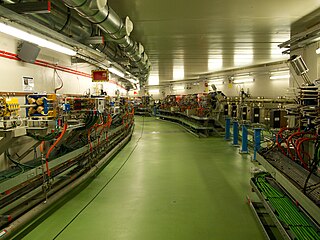
The hard X-ray nanoprobe at the Center for Nanoscale Materials (CNM), Argonne National Lab advanced the state of the art by providing a hard X-ray microscopy beamline with the highest spatial resolution in the world. It provides for fluorescence, diffraction, and transmission imaging with hard X-rays at a spatial resolution of 30nm or better. A dedicated source, beamline, and optics form the basis for these capabilities. This unique instrument is not only key to the specific research areas of the CNM; it will also be a general utility, available to the broader nanoscience community in studying nanomaterials and nanostructures, particularly for embedded structures.
The combination of diffraction, fluorescence, and transmission contrast in a single tool provides unique characterization capabilities for nanoscience. Current hard X-ray microprobes based on Fresnel zone plate optics have demonstrated a spatial resolution of 150nm at a photon energy of 8-10 keV. With advances in the fabrication of zone plate optics, coupled with an optimized beamline design, the performance goal is a spatial resolution of 30nm. The nanoprobe covers the spectral range of 3-30 keV, and the working distance between the focusing optics and the sample are typically in the range of 10–20mm.
Modes of operation
Transmission. In this mode, either attenuation or phase shift of the X-ray beam by the sample can be measured. Absorption contrast can be used to map the sample’s density. Particular elemental constituents can be located using measurements on each side of an absorption edge to give an element-specific difference image with moderate sensitivity. Phase-contrast imaging can be sensitive to internal structure even when absorption is low and can be enhanced by tuning the X-ray energy.
Fluorescence. Induced X-ray fluorescence reveals the spatial distribution of individual elements in a sample. Because an X-ray probe offers 1,000 times higher sensitivity than electron probes, the fluorescence technique is a powerful tool for quantitative trace element analysis, important for understanding material properties such as second-phase particles, defects, and interfacial segregation.
Spectroscopy. In spectroscopy mode, the primary X-ray beam’s energy is scanned across the absorption edge of an element, providing information on its chemical state (XANES) or its local environment (EXAFS), which allows the study of disordered samples.
Polarization. Both linearly and circularly polarized X-rays will be available. Contrast due to polarization is invaluable in distinguishing fluorescence and diffraction signals and imaging magnetic domain structure by using techniques such as linear and circular dichroism and magnetic diffraction.
Tomography. In X-ray tomography, one of these modes is combined with sample rotation to produce a series of two-dimensional projection images, to be used for reconstructing the sample’s internal three-dimensional structure. This will be particularly important for observing the morphology of complex nanostructures.
In summary, a hard X-ray nanoprobe provides advantages such as being noninvasive and quantitative, requiring minimal sample preparation, giving sub-optical spatial resolution, having the ability to penetrate inside a sample and study its internal structure, and having enhanced ability to study processes in situ. Another important distinction from charged-particle probes is that X-rays do not interact with applied electric or magnetic fields, which is an advantage for in-field studies. The design of the nanoprobe beamline aims to preserve these potential advantages.
General nanomaterials characterization with X-rays, including small-angle scattering (SAXS)
Related Research Articles
Microscopy is the technical field of using microscopes to view objects and areas of objects that cannot be seen with the naked eye. There are three well-known branches of microscopy: optical, electron, and scanning probe microscopy, along with the emerging field of X-ray microscopy.
A microscope is a laboratory instrument used to examine objects that are too small to be seen by the naked eye. Microscopy is the science of investigating small objects and structures using a microscope. Microscopic means being invisible to the eye unless aided by a microscope.
The optical microscope, also referred to as a light microscope, is a type of microscope that commonly uses visible light and a system of lenses to generate magnified images of small objects. Optical microscopes are the oldest design of microscope and were possibly invented in their present compound form in the 17th century. Basic optical microscopes can be very simple, although many complex designs aim to improve resolution and sample contrast.
Transmission electron microscopy (TEM) is a microscopy technique in which a beam of electrons is transmitted through a specimen to form an image. The specimen is most often an ultrathin section less than 100 nm thick or a suspension on a grid. An image is formed from the interaction of the electrons with the sample as the beam is transmitted through the specimen. The image is then magnified and focused onto an imaging device, such as a fluorescent screen, a layer of photographic film, or a sensor such as a scintillator attached to a charge-coupled device.
A synchrotron light source is a source of electromagnetic radiation (EM) usually produced by a storage ring, for scientific and technical purposes. First observed in synchrotrons, synchrotron light is now produced by storage rings and other specialized particle accelerators, typically accelerating electrons. Once the high-energy electron beam has been generated, it is directed into auxiliary components such as bending magnets and insertion devices in storage rings and free electron lasers. These supply the strong magnetic fields perpendicular to the beam which are needed to convert high energy electrons into photons.
An X-ray microscope uses electromagnetic radiation in the X-ray band to produce magnified images of objects. Since X-rays penetrate most objects, there is no need to specially prepare them for X-ray microscopy observations.
Phase-contrast imaging is a method of imaging that has a range of different applications. It measures differences in the refractive index of different materials to differentiate between structures under analysis. In conventional light microscopy, phase contrast can be employed to distinguish between structures of similar transparency, and to examine crystals on the basis of their double refraction. This has uses in biological, medical and geological science. In X-ray tomography, the same physical principles can be used to increase image contrast by highlighting small details of differing refractive index within structures that are otherwise uniform. In transmission electron microscopy (TEM), phase contrast enables very high resolution (HR) imaging, making it possible to distinguish features a few Angstrom apart.
High-energy X-rays or HEX-rays are very hard X-rays, with typical energies of 80–1000 keV (1 MeV), about one order of magnitude higher than conventional X-rays used for X-ray crystallography. They are produced at modern synchrotron radiation sources such as the beamline ID15 at the European Synchrotron Radiation Facility (ESRF). The main benefit is the deep penetration into matter which makes them a probe for thick samples in physics and materials science and permits an in-air sample environment and operation. Scattering angles are small and diffraction directed forward allows for simple detector setups.
Near-field scanning optical microscopy (NSOM) or scanning near-field optical microscopy (SNOM) is a microscopy technique for nanostructure investigation that breaks the far field resolution limit by exploiting the properties of evanescent waves. In SNOM, the excitation laser light is focused through an aperture with a diameter smaller than the excitation wavelength, resulting in an evanescent field on the far side of the aperture. When the sample is scanned at a small distance below the aperture, the optical resolution of transmitted or reflected light is limited only by the diameter of the aperture. In particular, lateral resolution of 6 nm and vertical resolution of 2–5 nm have been demonstrated.
X-ray optics is the branch of optics that manipulates X-rays instead of visible light. It deals with focusing and other ways of manipulating the X-ray beams for research techniques such as X-ray crystallography, X-ray fluorescence, small-angle X-ray scattering, X-ray microscopy, X-ray phase-contrast imaging, and X-ray astronomy.
ALBA is a third-generation synchrotron light source facility located in the Barcelona Synchrotron Park in Cerdanyola del Vallès near Barcelona, in Catalonia (Spain). It was constructed and is operated by CELLS, and co-financed by the Spanish central administration and regional Catalan Government.
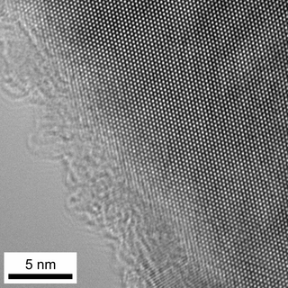
High-resolution transmission electron microscopy is an imaging mode of specialized transmission electron microscopes that allows for direct imaging of the atomic structure of samples. It is a powerful tool to study properties of materials on the atomic scale, such as semiconductors, metals, nanoparticles and sp2-bonded carbon. While this term is often also used to refer to high resolution scanning transmission electron microscopy, mostly in high angle annular dark field mode, this article describes mainly the imaging of an object by recording the two-dimensional spatial wave amplitude distribution in the image plane, in analogy to a "classic" light microscope. For disambiguation, the technique is also often referred to as phase contrast transmission electron microscopy. At present, the highest point resolution realised in phase contrast transmission electron microscopy is around 0.5 ångströms (0.050 nm). At these small scales, individual atoms of a crystal and its defects can be resolved. For 3-dimensional crystals, it may be necessary to combine several views, taken from different angles, into a 3D map. This technique is called electron crystallography.
An X-ray microscope uses electromagnetic radiation in the soft X-ray band to produce images of very small objects.
Diffraction topography is a quantum beam imaging technique based on Bragg diffraction. Diffraction topographic images ("topographies") record the intensity profile of a beam of X-rays diffracted by a crystal. A topography thus represents a two-dimensional spatial intensity mapping of reflected X-rays, i.e. the spatial fine structure of a Laue reflection. This intensity mapping reflects the distribution of scattering power inside the crystal; topographs therefore reveal the irregularities in a non-ideal crystal lattice. X-ray diffraction topography is one variant of X-ray imaging, making use of diffraction contrast rather than absorption contrast which is usually used in radiography and computed tomography (CT). Topography is exploited to a lesser extends with neutrons and other quantum beams. In the electron microscope community, such technique is called dark field imaging or diffraction contrast imaging.
The National Synchrotron Light Source II (NSLS-II) at Brookhaven National Laboratory (BNL) in Upton, New York is a national user research facility funded primarily by the U.S. Department of Energy's (DOE) Office of Science. NSLS-II is one of the world's most advanced synchrotron light sources, designed to produce x-rays 10,000 times brighter than BNL's original light source, the National Synchrotron Light Source (NSLS). NSLS-II supports basic and applied research in energy security, advanced materials synthesis and manufacturing, environment, and human health.
The technique of vibrational analysis with scanning probe microscopy allows probing vibrational properties of materials at the submicrometer scale, and even of individual molecules. This is accomplished by integrating scanning probe microscopy (SPM) and vibrational spectroscopy. This combination allows for much higher spatial resolution than can be achieved with conventional Raman/FTIR instrumentation. The technique is also nondestructive, requires non-extensive sample preparation, and provides more contrast such as intensity contrast, polarization contrast and wavelength contrast, as well as providing specific chemical information and topography images simultaneously.
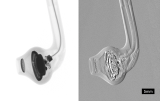
Phase-contrast X-ray imaging or phase-sensitive X-ray imaging is a general term for different technical methods that use information concerning changes in the phase of an X-ray beam that passes through an object in order to create its images. Standard X-ray imaging techniques like radiography or computed tomography (CT) rely on a decrease of the X-ray beam's intensity (attenuation) when traversing the sample, which can be measured directly with the assistance of an X-ray detector. However, in phase contrast X-ray imaging, the beam's phase shift caused by the sample is not measured directly, but is transformed into variations in intensity, which then can be recorded by the detector.
Super-resolution dipole orientation mapping (SDOM) is a form of fluorescence polarization microscopy (FPM) that achieved super resolution through polarization demodulation. It was first described by Karl Zhanghao and others in 2016. Fluorescence polarization (FP) is related to the dipole orientation of chromophores, making fluorescence polarization microscopy possible to reveal structures and functions of tagged cellular organelles and biological macromolecules. In addition to fluorescence intensity, wavelength, and lifetime, the fourth dimension of fluorescence—polarization—can also provide intensity modulation without the restriction to specific fluorophores; its investigation in super-resolution microscopy is still in its infancy.
Solaris is the only synchrotron in Central-Eastern Europe. Built in Poland in 2015, under the auspices of the Jagiellonian University, it is located on the Campus of the 600th Anniversary of the Jagiellonian University Revival, in the southern part of Krakow. It is the central facility of the National Center of Synchrotron Radiation SOLARIS.
This page is based on this Wikipedia article Text is available under the CC BY-SA 4.0 license; additional terms may apply. Images, videos and audio are available under their respective licenses.