
Histology, also known as microscopic anatomy or microanatomy, is the branch of biology which studies the microscopic anatomy of biological tissues. Histology is the microscopic counterpart to gross anatomy, which looks at larger structures visible without a microscope. Although one may divide microscopic anatomy into organology, the study of organs, histology, the study of tissues, and cytology, the study of cells, modern usage places all of these topics under the field of histology. In medicine, histopathology is the branch of histology that includes the microscopic identification and study of diseased tissue. In the field of paleontology, the term paleohistology refers to the histology of fossil organisms.
In chemistry, a salt is a chemical compound consisting of an ionic assembly of positively charged cations and negatively charged anions, which results in a compound with no net electric charge. A common example is table salt, with positively charged sodium ions and negatively charged chloride ions.
A primary standard in metrology is a standard that is sufficiently accurate such that it is not calibrated by or subordinate to other standards. Primary standards are defined via other quantities like length, mass and time. Primary standards are used to calibrate other standards referred to as working standards. See Hierarchy of Standards.

A corrosive substance is one that will damage or destroy other substances with which it comes into contact by means of a chemical reaction.

A mordant or dye fixative is a substance used to set dyes on fabrics by forming a coordination complex with the dye, which then attaches to the fabric. It may be used for dyeing fabrics or for intensifying stains in cell or tissue preparations. Although mordants are still used, especially by small batch dyers, it has been largely displaced in industry by directs.

Staining is a technique used to enhance contrast in samples, generally at the microscopic level. Stains and dyes are frequently used in histology, in cytology, and in the medical fields of histopathology, hematology, and cytopathology that focus on the study and diagnoses of diseases at the microscopic level. Stains may be used to define biological tissues, cell populations, or organelles within individual cells.
Inorganic ions in animals and plants are ions necessary for vital cellular activity. In body tissues, ions are also known as electrolytes, essential for the electrical activity needed to support muscle contractions and neuron activation. They contribute to osmotic pressure of body fluids as well as performing a number of other important functions. Below is a list of some of the most important ions for living things as well as examples of their functions:

Potassium dichromate, K2Cr2O7, is a common inorganic chemical reagent, most commonly used as an oxidizing agent in various laboratory and industrial applications. As with all hexavalent chromium compounds, it is acutely and chronically harmful to health. It is a crystalline ionic solid with a very bright, red-orange color. The salt is popular in the laboratory because it is not deliquescent, in contrast to the more industrially relevant salt sodium dichromate.

Murashige and Skoog medium is a plant growth medium used in the laboratories for cultivation of plant cell culture. MSO was invented by plant scientists Toshio Murashige and Folke K. Skoog in 1962 during Murashige's search for a new plant growth regulator. A number behind the letters MS is used to indicate the sucrose concentration of the medium. For example, MS0 contains no sucrose and MS20 contains 20 g/l sucrose. Along with its modifications, it is the most commonly used medium in plant tissue culture experiments in the laboratory, but according to recent scientific findings it should not be used as nutrient solution in deep water culture.

Paraformaldehyde (PFA) is the smallest polyoxymethylene, the polymerization product of formaldehyde with a typical degree of polymerization of 8–100 units. Paraformaldehyde commonly has a slight odor of formaldehyde due to decomposition. Paraformaldehyde is a poly-acetal.

Masson's trichrome is a three-colour staining procedure used in histology. The recipes evolved from Claude L. Pierre Masson's (1880–1959) original formulation have different specific applications, but all are suited for distinguishing cells from surrounding connective tissue.

Phosphotungstic acid haematoxylin (PTAH) is a mix of haematoxylin with phosphotungstic acid, used in histology for staining.
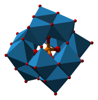
Phosphotungstic acid (PTA) or tungstophosphoric acid (TPA), is a heteropoly acid with the chemical formula H3PW12O40]. It forms hydrates H3[PW12O40]·nH2O. It is normally isolated as the n = 24 hydrate but can be desiccated to the hexahydrate. EPTA is the name of ethanolic phosphotungstic acid, its alcohol solution used in biology. It has the appearance of small, colorless-grayish or slightly yellow-green crystals, with melting point 89 °C. It is odorless and soluble in water. It is not especially toxic, but is a mild acidic irritant. The compound is known by a variety of names and acronyms.
This is the list of extremely hazardous substances defined in Section 302 of the U.S. Emergency Planning and Community Right-to-Know Act. The list can be found as an appendix to 40 C.F.R. 355. Updates as of 2006 can be seen on the Federal Register, 71 FR 47121.

Potassium acetate (CH3COOK) is the potassium salt of acetic acid. It is a hygroscopic solid at room temperature.
In the fields of histology, pathology, and cell biology, fixation is the preservation of biological tissues from decay due to autolysis or putrefaction. It terminates any ongoing biochemical reactions and may also increase the treated tissues' mechanical strength or stability. Tissue fixation is a critical step in the preparation of histological sections, its broad objective being to preserve cells and tissue components and to do this in such a way as to allow for the preparation of thin, stained sections. This allows the investigation of the tissues' structure, which is determined by the shapes and sizes of such macromolecules as proteins and nucleic acids.
Carnoy's solution is a fixative composed of 60% ethanol, 30% chloroform and 10% glacial acetic acid, 1 gram of ferric chloride.
Bouin solution, or Bouin's solution, is a compound fixative used in histology. It was invented by French biologist Pol Bouin and is composed of picric acid, acetic acid and formaldehyde in an aqueous solution. Bouin's fluid is especially useful for fixation of gastrointestinal tract biopsies because this fixative allows crisper and better nuclear staining than 10% neutral-buffered formalin. It is not a good fixative when tissue ultrastructure must be preserved for electron microscopy. However, it is a good fixative when tissue structure with a soft and delicate texture must be preserved. The acetic acid in this fixative lyses red blood cells and dissolves small iron and calcium deposits in tissue. A variant in which the acetic acid is replaced with formic acid can be used for both fixation of tissue and decalcification. The effects of the three chemicals in Bouin solution balance each other. Formalin causes cytoplasm to become basophilic but this effect is balanced by the effect of the picric acid. This results in excellent nuclear and cytoplasmic H&E staining. The tissue hardening effect of formalin is balanced by the soft tissue fixation of picric and acetic acids. The tissue swelling effect of acetic acid is balanced by the tissue shrinking effect of picric acid.