Related Research Articles

Dentures are prosthetic devices constructed to replace missing teeth, supported by the surrounding soft and hard tissues of the oral cavity. Conventional dentures are removable. However, there are many denture designs, some of which rely on bonding or clasping onto teeth or dental implants. There are two main categories of dentures, the distinction being whether they fit onto the mandibular arch or on the maxillary arch.

The gums or gingiva consist of the mucosal tissue that lies over the mandible and maxilla inside the mouth. Gum health and disease can have an effect on general health.

A bridge is a fixed dental restoration used to replace one or more missing teeth by joining an artificial tooth definitively to adjacent teeth or dental implants.

In dentistry, a crown or a dental cap is a type of dental restoration that completely caps or encircles a tooth or dental implant. A crown may be needed when a large dental cavity threatens the health of a tooth. A crown is typically bonded to the tooth by dental cement. They can be made from various materials, which are usually fabricated using indirect methods. Crowns are used to improve the strength or appearance of teeth and to halt deterioration. While beneficial to dental health, the procedure and materials can be costly.
Dental explorers, also known as sickle probes, are tools found in the dental arsenal that are frequently utilised. The explorer is designed with a sharp tip at the end to improve tactile perception.
Periodontology or periodontics is the specialty of dentistry that studies supporting structures of teeth, as well as diseases and conditions that affect them. The supporting tissues are known as the periodontium, which includes the gingiva (gums), alveolar bone, cementum, and the periodontal ligament. A periodontist is a dentist that specializes in the prevention, diagnosis and treatment of periodontal disease and in the placement of dental implants.
A removable partial denture (RPD) is a denture for a partially edentulous patient who desires to have replacement teeth for functional or aesthetic reasons and who cannot have a bridge for any reason, such as a lack of required teeth to serve as support for a bridge or financial limitations.

A periodontal probe is an instrument in dentistry commonly used in the dental armamentarium. It is usually long, thin, and blunted at the end. Its main function is to evaluate the depth of the pockets surrounding a tooth in order to determine the periodontium's overall health. For accuracy and readability, the instrument's head has markings written on it.

The maxillary central incisor is a human tooth in the front upper jaw, or maxilla, and is usually the most visible of all teeth in the mouth. It is located mesial to the maxillary lateral incisor. As with all incisors, their function is for shearing or cutting food during mastication (chewing). There is typically a single cusp on each tooth, called an incisal ridge or incisal edge. Formation of these teeth begins at 14 weeks in utero for the deciduous (baby) set and 3–4 months of age for the permanent set.

The mandibular second premolar is the tooth located distally from both the mandibular first premolars of the mouth but mesial from both mandibular first molars. The function of this premolar is assist the mandibular first molar during mastication, commonly known as chewing. Mandibular second premolars have three cusps. There is one large cusp on the buccal side of the tooth. The lingual cusps are well developed and functional. Therefore, whereas the mandibular first premolar resembles a small canine, the mandibular second premolar is more alike to the first molar. There are no deciduous (baby) mandibular premolars. Instead, the teeth that precede the permanent mandibular premolars are the deciduous mandibular molars.
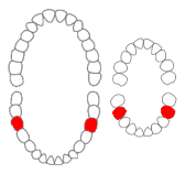
The mandibular first molar or six-year molar is the tooth located distally from both the mandibular second premolars of the mouth but mesial from both mandibular second molars. It is located on the mandibular (lower) arch of the mouth, and generally opposes the maxillary (upper) first molars and the maxillary 2nd premolar in normal class I occlusion. The function of this molar is similar to that of all molars in regard to grinding being the principal action during mastication, commonly known as chewing. There are usually five well-developed cusps on mandibular first molars: two on the buccal, two lingual, and one distal. The shape of the developmental and supplementary grooves, on the occlusal surface, are described as being 'M' shaped. There are great differences between the deciduous (baby) mandibular molars and those of the permanent mandibular molars, even though their function are similar. The permanent mandibular molars are not considered to have any teeth that precede it. Despite being named molars, the deciduous molars are followed by permanent premolars.

Toothlessness, or edentulism, is the condition of having no teeth. In organisms that naturally have teeth, it is the result of tooth loss.

A palatal lift prosthesis is a prosthesis that addresses a condition referred to as palatopharyngeal incompetence. Palatopharyngeal incompetence broadly refers to a muscular inability to sufficiently close the port between the nasopharynx and oropharynx during speech and/or swallowing. An inability to adequately close the palatopharyngeal port during speech results in hypernasalance that, depending upon its severity, can render speakers difficult to understand or unintelligible. The potential for compromised intelligibility secondary to hypernasalance is underscored when consideration is given to the fact that only three English language phonemes – /m/, /n/, and /ng/ – are pronounced with an open palatopharyngeal port. Furthermore, an impaired ability to effect a closure of the palatopharyngeal port while swallowing can result in the nasopharyngeal regurgitation of liquid or solid boluses.

The gingival sulcus is an area of potential space between a tooth and the surrounding gingival tissue and is lined by sulcular epithelium. The depth of the sulcus is bounded by two entities: apically by the gingival fibers of the connective tissue attachment and coronally by the free gingival margin. A healthy sulcular depth is three millimeters or less, which is readily self-cleansable with a properly used toothbrush or the supplemental use of other oral hygiene aids.
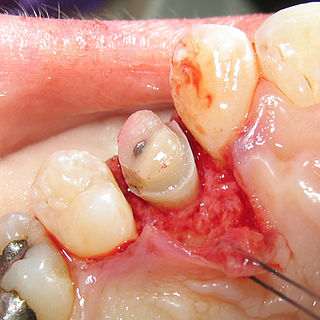
Crown lengthening is a surgical procedure performed by a dentist, or more frequently a periodontist, where more tooth is exposed by removing some of the gingival margin (gum) and supporting bone. Crown lengthening can also be achieved orthodontically by extruding the tooth.
A post and core crown is a type of dental restoration required where there is an inadequate amount of sound tooth tissue remaining to retain a conventional crown. A post is cemented into a prepared root canal, which retains a core restoration, which retains the final crown.
Gingivectomy is a dental procedure in which a dentist or oral surgeon cuts away part of the gums in the mouth.
A gum lift is a cosmetic dental procedure that raises and sculpts the gum line. This procedure involves reshaping the tissue and/or underlying bones to create the appearance of longer or symmetrical teeth, thereby making the smile more aesthetically pleasing. This procedure is typically done to reduce excessively gummy smiles or to balance out an asymmetrical gum line. The procedure, also known as crown-lengthening, has historically been used to treat gum disease. It is only within the past three to five years that dentists have commonly used this procedure for aesthetic purposes. The practice of cosmetic gum lifts was first developed in the late 1980s, but there were few oral surgeons and dental practitioners available to perform the procedures. Gum lifts can also include bone shaping to reduce the prominence of the upper jaw and even out the tooth and gum ratio. This method provides permanent results, while simple gum contouring may result in relapse or regrowth of the gingiva.
A complete denture is a removable appliance used when all teeth within a jaw have been lost and need to be prosthetically replaced. In contrast to a partial denture, a complete denture is constructed when there are no more teeth left in an arch, hence it is an exclusively tissue-supported prosthesis. A complete denture can be opposed by natural dentition, a partial or complete denture, fixed appliances or, sometimes, soft tissues.

Overdenture is any removable dental prosthesis that covers and rests on one or more remaining natural teeth, the roots of natural teeth, and/or dental implants. It is one of the most practical measures used in preventive dentistry. Overdentures can be either tooth supported or implant supported. It is found to help in the preservation of alveolar bone and delay the process of complete edentulism.
References
| | This dentistry article is a stub. You can help Wikipedia by expanding it. |