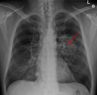
Lung cancer, also known as lung carcinoma, is a malignant lung tumor characterized by uncontrolled cell growth in tissues of the lung. This growth can spread beyond the lung by the process of metastasis into nearby tissue or other parts of the body. Most cancers that start in the lung, known as primary lung cancers, are carcinomas. The two main types are small-cell lung carcinoma (SCLC) and non-small-cell lung carcinoma (NSCLC). The most common symptoms are coughing, weight loss, shortness of breath, and chest pains.

The lungs are the primary organs of the respiratory system in humans and many other animals including a few fish and some snails. In mammals and most other vertebrates, two lungs are located near the backbone on either side of the heart. Their function in the respiratory system is to extract oxygen from the atmosphere and transfer it into the bloodstream, and to release carbon dioxide from the bloodstream into the atmosphere, in a process of gas exchange. Respiration is driven by different muscular systems in different species. Mammals, reptiles and birds use their different muscles to support and foster breathing. In early tetrapods, air was driven into the lungs by the pharyngeal muscles via buccal pumping, a mechanism still seen in amphibians. In humans, the main muscle of respiration that drives breathing is the diaphragm. The lungs also provide airflow that makes vocal sounds including human speech possible.

Pathology is the study of the causes and effects of disease or injury. The word pathology also refers to the study of disease in general, incorporating a wide range of bioscience research fields and medical practices. However, when used in the context of modern medical treatment, the term is often used in a more narrow fashion to refer to processes and tests which fall within the contemporary medical field of "general pathology," an area which includes a number of distinct but inter-related medical specialties that diagnose disease, mostly through analysis of tissue, cell, and body fluid samples. Idiomatically, "a pathology" may also refer to the predicted or actual progression of particular diseases, and the affix path is sometimes used to indicate a state of disease in cases of both physical ailment and psychological conditions. A physician practicing pathology is called a pathologist.
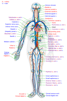
The circulatory system, also called the cardiovascular system or the vascular system, is an organ system that permits blood to circulate and transport nutrients, oxygen, carbon dioxide, hormones, and blood cells to and from the cells in the body to provide nourishment and help in fighting diseases, stabilize temperature and pH, and maintain homeostasis.

Cytopathology is a branch of pathology that studies and diagnoses diseases on the cellular level. The discipline was founded by George Nicolas Papanicolaou in 1928. Cytopathology is generally used on samples of free cells or tissue fragments, in contrast to histopathology, which studies whole tissues.

Berylliosis, or chronic beryllium disease (CBD), is a chronic allergic-type lung response and chronic lung disease caused by exposure to beryllium and its compounds, a form of beryllium poisoning. It is distinct from acute beryllium poisoning, which became rare following occupational exposure limits established around 1950. Berylliosis is an occupational lung disease.
Hemoptysis is the coughing up of blood or blood-stained mucus from the bronchi, larynx, trachea, or lungs. This can occur with lung cancer, infections such as tuberculosis, bronchitis, or pneumonia, and certain cardiovascular conditions. Hemoptysis is considered massive at 300 mL. In such cases, there are always severe injuries. The primary danger comes from choking, rather than blood loss.

Sputum is mucus and is the name used for the coughed-up material (phlegm) from the lower airways. In medicine, sputum samples are usually used for naked eye exam, microbiological investigations of respiratory infections, and cytological investigations of respiratory systems. It is critical that the patient not give a specimen that includes any mucoid material from the interior of the nose. Naked eye exam of sputum can be done at home by a patient in order to note the various colors. Any hint of yellow color suggests an airway infection. Such color hints are best detected when the sputum is viewed on a very white background such as white paper, a white pot, or a white sink surface. The more intense the yellow color, the more likely it is a bacterial infection.

A biopsy is a medical test commonly performed by a surgeon, interventional radiologist, or an interventional cardiologist involving extraction of sample cells or tissues for examination to determine the presence or extent of a disease. The tissue is generally examined under a microscope by a pathologist, and can also be analyzed chemically. When an entire lump or suspicious area is removed, the procedure is called an excisional biopsy. An incisional biopsy or core biopsy samples a portion of the abnormal tissue without attempting to remove the entire lesion or tumor. When a sample of tissue or fluid is removed with a needle in such a way that cells are removed without preserving the histological architecture of the tissue cells, the procedure is called a needle aspiration biopsy. Biopsies are most commonly performed for insight into possible cancerous and inflammatory conditions.

A pleural effusion is excess fluid that accumulates in the pleural cavity, the fluid-filled space that surrounds the lungs. This excess fluid can impair breathing by limiting the expansion of the lungs. Various kinds of pleural effusion, depending on the nature of the fluid and what caused its entry into the pleural space, are hydrothorax, hemothorax (blood), urinothorax (urine), chylothorax (chyle), or pyothorax (pus). A pneumothorax is the accumulation of air in the pleural space, and is commonly called a "collapsed lung".

A pulmonary artery is an artery in the pulmonary circulation that carries deoxygenated blood from the right side of the heart to the lungs. The largest pulmonary artery is the main pulmonary artery. or pulmonary trunk from the heart, and the smallest ones are the arterioles, which lead to the capillaries that surround the pulmonary alveoli.
Leukapheresis is a laboratory procedure in which white blood cells are separated from a sample of blood. It is a specific type of apheresis, the more general term for separating out one particular constituent of blood and returning the remainder to the circulation.
Mediastinoscopy is a procedure that enables visualization of the contents of the mediastinum, usually for the purpose of obtaining a biopsy. Mediastinoscopy is often used for staging of lymph nodes of lung cancer or for diagnosing other conditions affecting structures in the mediastinum such as sarcoidosis or lymphoma.

Respiratory disease is a medical term that encompasses pathological conditions affecting the organs and tissues that make gas exchange difficult in higher organisms, and includes conditions of the upper respiratory tract, trachea, bronchi, bronchioles, alveoli, pleura and pleural cavity, and the nerves and muscles of breathing. Respiratory diseases range from mild and self-limiting, such as the common cold, to life-threatening entities like bacterial pneumonia, pulmonary embolism, acute asthma and lung cancer.

Hemosiderosis is a form of iron overload disorder resulting in the accumulation of hemosiderin.

Geotrichosis is a mycosis caused by Geotrichum candidum.

Fetal adenocarcinoma (FA) of the lung is a rare subtype of pulmonary adenocarcinoma that exhibits tissue architecture and cell characteristics that resemble fetal lung tissue upon microscopic examination. It is currently considered a variant of solid adenocarcinoma with mucin production.

In medicine, sampling is gathering of matter from the body to aid in the process of a medical diagnosis and/or evaluation of an indication for treatment, further medical tests or other procedures. In this sense, the sample is the gathered matter, and the sampling tool or sampler is the person or material to collect the sample.
Trichomonas tenax, or oral trichomonas, is a species of Trichomonas commonly found in the oral cavity of humans, dogs and cats. Routine hygiene is generally not sufficient to eliminate the parasite, hence its Latin name, meaning "tenacious". The parasite is frequently encountered in periodontal infections, affecting more than 50% of the population in some areas, but it is usually considered insignificant. T. tenax is generally not found on the gums of healthy patients. It is known to play a pathonogenic role in necrotizing ulcerative gingivitis and necrotizing ulcerative periodontitis, worsening preexisting periodontal disease. This parasite is also implicated in some chronic lung diseases; in such cases, removal of the parasite is sufficient to allow recovery.
Diffuse idiopathic pulmonary neuroendocrine cell hyperplasia (DIPNECH) is a diffuse parenchymal lung disease which often presents with symptoms of cough and shortness of breath. The pathological definition published by the World Health Organization is “a generalized proliferation of scattered single cells, small nodules, or linear proliferations of pulmonary neuroendocrine (PNE) cells that may be confined to the bronchial and bronchiolar epithelium.” The true prevalence of this disease is not known. To date, just under 200 cases have been reported in the literature. However, with an increase in recognition of this disease by radiologists and pulmonologists, the number of cases has been increasing. DIPNECH predominantly affects middle-aged women with slowly progressive lung obstruction. DIPNECH is usually discovered in one of two ways: 1) as an unexpected finding following a lung surgery; or 2) by evaluation of a patient in a pulmonary clinic with longstanding, unexplained symptoms.
