Flexor digitorum superficialis is an extrinsic flexor muscle of the fingers at the proximal interphalangeal joints.
Osteophytes are exostoses that form along joint margins. They should not be confused with enthesophytes, which are bony projections that form at the attachment of a tendon or ligament. Osteophytes are not always distinguished from exostoses in any definite way, although in many cases there are a number of differences. Osteophytes are typically intra-articular.
Lateral collateral ligament can refer to:

The extensor digitorum muscle is a muscle of the posterior forearm present in humans and other animals. It extends the medial four digits of the hand. Extensor digitorum is innervated by the posterior interosseous nerve, which is a branch of the radial nerve.
The metacarpophalangeal joints (MCP) are situated between the metacarpal bones and the proximal phalanges of the fingers. These joints are of the condyloid kind, formed by the reception of the rounded heads of the metacarpal bones into shallow cavities on the proximal ends of the proximal phalanges. Being condyloid, they allow the movements of flexion, extension, abduction, adduction and circumduction at the joint.

A hinge joint is a bone joint in which the articular surfaces are molded to each other in such a manner as to permit motion only in one plane. According to one classification system they are said to be uniaxial. The direction which the distal bone takes in this motion is seldom in the same plane as that of the axis of the proximal bone; there is usually a certain amount of deviation from the straight line during flexion.

In human anatomy, the dorsal interossei (DI) are four muscles in the back of the hand that act to abduct (spread) the index, middle, and ring fingers away from hand's midline and assist in flexion at the metacarpophalangeal joints and extension at the interphalangeal joints of the index, middle and ring fingers.

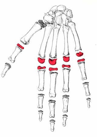
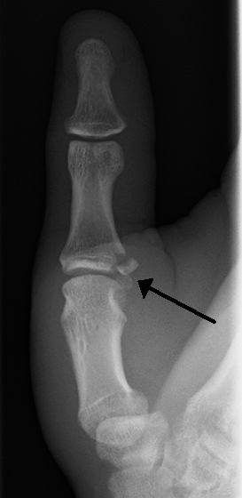
The interphalangeal joints of the hand are the hinge joints between the phalanges of the fingers that provide flexion towards the palm of the hand.

The interphalangeal joints of the foot are between the phalanx bones of the toes in the feet.
Ulnar collateral ligament may refer to:
Gamekeeper's thumb is a type of injury to the ulnar collateral ligament (UCL) of the thumb. The UCL may be merely stretched, or it may be torn from its insertion site into the proximal phalanx of the thumb; in approximately 90% of cases part of the bone is actually avulsed from the joint. This condition is commonly observed among gamekeepers and Scottish fowl hunters, as well as athletes. It also occurs among people who sustain a fall onto an outstretched hand while holding a rod, frequently skiers grasping ski poles.
Collateral ligament can refer to:

The collateral ligaments of interphalangeal joints are ligaments of the interphalangeal joints of the hand. They limit extension at these joints.

In the human hand, palmar or volar plates are found in the metacarpophalangeal (MCP) and interphalangeal (IP) joints, where they reinforce the joint capsules, enhance joint stability, and limit hyperextension. The plates of the MCP and IP joints are structurally and functionally similar, except that in the MCP joints they are interconnected by a deep transverse ligament. In the MCP joints, they also indirectly provide stability to the longitudinal palmar arches of the hand. The volar plate of the thumb MCP joint has a transverse longitudinal rectangular shape, shorter than those in the fingers.
The collateral ligaments of the interphalangeal joints of the foot are fibrous bands that are situated on both sides of the interphalangeal joints of the toes.
In the human foot, the plantar or volar plates are fibrocartilaginous structures found in the metatarsophalangeal (MTP) and interphalangeal (IP) joints. The anatomy and composition of the plantar plates are similar to the palmar plates in the metacarpophalangeal (MCP) and interphalangeal joints in the hand; the proximal origin is thin but the distal insertion is stout. Due to the weight-bearing nature of the human foot, the plantar plates are exposed to extension forces not present in the human hand.
Plantar ligament refer to ligaments in the sole of the foot:

Triphalangeal thumb (TPT) is a congenital malformation where the thumb has three phalanges instead of two. The extra phalangeal bone can vary in size from that of a small pebble to a size comparable to the phalanges in non-thumb digits. The true incidence of the condition is unknown, but is estimated at 1:25,000 live births. In about two-thirds of the patients with triphalangeal thumbs, there is a hereditary component. Besides the three phalanges, there can also be other malformations. It was first described by Columbi in 1559.

The extrinsic extensor muscles of the hand are located in the back of the forearm and have long tendons connecting them to bones in the hand, where they exert their action. Extrinsic denotes their location outside the hand. Extensor denotes their action which is to extend, or open flat, joints in the hand. They include the extensor carpi radialis longus (ECRL), extensor carpi radialis brevis (ECRB), extensor digitorum (ED), extensor digiti minimi (EDM), extensor carpi ulnaris (ECU), abductor pollicis longus (APL), extensor pollicis brevis (EPB), extensor pollicis longus (EPL), and extensor indicis (EI).
Collateral ligament of elbow joint may refer to:
This page is based on this
Wikipedia article Text is available under the
CC BY-SA 4.0 license; additional terms may apply.
Images, videos and audio are available under their respective licenses.