Digital Morphology (DigiMorph), part of the National Science Foundation Digital Libraries Initiative, creates and shares 2D and 3D visualizations of the internal and external structure of living and extinct vertebrates, and a growing number of 'invertebrates.'
The information core for the DigiMorph library is generated using a high-resolution X-ray computed tomographic (X-ray CT) scanner at the University of Texas at Austin. This instrument is comparable to a conventional medical diagnostic CAT scanner, but with greater resolution and penetrating power. The device uses X-rays to take images of thin slices through solid objects, such as bone and rock. Hundreds to thousands of slices are stacked up to create a three-dimensional model of the object, allowing researchers to peer inside without damaging it.
As of 2007, the DigiMorph library contains over a terabyte of imagery of natural history specimens that are important to education and research efforts. The DigiMorph library site now serves imagery, optimized for Web delivery, for over 475 specimens contributed by more than 125 collaborating researchers from natural history museums and universities worldwide.
The CT scanner is housed at the Jackson School of Geosciences at the University of Texas at Austin and is operated by scientists in The University of Texas High-Resolution X-ray Computed Tomography Facility (UTCT), a designated NSF-supported Multi-User Facility.
A computed tomography scan is a medical imaging technique used to obtain detailed internal images of the body. The personnel that perform CT scans are called radiographers or radiology technologists.
Tomography is imaging by sections or sectioning that uses any kind of penetrating wave. The method is used in radiology, archaeology, biology, atmospheric science, geophysics, oceanography, plasma physics, materials science, astrophysics, quantum information, and other areas of science. The word tomography is derived from Ancient Greek τόμος tomos, "slice, section" and γράφω graphō, "to write" or, in this context as well, "to describe." A device used in tomography is called a tomograph, while the image produced is a tomogram.
Technicare, formerly known as Ohio Nuclear, made CT, DR and MRI scanners and other medical imaging equipment. Its headquarters was in Solon, Ohio. Originally an independent company which became publicly traded, it was later purchased by Johnson & Johnson. At the time, Invacare was also owned by Technicare. A Harvard Business Case was written about the challenges that precipitated the transition. The company did not do well under Johnson & Johnson and in 1986, under economic pressure following unrelated losses from two Tylenol product tampering cases, J&J folded the company, selling the intellectual property and profitable service business to General Electric, a competitor.
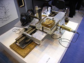
In radiography, X-ray microtomography uses X-rays to create cross-sections of a physical object that can be used to recreate a virtual model without destroying the original object. It is similar to tomography and X-ray computed tomography. The prefix micro- is used to indicate that the pixel sizes of the cross-sections are in the micrometre range. These pixel sizes have also resulted in creation of it's synonyms high-resolution X-ray tomography, micro-computed tomography, and similar terms. Sometimes the terms high-resolution computed tomography (HRCT) and micro-CT are differentiated, but in other cases the term high-resolution micro-CT is used. Virtually all tomography today is computed tomography.
Nanotomography, much like its related modalities tomography and microtomography, uses x-rays to create cross-sections from a 3D-object that later can be used to recreate a virtual model without destroying the original model, applying Nondestructive testing. The term nano is used to indicate that the pixel sizes of the cross-sections are in the nanometer range
The Texas Advanced Computing Center (TACC) at the University of Texas at Austin, United States, is an advanced computing research center that is based on comprehensive advanced computing resources and supports services to researchers in Texas and across the US. The mission of TACC is to enable discoveries that advance science and society through the application of advanced computing technologies. Specializing in high performance computing, scientific visualization, data analysis & storage systems, software, research & development and portal interfaces, TACC deploys and operates advanced computational infrastructure to enable the research activities of faculty, staff, and students of UT Austin. TACC also provides consulting, technical documentation, and training to support researchers who use these resources. TACC staff members conduct research and development in applications and algorithms, computing systems design/architecture, and programming tools and environments.
Quantitative computed tomography (QCT) is a medical technique that measures bone mineral density (BMD) using a standard X-ray Computed Tomography (CT) scanner with a calibration standard to convert Hounsfield Units (HU) of the CT image to bone mineral density values. Quantitative CT scans are primarily used to evaluate bone mineral density at the lumbar spine and hip.
The Jackson School of Geosciences at The University of Texas at Austin unites the Department of Geological Sciences with two research units, the Institute for Geophysics and the Bureau of Economic Geology.
Willi A. Kalender is a German medical physicist and professor and former chairman of the Institute of Medical Physics of the University of Erlangen-Nuremberg. Kalender has produced several new technologies in the field of diagnostic radiology imaging.
Bernard Marshall Gordon is an American engineer, inventor, entrepreneur, and philanthropist. He is considered "the father of high-speed analog-to-digital conversion".
High-resolution computed tomography (HRCT) is a type of computed tomography (CT) with specific techniques to enhance image resolution. It is used in the diagnosis of various health problems, though most commonly for lung disease, by assessing the lung parenchyma. On the other hand, HRCT of the temporal bone is used to diagnose various middle ear diseases such as otitis media, cholesteatoma, and evaluations after ear operations.
Positron emission tomography–computed tomography is a nuclear medicine technique which combines, in a single gantry, a positron emission tomography (PET) scanner and an x-ray computed tomography (CT) scanner, to acquire sequential images from both devices in the same session, which are combined into a single superposed (co-registered) image. Thus, functional imaging obtained by PET, which depicts the spatial distribution of metabolic or biochemical activity in the body can be more precisely aligned or correlated with anatomic imaging obtained by CT scanning. Two- and three-dimensional image reconstruction may be rendered as a function of a common software and control system.
Neutron tomography is a form of computed tomography involving the production of three-dimensional images by the detection of the absorbance of neutrons produced by a neutron source. It creates a three-dimensional image of an object by combining multiple planar images with a known separation. It has a resolution of down to 25 μm. Whilst its resolution is lower than that of X-ray tomography, it can be useful for specimens containing low contrast between the matrix and object of interest; for instance, fossils with a high carbon content, such as plants or vertebrate remains.
Cone beam computed tomography is a medical imaging technique consisting of X-ray computed tomography where the X-rays are divergent, forming a cone.
Coronary CT angiography is the use of computed tomography (CT) angiography to assess the coronary arteries of the heart. The patient receives an intravenous injection of radiocontrast and then the heart is scanned using a high speed CT scanner, allowing physicians to assess the extent of occlusion in the coronary arteries, usually in order to diagnose coronary artery disease.
Gustafsonia is an extinct genus of carnivoran belonging to the family Amphicyonidae. The type species, Gustafsonia cognita, was described in 1986 by Eric Paul Gustafson, who originally interpreted it as a miacid and named it Miacis cognitus. It was subsequently considered to be the only species of the diverse genus Miacis that belonged to the crown-group Carnivora, within the Caniformia, and it was ultimately assigned to the family Amphicyonidae. The type specimen or holotype was discovered in Reeve's bonebed, western Texas, in the Chambers Tuff Formation in 1986. The University of Texas holds this specimen. It is the only confirmed fossil of this species.
Cardiac imaging refers to minimally invasive imaging of the heart using ultrasound, magnetic resonance imaging (MRI), computed tomography (CT), or nuclear medicine (NM) imaging with PET or SPECT. These cardiac techniques are otherwise referred to as echocardiography, Cardiac MRI, Cardiac CT, Cardiac PET and Cardiac SPECT including myocardial perfusion imaging.
A digital autopsy is a non-invasive autopsy in which digital imaging technology, such as with Computerized Tomography (CT) or Magnetic Resonance Imaging (MRI) scans, is used to develop three-dimensional images for a virtual exploration of a human body.
X-ray computed tomography operates by using an X-ray generator that rotates around the object; X-ray detectors are positioned on the opposite side of the circle from the X-ray source.
The history of X-ray computed tomography dates back to at least 1917 with the mathematical theory of the Radon transform In October 1963, William H. Oldendorf received a U.S. patent for a "radiant energy apparatus for investigating selected areas of interior objects obscured by dense material". The first clinical CT scan was performed in 1971 using a scanner invented by Sir Godfrey Hounsfield.
This page is based on this Wikipedia article Text is available under the CC BY-SA 4.0 license; additional terms may apply. Images, videos and audio are available under their respective licenses.